Abstract
In Saccharomyces cerevisiae, the metaphase-anaphase transition is initiated by the anaphase-promoting complex-dependent degradation of Pds1, whereby Esp1 is activated to promote sister chromatid separation. Although this is a fundamental step in the cell cycle, little is known about the regulation of Esp1 and how loss of cohesion is coordinated with movement of the anaphase spindle. Here, we show that Esp1 has a novel role in promoting anaphase spindle elongation. The localization of Esp1 to the spindle apparatus, analyzed by live cell imaging, is regulated in a manner consistent with a function during anaphase B. The protein accumulates in the nucleus in G2 and is mobilized onto the spindle pole bodies and spindle midzone at anaphase onset, where it persists into midanaphase. Association with Pds1 occurs during S phase and is required for efficient nuclear targeting of Esp1. Spindle association is not fully restored in pds1 mutants expressing an Esp1-nuclear localization sequence fusion protein, suggesting that Pds1 is also required to promote Esp1 spindle binding. In agreement, Pds1 interacts with the spindle at the metaphase-anaphase transition and a fraction remains at the spindle pole bodies and the spindle midzone in anaphase cells. Finally, mutational analysis reveals that the conserved COOH-terminal region of Esp1 is important for spindle interaction.
🔬 Techniques
🧬 Organisms
✨ Fluorophores
🧪 Sample Preparation
🏭 Microscope Brands
🧪 Reagent Suppliers
📷 Detectors
🏛️ Research Organizations (ROR)
Affiliated research institutions:
📋 Methods
Yeast Strains and Methods
The relevant genotypes of the yeast strains used in this study are listed in Table . All strains are isogenic derivatives of BF264-15 15DU: a ura3 Δns ade1 his2 leu2-3,112 trp1-1 a ( Richardson et al. 1989 ). Yeast media and genetic procedures were performed according to Guthrie and Fink 1991 . Gene disruptions were performed by PCR-targeting technique ( Wach et al. 1994 ).
Induction of integrated
ESP1GFP from the GAL1 promoter was performed as follows. Approximately 10 7 cells of a YEPRaffinose culture was filtered onto 47-mm 0.45 μm GN-6 Metricel membrane (Gelman Sciences). Filters were placed on YEPGalactose plates for 15–30 min at room temperature. Cells were eluted into 1 M sorbitol and Esp1GFP fluorescence visualized by microscopy. For induction of Esp1GFP in time-course experiments, cells were diluted in YEPRaffinose to an OD 600 = 0.15. α-factor (200 ng/ml) was added and cells were incubated at 30°C for 1 h, 2% galactose was added, and incubation was continued for 1 h. Cells were released in YEPDextrose at 30°C and samples were collected for analysis of cell-cycle progression by DAPI and protein fluorescence by microscopy. For loss of cohesion assays, the region adjacent to the centromere of chromosome IV was visualized by using the binding of tetR-GFP fusion proteins expressed from the CUP1 promoter (by addition of 0.25 mM CuSO 4 to the growth medium) to tandemly integrated tetO sequences at the TRP1 locus ( Clarke et al. 1999 ).
Plasmids
ESP1-pRSG is an integrative plasmid carrying the ESP1 open reading frame in addition to 200 bases of the 5′ flanking sequence under the control of the GAL1 promoter. ESP1GFP-pRSG is derived from this plasmid by inserting a PCR-generated sequence encoding the GFP epitope (F64L, S65T, Q80R mutant) into a SmaI site introduced just before the stop codon. All pRSG-derived plasmids are linearized with StuI and integrants isolated by selecting for growth on dex-ura media. The parental pRSG plasmid is a derivative of pRS406 ( Sikorski and Hieter 1989 ), where the NaeI-PvuI fragment spanning the multiple cloning site (MCS) linker has been substituted by the NaeI-PvuI fragment from pYES2 (Invitrogen) containing a MCS and the GAL1 promoter. The ESP1myc18-pTRP1 plasmid was used to tag endogenous Esp1 protein with 18 myc epitopes at the COOH terminus. The integrative pTRP1 plasmid carries the sequence encoding six myc epitopes, which can be fused to a protein of interest at the NotI site ( Mondesert et al. 1997 ). A SalI-NotI fragment spanning the last 480 bases of the ESP1 gene was cloned into this plasmid. An additional fragment encoding 12 myc epitopes was subsequently introduced into the NotI site generating the final ESP1myc18-pTRP1 construct. The plasmid was linearized with XbaI and integrants isolated by selecting for growth on dex-trp. The vector pOC78 ( Cohen-Fix et al. 1996 ) was employed for tagging endogenous Pds1 internally with three hemagglutinin (HA) epitopes. To tag the Pds1-128 protein in a similar fashion, the XbaI-AvrII fragment of pds1-128 YEp24 constituting the 3′ end of the gene with the mutation was cloned into the XbaI/AvrII sites of a pBSII-derived plasmid containing the SacI-ApaI Pds1(HA)3 fragment from pOC78. The AvrII-ApaI pds1-128 (HA)3 fragment was used for transformation, Leu + colonies were isolated, and the presence of HA epitopes was verified by PCR. The integrative pGAL1-Pds1Δdb plasmid described previously ( Kaiser et al. 1999 ) was used to construct the strain expressing the nondegradable version of Pds1. The construct expressing galactose-inducible Scc1 was generated by cloning the SCC1 ORF into YIplac128(LEU2)GAL1 using BamHI and XhoI. The CFP-TUB1 plasmid was constructed by replacing the GFP encoding XhoI-EcoRI fragment in pAFS91 ( Straight et al.., 1997 ) with a PCR-generated cyan fluorescent protein (CFP) fragment cut with XhoI and EcoRI. The resultant CFP-Tub1 fusion was integrated at the URA3 locus after StuI digestion. A panel of esp1 ts alleles was generated by error-prone PCR followed by in vivo gap repair as described previously ( Tang and Reed 1993 ). PCR mutagenesis was performed on separate ESP1 fragments to isolate temperature-sensitive alleles mapping to the NH 2 -terminal, central, and COOH-terminal region of Esp1. All esp1 ts alleles exhibited similar phenotypes. Alleles esp1-N5 and esp1-B3 have restrictive temperatures of 35° and 30°C, respectively. Due to the lower temperature, the esp1-B3 mutant is more suitable for kinetic experiments. The integrative pKGFP plasmid, which carries the KAN R marker for G418 resistance was used to fuse the endogenous Esp1 protein to GFP. pKGFP was designed with the GFP-encoding sequence inserted after NotI, allowing in frame fusion to any protein of interest. The ESP1 SalI-NotI fragment used to construct ESP1myc18-pTRP1 was inserted in pKGFP cut with SalI and NotI. The plasmid was linearized with XbaI and integrants were isolated by selecting for G418 resistance. pKGFP2 was used to tag endogenous Pds1 protein at the COOH terminus. This plasmid carries a GFP sequence with additional mutations: V163A and S175G, which produces a brighter GFP fluorescent signal. To tag Pds1, a PCR-derived SalI-NotI fragment covering the last 500 bases of the PDS1 gene was introduced into pKGFP2 at SalI-NotI. The Pds1-pKGFP2 plasmid was linearized with StuI and integrants selected by growth on G418 plates. Plasmids ESP1(1-1568)-pRSG, ESP1(D1568A)-pRSG, and ESP1 (D1568A/D1570A)-pRSG were used to produce strains expressing the COOH-terminal truncation mutant and mutants in the putative calcium-binding site of Esp1 from the GAL1 promoter, respectively. To generate ESP1(1-1568)-pRSG, a SalI-SmaI PCR fragment spanning the region from the internal SalI site in ESP1 to the sequence encoding residue 1568 was introduced into the ESP1-pRSG plasmid digested with SalI and SmaI, thereby replacing the 3′ region of the ESP1 gene. Site-directed PCR was employed to make ESP1(D1568A)-pRSG. Primers 5′ CCGAACGGAGATTTGTCC 3′ and 5′ GCTAAATTTATCG*ATATCTTTG G CAGTTAC-ATCCC 3′ were used to generate a mutated fragment with a single amino acid substitution (bold) and a silent mutation generating an EcoRV restriction site (underlined). This PCR product was used in a second reaction with primer 5′ GACGAGATCTTTA CCCGGG TGAT-ACGAACTTGATCGG 3′, where the SmaI site has been underlined. The cut fragment was introduced into the ESP1-pRSG vector digested with SalI and SmaI to remove the corresponding wild-type fragment. The double mutant ESP1(D1568A/D1570A)-pRSG was created by a similar PCR scheme using the following primers: 5′ CCGAACGGAGATTTGTCC3′, 5′ GCTAAATTTATCG*ATAG*CTTTGGCAGTT-ACATCCC 3′ (the modified EcoRV site is underlined and the mutation resulting in the second amino acid substitution shown in bold), and 5′GACGAGATCTTTA- CCCGGG TGATACGAACTTGATCGG 3′ (SmaI site is underlined). The cut fragment was introduced into the cut ESP1(D1568A)-pRSG vector to restore the ESP1 gene to its full length. Clones carrying the double mutation were selected by screening for loss of the EcoRV site. GFP-tagged versions of the constructs were made by inserting a SmaI fragment encoding the GFP epitope into the respective ESP1-pRSG plasmids opened by SmaI. The integrative plasmid ESP1GFPNLS-pRSG was used to make strains expressing Esp1GFP fused at the COOH terminus to the SV40 NLS from the GAL1 promoter. A PCR fragment produced with following primers: 5′ CTAG CCCGGG AAGAAAAAGCGAAAGGTCG-GCCGCATGAGTAAAG 3′ and 5′ GCTA CCCGGG GACCTTTCGCTTCTTCTTGGG TTTGTATAGTTCATCCATGC 3′, where SmaI sites are underlined and the SV40 NLS sequence is shown in bold, was inserted into the ESP1-pRSG plasmid digested with SmaI. Plasmid ESP1-pRS415 was made by inserting the ESP1 promoter sequence and ORF into the ARS/CEN plasmid pRS415 digested with SalI and SacI ( Sikorski and Hieter 1989 ). To generate ESP1(1-1568)-pRS415, the SpeI fragment from ESP1(1-1568)-pRSG containing the 3′ end of the ESP1 gene was introduced into the ESP1-pRS415 plasmid cut with SpeI. The ESP1 calcium-binding site mutants were cloned into pRS415 by substituting the NdeI fragment derived from ESP1(D1568A)- and ESP1(D1568A/D1570A)-pRSG with the corresponding fragment in ESP1-pRS415. All plasmids were subjected to sequencing to verify their integrity.
Show full methods section
Yeast Strains and Methods
The relevant genotypes of the yeast strains used in this study are listed in Table . All strains are isogenic derivatives of BF264-15 15DU: a ura3 Δns ade1 his2 leu2-3,112 trp1-1 a ( Richardson et al. 1989 ). Yeast media and genetic procedures were performed according to Guthrie and Fink 1991 . Gene disruptions were performed by PCR-targeting technique ( Wach et al. 1994 ).
Induction of integrated
ESP1GFP from the GAL1 promoter was performed as follows. Approximately 10 7 cells of a YEPRaffinose culture was filtered onto 47-mm 0.45 μm GN-6 Metricel membrane (Gelman Sciences). Filters were placed on YEPGalactose plates for 15–30 min at room temperature. Cells were eluted into 1 M sorbitol and Esp1GFP fluorescence visualized by microscopy. For induction of Esp1GFP in time-course experiments, cells were diluted in YEPRaffinose to an OD 600 = 0.15. α-factor (200 ng/ml) was added and cells were incubated at 30°C for 1 h, 2% galactose was added, and incubation was continued for 1 h. Cells were released in YEPDextrose at 30°C and samples were collected for analysis of cell-cycle progression by DAPI and protein fluorescence by microscopy. For loss of cohesion assays, the region adjacent to the centromere of chromosome IV was visualized by using the binding of tetR-GFP fusion proteins expressed from the CUP1 promoter (by addition of 0.25 mM CuSO 4 to the growth medium) to tandemly integrated tetO sequences at the TRP1 locus ( Clarke et al. 1999 ).
Plasmids
ESP1-pRSG is an integrative plasmid carrying the ESP1 open reading frame in addition to 200 bases of the 5′ flanking sequence under the control of the GAL1 promoter. ESP1GFP-pRSG is derived from this plasmid by inserting a PCR-generated sequence encoding the GFP epitope (F64L, S65T, Q80R mutant) into a SmaI site introduced just before the stop codon. All pRSG-derived plasmids are linearized with StuI and integrants isolated by selecting for growth on dex-ura media. The parental pRSG plasmid is a derivative of pRS406 ( Sikorski and Hieter 1989 ), where the NaeI-PvuI fragment spanning the multiple cloning site (MCS) linker has been substituted by the NaeI-PvuI fragment from pYES2 (Invitrogen) containing a MCS and the GAL1 promoter. The ESP1myc18-pTRP1 plasmid was used to tag endogenous Esp1 protein with 18 myc epitopes at the COOH terminus. The integrative pTRP1 plasmid carries the sequence encoding six myc epitopes, which can be fused to a protein of interest at the NotI site ( Mondesert et al. 1997 ). A SalI-NotI fragment spanning the last 480 bases of the ESP1 gene was cloned into this plasmid. An additional fragment encoding 12 myc epitopes was subsequently introduced into the NotI site generating the final ESP1myc18-pTRP1 construct. The plasmid was linearized with XbaI and integrants isolated by selecting for growth on dex-trp. The vector pOC78 ( Cohen-Fix et al. 1996 ) was employed for tagging endogenous Pds1 internally with three hemagglutinin (HA) epitopes. To tag the Pds1-128 protein in a similar fashion, the XbaI-AvrII fragment of pds1-128 YEp24 constituting the 3′ end of the gene with the mutation was cloned into the XbaI/AvrII sites of a pBSII-derived plasmid containing the SacI-ApaI Pds1(HA)3 fragment from pOC78. The AvrII-ApaI pds1-128 (HA)3 fragment was used for transformation, Leu + colonies were isolated, and the presence of HA epitopes was verified by PCR. The integrative pGAL1-Pds1Δdb plasmid described previously ( Kaiser et al. 1999 ) was used to construct the strain expressing the nondegradable version of Pds1. The construct expressing galactose-inducible Scc1 was generated by cloning the SCC1 ORF into YIplac128(LEU2)GAL1 using BamHI and XhoI. The CFP-TUB1 plasmid was constructed by replacing the GFP encoding XhoI-EcoRI fragment in pAFS91 ( Straight et al.., 1997 ) with a PCR-generated cyan fluorescent protein (CFP) fragment cut with XhoI and EcoRI. The resultant CFP-Tub1 fusion was integrated at the URA3 locus after StuI digestion. A panel of esp1 ts alleles was generated by error-prone PCR followed by in vivo gap repair as described previously ( Tang and Reed 1993 ). PCR mutagenesis was performed on separate ESP1 fragments to isolate temperature-sensitive alleles mapping to the NH 2 -terminal, central, and COOH-terminal region of Esp1. All esp1 ts alleles exhibited similar phenotypes. Alleles esp1-N5 and esp1-B3 have restrictive temperatures of 35° and 30°C, respectively. Due to the lower temperature, the esp1-B3 mutant is more suitable for kinetic experiments. The integrative pKGFP plasmid, which carries the KAN R marker for G418 resistance was used to fuse the endogenous Esp1 protein to GFP. pKGFP was designed with the GFP-encoding sequence inserted after NotI, allowing in frame fusion to any protein of interest. The ESP1 SalI-NotI fragment used to construct ESP1myc18-pTRP1 was inserted in pKGFP cut with SalI and NotI. The plasmid was linearized with XbaI and integrants were isolated by selecting for G418 resistance. pKGFP2 was used to tag endogenous Pds1 protein at the COOH terminus. This plasmid carries a GFP sequence with additional mutations: V163A and S175G, which produces a brighter GFP fluorescent signal. To tag Pds1, a PCR-derived SalI-NotI fragment covering the last 500 bases of the PDS1 gene was introduced into pKGFP2 at SalI-NotI. The Pds1-pKGFP2 plasmid was linearized with StuI and integrants selected by growth on G418 plates. Plasmids ESP1(1-1568)-pRSG, ESP1(D1568A)-pRSG, and ESP1 (D1568A/D1570A)-pRSG were used to produce strains expressing the COOH-terminal truncation mutant and mutants in the putative calcium-binding site of Esp1 from the GAL1 promoter, respectively. To generate ESP1(1-1568)-pRSG, a SalI-SmaI PCR fragment spanning the region from the internal SalI site in ESP1 to the sequence encoding residue 1568 was introduced into the ESP1-pRSG plasmid digested with SalI and SmaI, thereby replacing the 3′ region of the ESP1 gene. Site-directed PCR was employed to make ESP1(D1568A)-pRSG. Primers 5′ CCGAACGGAGATTTGTCC 3′ and 5′ GCTAAATTTATCG*ATATCTTTG G CAGTTAC-ATCCC 3′ were used to generate a mutated fragment with a single amino acid substitution (bold) and a silent mutation generating an EcoRV restriction site (underlined). This PCR product was used in a second reaction with primer 5′ GACGAGATCTTTA CCCGGG TGAT-ACGAACTTGATCGG 3′, where the SmaI site has been underlined. The cut fragment was introduced into the ESP1-pRSG vector digested with SalI and SmaI to remove the corresponding wild-type fragment. The double mutant ESP1(D1568A/D1570A)-pRSG was created by a similar PCR scheme using the following primers: 5′ CCGAACGGAGATTTGTCC3′, 5′ GCTAAATTTATCG*ATAG*CTTTGGCAGTT-ACATCCC 3′ (the modified EcoRV site is underlined and the mutation resulting in the second amino acid substitution shown in bold), and 5′GACGAGATCTTTA- CCCGGG TGATACGAACTTGATCGG 3′ (SmaI site is underlined). The cut fragment was introduced into the cut ESP1(D1568A)-pRSG vector to restore the ESP1 gene to its full length. Clones carrying the double mutation were selected by screening for loss of the EcoRV site. GFP-tagged versions of the constructs were made by inserting a SmaI fragment encoding the GFP epitope into the respective ESP1-pRSG plasmids opened by SmaI. The integrative plasmid ESP1GFPNLS-pRSG was used to make strains expressing Esp1GFP fused at the COOH terminus to the SV40 NLS from the GAL1 promoter. A PCR fragment produced with following primers: 5′ CTAG CCCGGG AAGAAAAAGCGAAAGGTCG-GCCGCATGAGTAAAG 3′ and 5′ GCTA CCCGGG GACCTTTCGCTTCTTCTTGGG TTTGTATAGTTCATCCATGC 3′, where SmaI sites are underlined and the SV40 NLS sequence is shown in bold, was inserted into the ESP1-pRSG plasmid digested with SmaI. Plasmid ESP1-pRS415 was made by inserting the ESP1 promoter sequence and ORF into the ARS/CEN plasmid pRS415 digested with SalI and SacI ( Sikorski and Hieter 1989 ). To generate ESP1(1-1568)-pRS415, the SpeI fragment from ESP1(1-1568)-pRSG containing the 3′ end of the ESP1 gene was introduced into the ESP1-pRS415 plasmid cut with SpeI. The ESP1 calcium-binding site mutants were cloned into pRS415 by substituting the NdeI fragment derived from ESP1(D1568A)- and ESP1(D1568A/D1570A)-pRSG with the corresponding fragment in ESP1-pRS415. All plasmids were subjected to sequencing to verify their integrity.
Cell Biology Protocols Fluorescence and differential interference contrast
(DIC) microscopy was performed using an Eclipse E800 microscope (Nikon) with a 100× objective. Cell images were captured with a Quantix CCD (Photometrics) camera using IPlab Spectrum software (Signal Analytics Co.). Spindle measurements were performed on captured images using the NIH Image measuring tool calibrated with a stage micrometer. For microscopy of live cells expressing either wild-type/mutant Esp1GFP or Pds1GFP, cells were grown in YEPRaffinose/YEPGalactose or YEPDextrose, respectively, containing extra supplement of adenine (0.2 mg/ml). Images were acquired using 500-ms exposures. For simultaneous detection of GFP- and CFP-labeled proteins, images were captured using a Photometrics CH350L CCD camera on an Olympus IX70 inverted microscope with a 100× magnification. Images were taken of a single focal plane and later manipulated using SoftWoRx software (Applied Precision Inc.). Cross bleeding of GFP and CFP signals did not occur as there was no CFP signal in the GFP channel and vice-versa in singly tagged strains. Nuclei were visualized with DAPI as described previously ( Mondesert et al. 1997 ). Cell cultures were analyzed for DNA content using flow cytometry as described previously (Mondesert et al.. 1997).
Immunoprecipitation and Immunostaining
Protein isolation was essentially as previously described ( Kaiser et al. 1999 ). Cells were broken by glass beads in NP-40 buffer (50 mM Tris-HCl, pH 7.5, 150 mM NaCl, 0.1% NP-40, 10 mM sodium pyrophosphate, 5 mM EDTA, 5 mM EGTA, 0.1 mM orthovanadate, 1 mM PMSF, 2 mg/ml aprotinin, leupeptin, and pepstatin A). A total of 750 μg of extract was incubated with myc9E10 antibody prebound to protein A sepharose or with 12CA5 antibody cross linked to protein A sepharose for 2 h, and immunocomplexes were washed four times with 1 ml of extraction buffer. Bound proteins were eluted by boiling in 2× SDS sample buffer, separated by SDS-PAGE (7.5% protein gels), and analyzed by immunostaining with anti–HA antibody (12CA5, BabCO), anti–myc antibody (9E10) and anti–GFP antibody (CLONTECH Laboratories, Inc.). Extracts prepared solely for immunostaining were separated on 8.5% SDS-polyacrylamide gels. Cdc28 protein serving as a loading control was recognized by the anti–PSTAIRE antibody.
Yeast Strains and Methods
The relevant genotypes of the yeast strains used in this study are listed in Table . All strains are isogenic derivatives of BF264-15 15DU: a ura3 Δns ade1 his2 leu2-3,112 trp1-1 a ( Richardson et al. 1989 ). Yeast media and genetic procedures were performed according to Guthrie and Fink 1991 . Gene disruptions were performed by PCR-targeting technique ( Wach et al. 1994 ).
Induction of integrated
ESP1GFP from the GAL1 promoter was performed as follows. Approximately 10 7 cells of a YEPRaffinose culture was filtered onto 47-mm 0.45 μm GN-6 Metricel membrane (Gelman Sciences). Filters were placed on YEPGalactose plates for 15–30 min at room temperature. Cells were eluted into 1 M sorbitol and Esp1GFP fluorescence visualized by microscopy. For induction of Esp1GFP in time-course experiments, cells were diluted in YEPRaffinose to an OD 600 = 0.15. α-factor (200 ng/ml) was added and cells were incubated at 30°C for 1 h, 2% galactose was added, and incubation was continued for 1 h. Cells were released in YEPDextrose at 30°C and samples were collected for analysis of cell-cycle progression by DAPI and protein fluorescence by microscopy. For loss of cohesion assays, the region adjacent to the centromere of chromosome IV was visualized by using the binding of tetR-GFP fusion proteins expressed from the CUP1 promoter (by addition of 0.25 mM CuSO 4 to the growth medium) to tandemly integrated tetO sequences at the TRP1 locus ( Clarke et al. 1999 ).
Cell Biology Protocols Fluorescence and differential interference contrast
(DIC) microscopy was performed using an Eclipse E800 microscope (Nikon) with a 100× objective. Cell images were captured with a Quantix CCD (Photometrics) camera using IPlab Spectrum software (Signal Analytics Co.). Spindle measurements were performed on captured images using the NIH Image measuring tool calibrated with a stage micrometer. For microscopy of live cells expressing either wild-type/mutant Esp1GFP or Pds1GFP, cells were grown in YEPRaffinose/YEPGalactose or YEPDextrose, respectively, containing extra supplement of adenine (0.2 mg/ml). Images were acquired using 500-ms exposures. For simultaneous detection of GFP- and CFP-labeled proteins, images were captured using a Photometrics CH350L CCD camera on an Olympus IX70 inverted microscope with a 100× magnification. Images were taken of a single focal plane and later manipulated using SoftWoRx software (Applied Precision Inc.). Cross bleeding of GFP and CFP signals did not occur as there was no CFP signal in the GFP channel and vice-versa in singly tagged strains. Nuclei were visualized with DAPI as described previously ( Mondesert et al. 1997 ). Cell cultures were analyzed for DNA content using flow cytometry as described previously (Mondesert et al.. 1997).
📊 Figures
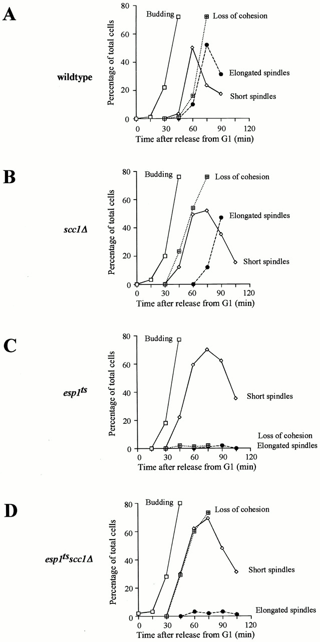
Figure 3
Esp1 is essential for spindle elongation. Cells of wild-type (A), scc1 u0394 (SY118) (B), esp1-B3 (SY119) (C), and scc1 u0394 esp1-B3 (SY120) (D) strains containing CFP-tubulin and GFP-labeled centrom...
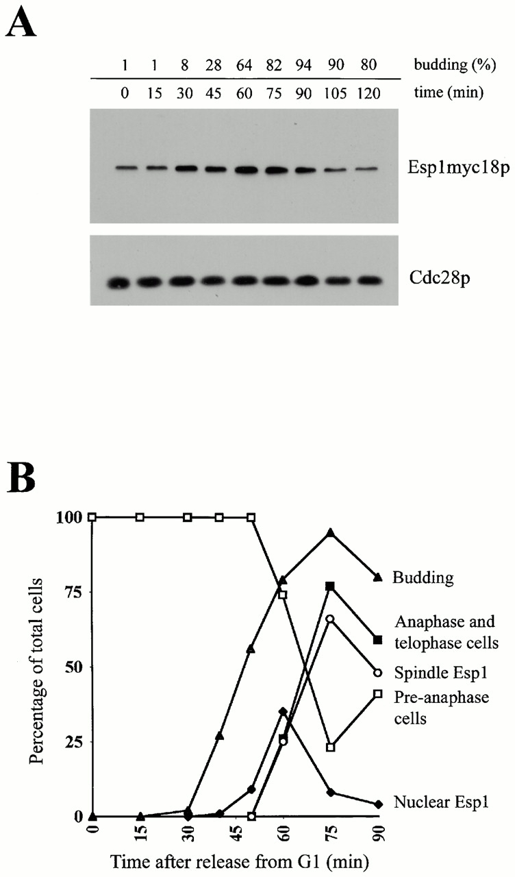
Figure 1
Cell cycleu2013dependent regulation of Esp1. (A) Esp1 protein level during the cell cycle. Strain carrying epitope-tagged Esp1 integrated at the chromosomal locus (SY108) was arrested in G1 with u03b1...
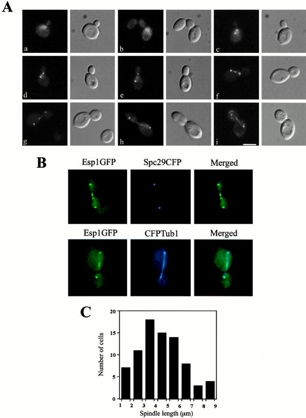
Figure 2
Localization of Esp1GFP at different stages of the cell cycle. (A) Diploid cells carrying an integrated ESP1GFP allele (SY203) grown in YEPRaffinose were collected on a nitrocellulose filter and induc...
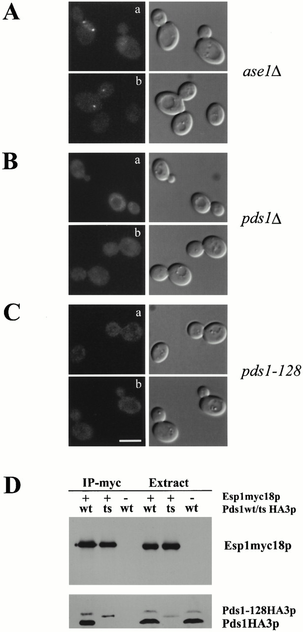
Figure 4
Proper Esp1 localization depends on Pds1. (A, a and b) Esp1 signal at the spindle midzone is absent in ase1 u0394 mutant. A diploid ase1 u0394 strain carrying GAL1 -inducible ESP1 GFP (SY204) was exam...
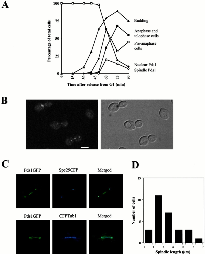
Figure 6
Pds1 associates with the mitotic spindle apparatus. (A) A strain carrying PDS1 fused at the COOH-terminal end to GFP integrated at the chromosomal locus (SY116) was arrested in G1 with u03b1-factor. A...
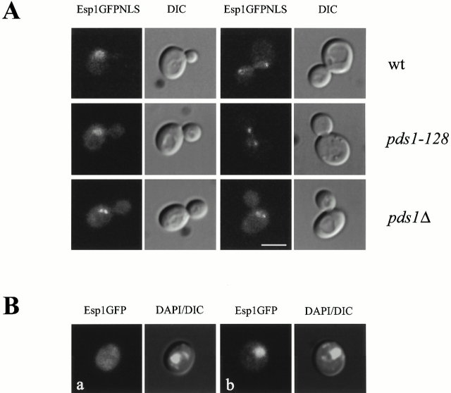
Figure 5
Pds1 is required to load Esp1 onto the spindle. (A) A version of Esp1GFP fused at its COOH terminus to the SV40 NLS was integrated into a wild-type strain (SY105, top), a pds1-128 mutant (SY107, middl...
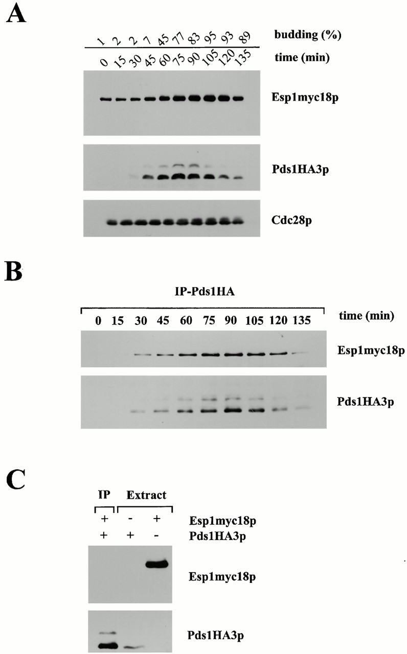
Figure 7
Complex formation between Esp1 and Pds1. (A) A strain expressing endogenous myc18-tagged Esp1 and HA3-tagged Pds1 (SY109) was arrested in G1 by u03b1-factor. After release into YEPDextrose at 22u00b0C...
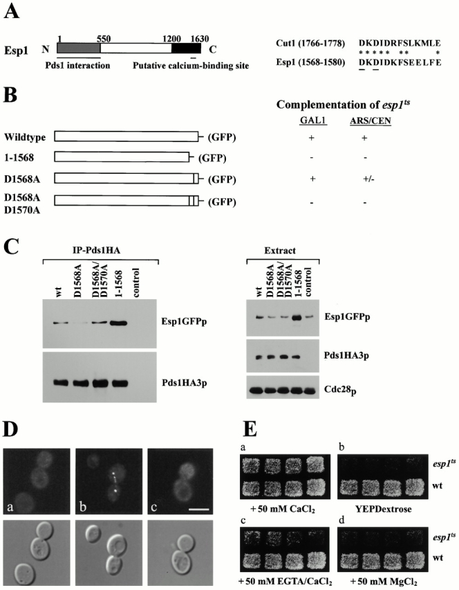
Figure 8
The role of the putative calcium-binding site in the conserved COOH-terminal domain of Esp1. (A) A schematic representation of the domain structure of Esp1. The NH 2 -terminal domain constituting 1/3 ...
Figure images are served from the NIH/NLM PubMed Central Open Access Subset or Europe PMC; copyright remains with the publishers and authors.
💬 Discussion
0 commentsNo comments yet. Be the first to start a discussion!
Leave a Comment