Abstract
The ClpXP degradation machine consists of a hexameric AAA+ unfoldase (ClpX) and a pair of heptameric serine protease rings (ClpP) that unfold, translocate, and subsequently degrade client proteins. ClpXP is an important target for drug development against infectious diseases. Although structures are available for isolated ClpX and ClpP rings, it remains unknown how symmetry mismatched ClpX and ClpP work in tandem for processive substrate translocation into the ClpP proteolytic chamber. Here, we present cryo-EM structures of the substrate-bound ClpXP complex from Neisseria meningitidis at 2.3 to 3.3 Å resolution. The structures allow development of a model in which the sequential hydrolysis of ATP is coupled to motions of ClpX loops that lead to directional substrate translocation and ClpX rotation relative to ClpP. Our data add to the growing body of evidence that AAA+ molecular machines generate translocating forces by a common mechanism.
🔬 Techniques
🔭 Microscopes
🧬 Organisms
💻 Software
✨ Fluorophores
🔬 Cell Lines
🏭 Microscope Brands
🧪 Reagent Suppliers
📷 Detectors
💻 Software Details
💾 Data Repositories
🏷️ Research Resource Identifiers (RRIDs)
Verified research resources used in this paper:
🏛️ Research Organizations (ROR)
Affiliated research institutions:
📋 Methods
Key resources table
Reagent type (species) or resource Designation Source or reference Identifiers Additional information Gene ( Neisseria meningitidis ) clpX UniProtKB - Q9JYY3 Gene ( Neisseria meningitidis ) clpP UniProtKB - Q9JZ38 Strain, strain background ( Escherichia coli ) BL21(DE3) Sigma-Aldrich CMC0016 Chemically competent cells Recombinant DNA reagent pET28-NmClpP Synthetic (GenScript) UniProtKB - Q9JYY3 Plasmid containing ClpP Recombinant DNA reagent pET28a-NmClpX Synthetic (GenScript) UniProtKB - Q9JZ38 Plasmid containing ClpX Recombinant DNA reagent pET28a- GFP-SsrA ( Ripstein et al., 2017 ) Plasmid containing GFP-SsrA Chemical compound, drug PKM-AMC GenScript Fluorogenic peptide for protease assays Software, algorithm EPU Thermo Fischer Scientific EM imaging software Software, algorithm cryoSPARC v2 ( Punjani et al., 2017 ) RRID: SCR_016501 EM reconstruction software Software, algorithm UCSF Chimera ( Pettersen et al., 2004 ) RRID: SCR_004097 Molecular Visualization Software Software, algorithm UCSF ChimeraX ( Goddard et al., 2018 ) RRID: SCR_015872 Molecular Visualization Software Software, algorithm Coot ( Emsley and Cowtan, 2004 ) RRID: SCR_014222 Protein Model Building Software Software, algorithm Phyre2 ( Kelley et al., 2015 ) RRID: SCR_010270 Protein Model Building Software Software, algorithm Rosetta ( Wang et al., 2015 ) RRID: SCR_015701 Protein Model Building Software Software, algorithm Phenix ( Adams et al., 2010 ) RRID: SCR_014224 Protein Model Building Software Software, algorithm Molprobity ( Arendall III et al., 2010 ) RRID: SCR_014226 Protein Model Evaluation Software Software, algorithm EMRinger ( Barad et al., 2015 ) Protein Model Evaluation Software Plasmids and constructs Codon-optimized genes encoding NmClpX (Uniprot entry: Q9JYY3 ) bearing an N-terminal His 6 -TEV affinity tag and ClpP (Uniprot entry: Q9JZ38 ) with an N-terminal His 6 -SUMO tag were synthesized by GenScript (Piscataway, NJ) and cloned into the NdeI and BamHI sites of pET28a+ (Novagen, Madison, WI). Point mutations were introduced with the Quikchange mutagenesis method (Agilent, Santa Clara, CA).
Show full methods section
Key resources table
Reagent type (species) or resource Designation Source or reference Identifiers Additional information Gene ( Neisseria meningitidis ) clpX UniProtKB - Q9JYY3 Gene ( Neisseria meningitidis ) clpP UniProtKB - Q9JZ38 Strain, strain background ( Escherichia coli ) BL21(DE3) Sigma-Aldrich CMC0016 Chemically competent cells Recombinant DNA reagent pET28-NmClpP Synthetic (GenScript) UniProtKB - Q9JYY3 Plasmid containing ClpP Recombinant DNA reagent pET28a-NmClpX Synthetic (GenScript) UniProtKB - Q9JZ38 Plasmid containing ClpX Recombinant DNA reagent pET28a- GFP-SsrA ( Ripstein et al., 2017 ) Plasmid containing GFP-SsrA Chemical compound, drug PKM-AMC GenScript Fluorogenic peptide for protease assays Software, algorithm EPU Thermo Fischer Scientific EM imaging software Software, algorithm cryoSPARC v2 ( Punjani et al., 2017 ) RRID: SCR_016501 EM reconstruction software Software, algorithm UCSF Chimera ( Pettersen et al., 2004 ) RRID: SCR_004097 Molecular Visualization Software Software, algorithm UCSF ChimeraX ( Goddard et al., 2018 ) RRID: SCR_015872 Molecular Visualization Software Software, algorithm Coot ( Emsley and Cowtan, 2004 ) RRID: SCR_014222 Protein Model Building Software Software, algorithm Phyre2 ( Kelley et al., 2015 ) RRID: SCR_010270 Protein Model Building Software Software, algorithm Rosetta ( Wang et al., 2015 ) RRID: SCR_015701 Protein Model Building Software Software, algorithm Phenix ( Adams et al., 2010 ) RRID: SCR_014224 Protein Model Building Software Software, algorithm Molprobity ( Arendall III et al., 2010 ) RRID: SCR_014226 Protein Model Evaluation Software Software, algorithm EMRinger ( Barad et al., 2015 ) Protein Model Evaluation Software Plasmids and constructs Codon-optimized genes encoding NmClpX (Uniprot entry: Q9JYY3 ) bearing an N-terminal His 6 -TEV affinity tag and ClpP (Uniprot entry: Q9JZ38 ) with an N-terminal His 6 -SUMO tag were synthesized by GenScript (Piscataway, NJ) and cloned into the NdeI and BamHI sites of pET28a+ (Novagen, Madison, WI). Point mutations were introduced with the Quikchange mutagenesis method (Agilent, Santa Clara, CA).
Expression and purification of NmClpP and NmClpX Transformed Codon+
E. coli BL21(DE3) cells were grown in LB media at 37°C. Protein over-expression was induced by addition of 0.2 mM IPTG at OD 600 = 1.0 and was allowed to proceed overnight at 18°C. Cells were lysed in buffer containing 50 mM Tris, 300 KCl, 10 mM imidazole, 10% glycerol, pH 7.0 and NmClpP and NmClpX proteins purified by Ni-affinity chromatography [HisTrap HP (GE)] in lysis buffer. Bound proteins were eluted from the Ni column by increasing the imidazole concentration to 500 mM. The affinity tag was removed by the addition of TEV (for ClpX) or Ulp1 (for ClpP) protease followed by dialysis against lysis/wash buffer that included 5 mM DTT. Following a reverse Ni-affinity chromatography step, the flow-through, free from the cleaved tag and other impurities, was concentrated with an Amicon Ultra-15 50K MWCO (Millipore) concentrator and subjected to size exclusion chromatography (SEC) with a Superdex 200 Increase 10/300 (GE) column in SEC buffer (50 mM imidazole, 100 mM KCl, 5 mM DTT, pH 7.0). Fractions corresponding to NmClpX and NmClpP were pooled and stored at 4°C in the SEC buffer until further use. Salts containing magnesium were avoided during the purification of NmClpX to prevent protein aggregation. Protein concentrations were determined in 8 M GdnCl using extinction coefficient values (7450 M −1 cm −1 for ClpP, 8940 M −1 cm −1 for ClpX) determined with ExPASy’s ProtParam ( Gasteiger et al., 2005 ).
Expression and purification of GFP-SsrA Green fluorescent protein
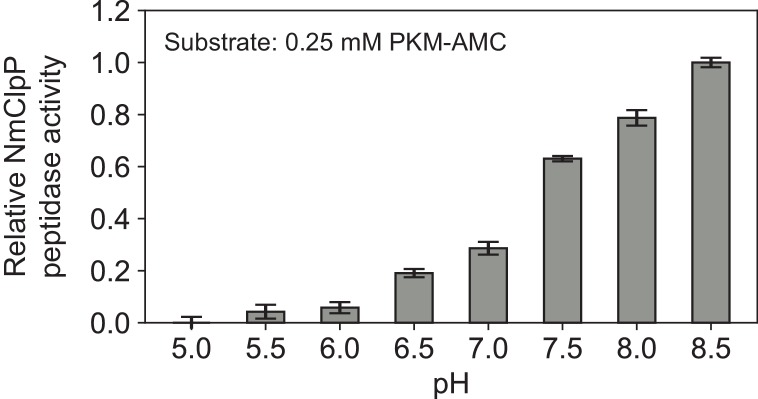
(GFP) bearing an 11-residue SsrA degradation tag at its carboxyl terminus and a non-cleavable N-terminal His × 6 tag was purified by Ni affinity chromatography followed by SEC on a HiLoad 16/60 Superdex 75 pg column (GE). Peptidase rate measurements of NmClpP as a function of pH The peptidase activity of NmClpP was measured at 37°C with Acetyl-L-Pro-L-Lys-L-Met bearing a C-terminal fluorogenic 7-amino-4-methylcoumarin group (PKM-AMC) as substrate. The reaction was followed with a Synergy Neo2 96-well microplate reader making a measurement every 21 s for 60 min at λ ex : 355 nm, λ em : 460 nm. Each well contained 1 μM NmClpP (monomer concentration), 250 μM PKM-AMC, 50 mM citrate, 50 mM phosphate, 50 mM Tris, 100 mM KCl in a total volume of 100 μL adjusted to the appropriate pH. Activities are derived from initial rates extracted and analyzed using a python script written in-house. Standard errors are calculated from repeating each reaction in triplicate.
GFP-SsrA degradation assays
Degradation of 1 μM samples of GFP-SsrA was followed by the loss of GFP fluorescence (λ ex : 480 nm, λ em : 508 nm) with a Synergy Neo2 96-well microplate reader at 25°C. The wells included an ATP-regeneration system ( Nørby, 1988 ) that contained 1.5 mM phosphoenolpyruvate, 0.2 mM NADH, 40 μg/mL pyruvate kinase, 40 μg/mL lactate dehydrogenase, and 2 mM MgATP at pH 8.2. In some assays, solutions also contained WT NmClpX at 0.5 μM (hexamer) and/or WT ClpP at a 0.25 μM (tetradecamer), as indicated in Figure 1B . All assays were performed in triplicate. Preparation of samples for cryo-EM A 1 mL mixture containing 10 μM (tetradecamer) NmClpP and 20 μM NmClpX together with 200 μM GFP-SsrA was incubated with 20 mM MgATP for 10 min at room temperature. This mixture was applied to a Superdex 200 Increase 10/300 (GE) column equilibrated with 50 mM bicine, 100 mM KCl, 2 mM MgATP, pH adjusted to 8.2 at room temperature (equivalent to pH 8.5 at 4°C – see Appendix 1—figure 1 ), as the running buffer. Following SEC, a 0.5 mL fraction (denoted with a * in Figure 1—figure supplement 1 ) containing doubly capped ClpXP bound to GFP-SsrA was supplemented with 20 μM GFP-SsrA and vitrified immediately without the addition of any cross-linking agent or detergent. Sample vitrification 2.5 μL of the sample mixtures were applied to nanofabricated holey gold grids ( Marr et al., 2014 ; Russo and Passmore, 2014 ; Meyerson et al., 2015 ) with a hole size of ~1 μm, that had been glow discharged in air for 15 s. Grids were blotted on both sides using a FEI Vitrobot mark III for 15 s at 4°C and ~100% relative humidity before freezing in a liquid ethane/propane mixture ( Tivol et al., 2008 ).
Electron microscopy
NmClpXP was imaged with a Thermo Fisher Scientific Titan Krios G3 microscope operating at 300 kV and equipped with a FEI Falcon III DDD camera. Structures were calculated from counting mode movies consisting of 30 frames, obtained over a 60 s exposure with defocuses ranging from 0.9 to 1.7 μm. Movies were at a nominal magnification of 75,000 × corresponding to a calibrated pixel size of 1.06 Å and with an exposure of 0.8 electrons/pixel/s, giving a total exposure of 43 electrons/Å 2 . 2680 movies were collected using the microscope’s EPU software. The Apo-NmClpP structure (with no ClpX or GFP present) was calculated from data obtained using a FEI Tecnai F20 electron microscope operating at 200 kV and equipped with a Gatan K2 Summit direct detector device camera. Movies consisting of 30 frames over a 15 s exposure were obtained with defocuses ranging from 1.7 to 2.9 μm. Movies were collected in counting mode at a nominal magnification of 25,000 × corresponding to a calibrated pixel size of 1.45 Å and with an exposure of 5 electrons/pixel/s, and a total exposure of 35 electrons/Å 2 . 122 movies were collected using Digital Micrograph software.
EM image analysis
Patch based whole frame alignment and exposure weighting was performed in cryoSPARC v2 ( Punjani et al., 2017 ) with a 10 × 10 grid and the resulting averages of frames were used for patch based contrast transfer function (CTF) determination. Templates for particle selection were generated by 2D classification of manually selected particles. Particle images were extracted in 300 × 300 pixel boxes for further analysis. Ab inito map calculation was performed on a random subset of 30,000 particle images, generating an initial map showing density for the complex of a ClpP tetradecamer bound to two ClpX hexamers. A single round of 2D classification was used to remove images of damaged particles and other contaminants from a dataset of 466,549 particle images, with selected classes leaving 377,234 particle images for further analysis. Homogeneous refinement of these particle images using D7 symmetry yielded a map of the complex, with good density for only the ClpP portion, at 2.3 Å resolution. To improve the density of the ClpX portion of the map, a round of Ab initio classification was performed using three classes, of which two classes containing 289,144 particle images had good density for ClpX, with the remaining class containing mostly density for ClpP. Refinement of these particle images with C1 symmetry resulted in a map of the ClpXP complex at a nominal resolution of 2.8 Å, but with poorly defined density for much of ClpX. To improve the interpretability of the map in the ClpX region, local refinement was performed with a mask around the six ClpX subunits without performing signal subtraction for ClpP. This refinement greatly improved the map in the ClpX region, while blurring the density at the distal ClpP ring (indicative of flexibility between ClpX and ClpP). However, density for two of the ClpX subunits remained fragmented and at lower resolution. To help resolve the heterogeneity of this region ‘3D variability analysis’ was performed, which utilizes principle component analysis to separate conformations. Clustering was performed along three eigenvectors. Two clusters were identified along a single eigenvector corresponding to two conformations of ClpX bound to substrate. From the trajectory identified, the two endpoints were used to seed a heterogeneous classification in which the O-EM learning rate was reduced 10-fold to preserve the original character of the seeds yielding two classes split ~40%:60% with 110,696 and 178,448 particle images for Conformations A and B respectively. Subsequent non-uniform refinement yielded maps at 3.3 Å and 2.9 Å respectively, which were then used for model building. For Apo-NmClpP the same preprocessing steps were applied as described above, and 100,132 particle images were extracted in 160 × 160 pixel boxes for further analysis. Rounds of 2D classification and ab initio classification led to a subset of 50,400 particle images that were used to refined a map to 4.1 Å resolution.
Atomic model building and refinement
To model NmClpP, the crystal structure of NmClpP (PDBID: 5DKP) ( Goodreid et al., 2016 ) was rigidly fit with UCSF Chimera ( Pettersen et al., 2004 ) into the 2.3 Å D7 symmetric map, followed by relaxation with Rosetta using the density map as an additional term in the scoring function ( Wang et al., 2015 ), and utilizing D7 non crystallographic symmetry. The best scoring model was then rigidly fit into the C1 symmetry focussed maps for Conformations A and B. Visual inspection and real space refinement in Coot ( Emsley and Cowtan, 2004 ) was then used to better fit the model into the density where it deviated from the ideal symmetry version, specifically in areas where ClpX contacted ClpP, and the apical loops (N-terminal β-hairpins). For NmClpX, Phyre2 ( Kelley et al., 2015 ) was used to perform one-to-one threading onto the previous crystal structure of ClpX from E. coli (PDBID 3HWS chain A) ( Glynn et al., 2009 ). A single chain was then rigidly docked into the X3 position of the 2.9 Å map, and real space refinement and Ab initio model building of regions that poorly fit the density, as well as for regions missing from the homology model was performed in Coot ( Emsley and Cowtan, 2004 ). This model was then relaxed with Rosetta ( Wang et al., 2015 ), and rigidly fit into the density for the other five ClpX protomers. Iterative rounds of real space refinement and Ab initio model building in Coot ( Emsley and Cowtan, 2004 ), relaxation in Rosetta ( Wang et al., 2015 ), and real space refinement in Phenix ( Adams et al., 2010 ), were then used to create the final model. To model Conformation A, the model for Conformation B was used as a starting point before iterative rounds of real space refinement and Ab initio model building in Coot , relaxation in Rosetta, and real space refinement in Phenix. While the experimental density for the substrate showed some bulky side chains, attempts to register the SsrA sequence in the density were unsuccessful and the substrate was modeled as polyalanine. Models were evaluated with Molprobity ( Arendall III et al., 2010 ) and EMRinger ( Barad et al., 2015 ; Table 1 ). Figures and movies were generated in UCSF Chimera ( Pettersen et al., 2004 ) and UCSF ChimeraX ( Goddard et al., 2018 ), and colors chosen with ColorBrewer ( Harrower and Brewer, 2003 ).
Additional files Transparent reporting form
📊 Figures
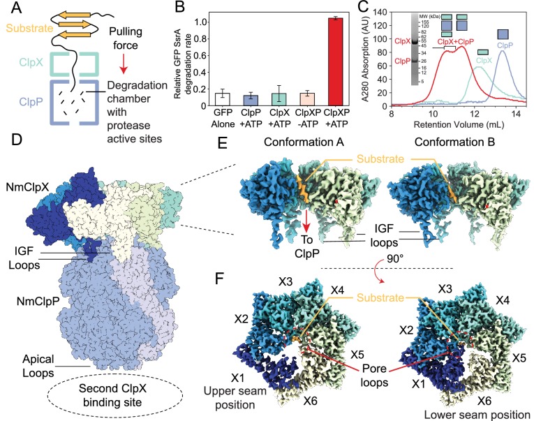
Figure 1.
Functional and structural characterization of ClpXP from N.u00a0meningitidis .
( A ) Schematic representation of the ClpXP degradation machinery. The overall positions of the substrate (orange), ClpX unfoldase (green), and the ClpP protease (blue) are shown; ( B ) GFP-SsrA degra...
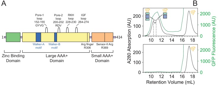
Figure 1u2014figure supplement 1.
Sequence map of NmClpX and SEC analysis of substrate binding by NmClpXP.
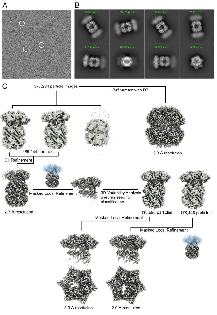
Figure 1u2014figure supplement 2.
Cryo-EM image processing.
( A ) Representative cryo-EM micrograph of NmClpXP bound to GFP-SsrA. Example complexes are circled in white. ( B ) Representative 2D class averages of NmClpXP with GFP-SsrA. ( C ) Image processing wo...
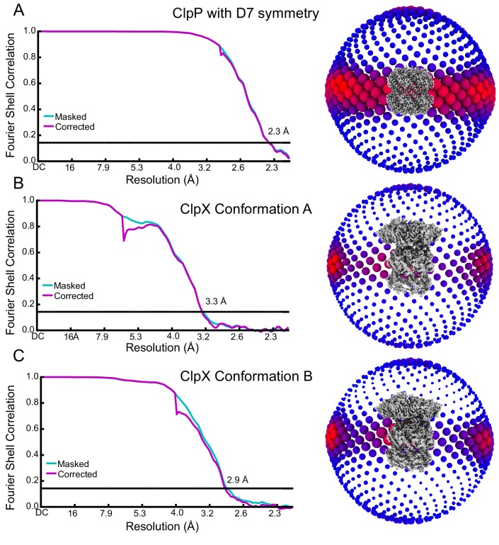
Figure 1u2014figure supplement 3.
Cryo-EM map validation.
( A to C ) Fourier Shell Correlation (FSC) plots (left) and orientation distribution (right). Resolution values reported are for FSCu00a0=u00a00.143.
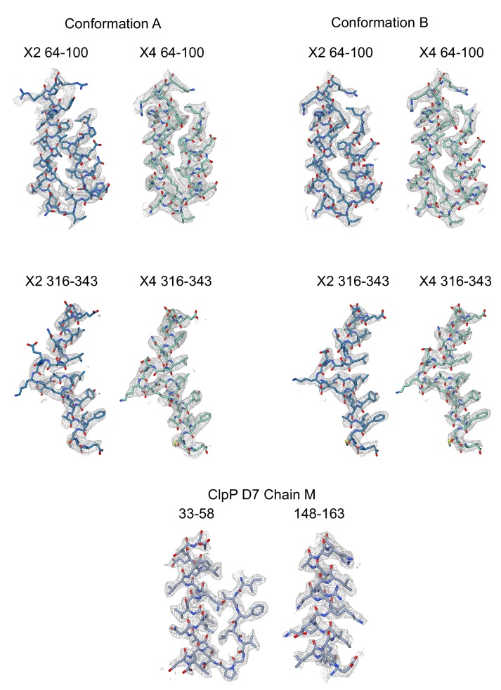
Figure 1u2014figure supplement 4.
Examples of regions of the atomic models built into the experimental cryo-EM maps.
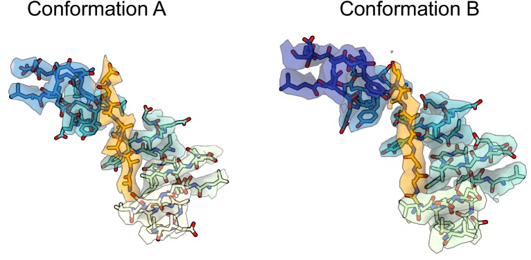
Figure 1u2014figure supplement 5.
Experimental density maps and models for the substrate pore-1 loop interaction interface.
Video 1.
Overview of the substrate-bound ClpXP complex (Conformation B).
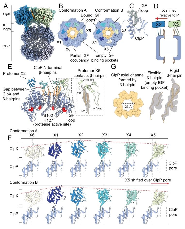
Figure 2.
The interaction interface between ClpX and ClpP.
Protomer X1 occupies the US position in Conformation A and protomer X6 the LS position in Conformation B. ( A ) Cutaway density map of the overall architecture of the ClpXP interaction interface. ( B ...
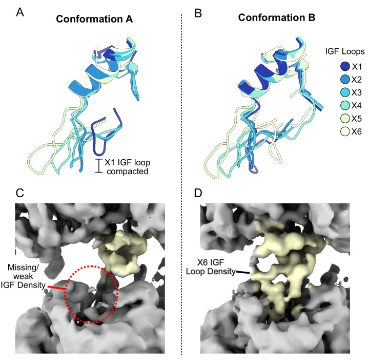
Figure 2u2014figure supplement 1.
IGF loop flexibility.
( A and B ) Large domains of ClpX were aligned and models for the IGF loops are shown. ( C and D ) Unsharpened density map for Conformations A and B looking at protomer X6.
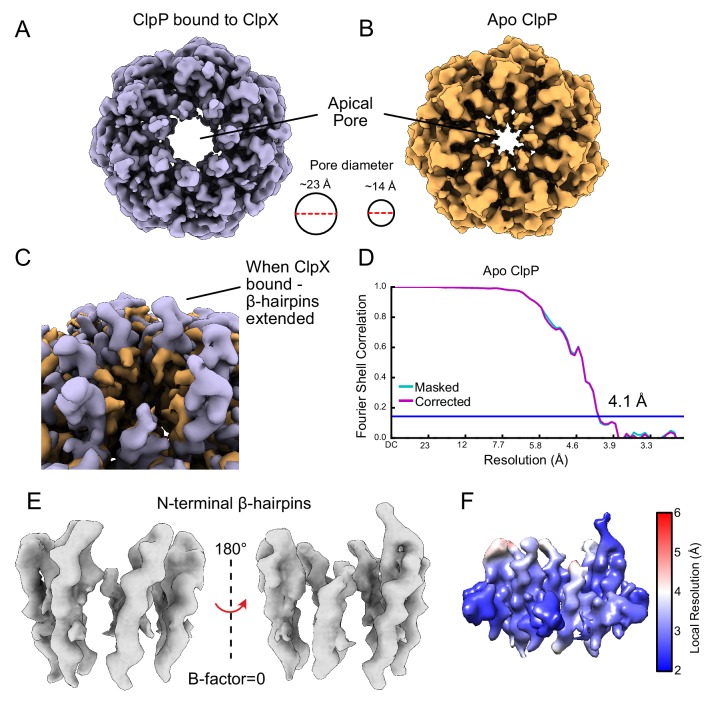
Figure 2u2014figure supplement 2.
The ClpP apical loops extend upwards from the ring surface.
( A ) Unsharpened density map of the ClpP N-terminal u03b2-hairpins when bound to ClpX. ( B ) Cryo-EM map of apo ClpP from N. meningitidis showing that the apical loops no longer extend u2018upu2019. ...
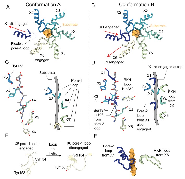
Figure 3.
Substrate engagement by the ClpX pore loops.
( A and B ) Pore-1 loop residues grip the substrate, as observed in this view looking down the axial channel. The substrate is modeled as poly-Ala. ( A ) In Conformation A, the X1 protomer is disengag...
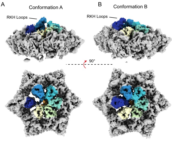
Figure 3u2014figure supplement 1.
RKH loop positions.
( A and B ) Unsharpened maps for both conformations A and B of ClpX are shown, with RKH loops highlighted in color (coloring done by a radius of 10 u00c5 around residues 220u2013240).
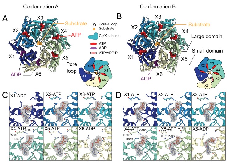
Figure 4.
Nucleotide occupancy and interactions with ClpX.
( A and B ) Model of ClpX in conformations A and B, looking into the ClpX pore. Nucleotides are shown and color-coded, with ATP red and ADP purple, and bind between the large and small domains (boxed)...
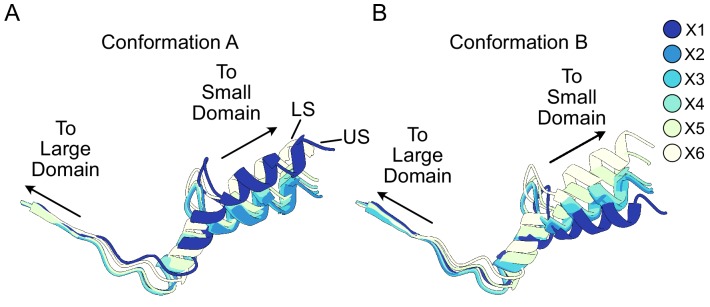
Figure 4u2014figure supplement 1.
Relative orientation between large and small domains of ClpX.
( A and B ) Residues 308u2013343 are shown. Note that protomers X1 and X6 in Conformations A and B, respectively, are displaced from substrate. These show among the largest differences in domain orien...
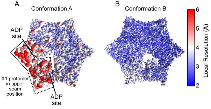
Figure 4u2014figure supplement 2.
Local resolution maps of ClpX.
( A and B ) Density maps colored by local resolution for Conformations A and B. In Conformation A the US protomer is highlighted with a dashed line.
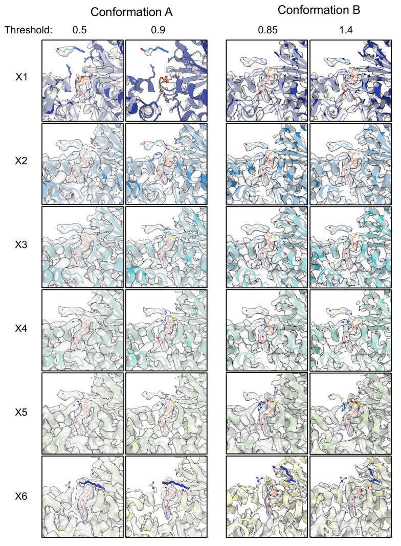
Figure 4u2014figure supplement 3.
ATP-binding pocket densities.
Experimental density maps and models are shown for the ATP-binding pockets of all protomers. Two numerical thresholds are shown to highlight differences in density.
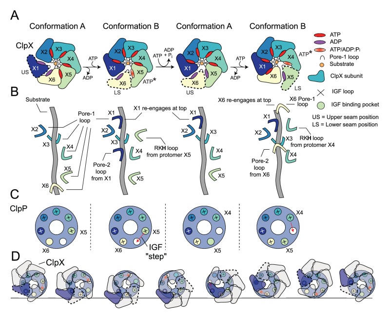
Figure 5.
Translocation model for ClpXP.
( A ) Schematic of the ClpX-nucleotide-dependent conformational cycle. The transition from Conformation A to Conformation B is mediated through nucleotide exchange in the US position (1st to 2nd ring)...
Video 2.
Hand-over-hand translocation cycle of the ClpXP complex.
Interpolation between states A u2192 B u2192 A u2192 B. Side and top views of the complex are shown (top), along with the pore-1 loop Tyr153 and substrate (bottom left) as well as IGF loops (bottom ri...
Video 3.
ClpX precession on the ClpP apical surface.
Repeated interpolation between states reveals a precession of the ClpX ring as its IGF loops are placed successively in different ClpP binding sites. The position of the empty binding pocket on ClpP u...
Appendix 1u2014figure 1.
Peptidase rate measurements of NmClpP as a function of pH, monitored using the fluorogenic substrate PKM-AMC.
Figure images are served from the NIH/NLM PubMed Central Open Access Subset or Europe PMC; copyright remains with the publishers and authors.
💬 Discussion
0 commentsNo comments yet. Be the first to start a discussion!
Leave a Comment