Abstract
Elucidation of neural circuit functions requires visualization of the fine structure of neurons in the inner regions of thick brain specimens. However, the tissue penetration depth of laser scanning microscopy is limited by light scattering and/or absorption by the tissue. Recently, several optical clearing reagents have been proposed for visualization in fixed specimens. However, they require complicated protocols or long treatment times. Here we report the effects of 2,2'-thiodiethanol (TDE) solutions as an optical clearing reagent for fixed mouse brains expressing a yellow fluorescent protein. Immersion of fixed brains in TDE solutions rapidly (within 30 min in the case of 400-µm-thick fixed brain slices) increased their transparency and enhanced the penetration depth in both confocal and two-photon microscopy. In addition, we succeeded in visualizing dendritic spines along single dendrites at deep positions in fixed thick brain slices. These results suggest that our proposed protocol using TDE solution is a rapid and useful method for optical clearing of fixed specimens expressing fluorescent proteins.
🔬 Techniques
🔭 Microscopes
✨ Fluorophores
🧪 Sample Preparation
🏭 Microscope Brands
🧪 Reagent Suppliers
🔴 Lasers
🏛️ Research Organizations (ROR)
Affiliated research institutions:
📋 Methods
Ethics Statement
All protocols were approved by the Institutional Animal Care and Use Committee of National University Corporation Hokkaido University (Permit Number: 10-0119). All experiments were performed under general anesthesia, and all efforts were made to minimize suffering.
Animals Adult thy1-YFP-H transgenic mice expressing enhanced yellow fluorescent protein
(EYFP) in subsets of hippocampal pyramidal neurons and layer V cortical pyramidal neurons [ 16 ], were used for fluorescent imaging and for preparing the fixed brain photograms shown in Figs. 1 , S2 , and S3 . Adult C57BL/6 mice were used for the measurement of light transmittance. 10.1371/journal.pone.0116280.g001 Figure 1 Fixed brain tissue clearing with TDE solutions. (a, b) Fixed adult mouse brains after treatment with different concentrations of TDE solutions. Whole brains (a) and brain slices (400 µm in thickness) (b) were immersed in each TDE solution for 2 days and 1 h, respectively. The photograms were taken under backlighting. (c) Transmission curves of fixed brain slices (400 µm in thickness, n = 3) after 2 h of immersion in each TDE solution. Data shown represent the average ± SEM.
Preparation of fixed brain slices
For preparing the fixed brain photograms shown in Figs. 1 , S2 , and S3 , the images of dendritic spines in a single hippocampal neuron, mice were anesthetized with pentobarbital sodium. They were transcardially perfused with phosphate-buffered saline (PBS) followed by 4% formaldehyde in PBS, and their brains were removed. Slices (400 or 500 µm in thickness) were prepared using a vibratome (7000smz, Campden, Instruments Ltd, UK). For the measurement of light transmittance, evaluation of the penetration depth in fixed hippocampal slices, and comparison of the combination of TDE and the objective lens, mice were anesthetized with isoflurane and their brains were removed quickly. Slices (400 µm in thickness) were cut using the vibratome in ice-cold artificial cerebrospinal fluid (containing 119 mM NaCl, 2.5 mM KCl, 1.0 mM NaH 2 PO 4 , 1.3 mM MgSO 4 , 2.5 mM CaCl 2 , 26 mM NaHCO 3 , and 10 mM glucose, bubbled with O 2 /CO 2 : 95%/5%) and incubated for 1 h at room temperature, followed by fixation with 4% formaldehyde in PBS.
Show full methods section
Ethics Statement
All protocols were approved by the Institutional Animal Care and Use Committee of National University Corporation Hokkaido University (Permit Number: 10-0119). All experiments were performed under general anesthesia, and all efforts were made to minimize suffering.
Animals Adult thy1-YFP-H transgenic mice expressing enhanced yellow fluorescent protein
(EYFP) in subsets of hippocampal pyramidal neurons and layer V cortical pyramidal neurons [ 16 ], were used for fluorescent imaging and for preparing the fixed brain photograms shown in Figs. 1 , S2 , and S3 . Adult C57BL/6 mice were used for the measurement of light transmittance. 10.1371/journal.pone.0116280.g001 Figure 1 Fixed brain tissue clearing with TDE solutions. (a, b) Fixed adult mouse brains after treatment with different concentrations of TDE solutions. Whole brains (a) and brain slices (400 µm in thickness) (b) were immersed in each TDE solution for 2 days and 1 h, respectively. The photograms were taken under backlighting. (c) Transmission curves of fixed brain slices (400 µm in thickness, n = 3) after 2 h of immersion in each TDE solution. Data shown represent the average ± SEM.
Preparation of fixed brain slices
For preparing the fixed brain photograms shown in Figs. 1 , S2 , and S3 , the images of dendritic spines in a single hippocampal neuron, mice were anesthetized with pentobarbital sodium. They were transcardially perfused with phosphate-buffered saline (PBS) followed by 4% formaldehyde in PBS, and their brains were removed. Slices (400 or 500 µm in thickness) were prepared using a vibratome (7000smz, Campden, Instruments Ltd, UK). For the measurement of light transmittance, evaluation of the penetration depth in fixed hippocampal slices, and comparison of the combination of TDE and the objective lens, mice were anesthetized with isoflurane and their brains were removed quickly. Slices (400 µm in thickness) were cut using the vibratome in ice-cold artificial cerebrospinal fluid (containing 119 mM NaCl, 2.5 mM KCl, 1.0 mM NaH 2 PO 4 , 1.3 mM MgSO 4 , 2.5 mM CaCl 2 , 26 mM NaHCO 3 , and 10 mM glucose, bubbled with O 2 /CO 2 : 95%/5%) and incubated for 1 h at room temperature, followed by fixation with 4% formaldehyde in PBS.
TDE treatment
TDE was purchased from Sigma-Aldrich (St. Louis, MO). Solutions of 0% (PBS only), 30%, 60%, and 97% TDE were prepared by mixing TDE, Milli-Q water, and PBS powder (Kohjin-Bio, Saitama, Japan). The refractive index of each TDE solution was measured with a pocket refractometer (PAL-RI, Atago, Tokyo, Japan) ( S1 Fig. ). The fixed slices and whole brains were immersed in the indicated concentrations of TDE solutions. Transparency was confirmed using a stereomicroscope (SZ61/SZ2-ILST, Olympus, Tokyo, Japan) and photos were taken using a camera (DP21, Olympus, Tokyo, Japan).
Measurement of light transmittance
Light transmittance by fixed brain slices was recorded using a UV/visible/near infrared (NIR) spectrophotometer (U-2900, HITACHI, Tokyo, Japan). Wild-type mice were used to measure light transmittance. Thy1-YFP-H mice were not used for avoiding EYFP light excitation and emission. The hippocampal region of the fixed slices (400 µm in thickness) was superposed on a window in thick paper to measure light transmittance by only the hippocampal region.
Imaging EYFP-expressing brain
All observations of EYFP-expressing brain for the measurement of the penetration depth were performed with an upright confocal and two-photon microscope system (A1R-MP and FN1, Nikon, Tokyo, Japan) equipped with a Ti: sapphire laser (MaiTai eHP DeepSee, SpectraPhysics, Santa Clara, CA). For observing brain slices, a 20× multi-immersion objective lens [Plan Fluor, numerical aperture (NA): 0.75, working distance (WD): 0.35 mm; Nikon, Tokyo, Japan] was used. The excitation wavelength was 488 nm (confocal) or 950 nm (two-photon). For imaging the whole brain, images were acquired with a 25× water-immersion objective lens (CFI Apo LWD, NA: 1.1, WD: 2 mm; Nikon, Tokyo, Japan). The excitation wavelength was 1010 nm. For observation of dendritic spines in the hippocampus, 60% TDE-immersed hippocampal slices were placed on a 0.06–0.08-mm-thick glass-bottom dish and covered with a paper wetted with 60% TDE. Images were acquired with the inverted confocal microscope system (A1R and Ti-E, Nikon, Tokyo, Japan). A 60× water-immersion objective lens (Plan Apo, NA: 1.2, WD: 0.27 mm; Nikon, Tokyo, Japan) or a 60× oil-immersion objective lens (Apo-TIRF, NA: 1.49, WD: 0.12 mm; Nikon, Tokyo, Japan) was used. The excitation wavelength was 514.5 nm. The emitted fluorescence signals within in the range of 525–555 nm, split with dichroic mirrors, were detected.
Supporting Information S1 Fig Refractive index of TDE solutions. Plot of the refractive index against the concentration of TDE solution (n = 3). Data represent the average ± SEM. As previously reported [ 13 ], the refractive index increased in a concentration-dependent manner. The refractive index of the TDE solution is different from that of water (1.33) and standard immersion oil (1.52). (TIF) Click here for additional data file. S2 Fig Time changes in fixed whole brains after immersion in each TDE solution. Photograms of fixed whole brains before and after immersion in 30%, 60%, and 97% TDE solutions for 6 h, 1 day, 5 days, and 7 days. The photograms at 2 days are shown in Fig. 1a . They were taken under backlighting. (TIF) Click here for additional data file. S3 Fig Fixed brain slices before and after immersion in each TDE solution. (a) Photograms of fixed brain slices before and after immersion in each TDE solution for 3 h. (b) Photograms of the same fixed brain slice that was immersed in increasing concentrations of TDE (30%, 60%, 97%) every hour in a stepwise manner. Data represent the average ± SEM. (TIF) Click here for additional data file. S4 Fig Time changes in light transmittance by hippocampal slices in TDE solutions. Plot of light transmittance by hippocampal slices (400 µm in thickness, n = 3) against treatment time. Transmittances at light wavelengths of 488, 527, and 950 nm are presented according to EYFP excitation and emission: 488 nm for confocal and 950 nm for two-photon microscopy excitation; 527 nm for detecting EYFP emission. (TIF) Click here for additional data file. S5 Fig Time-lapse images of fixed brain slices after immersion in 97% TDE. (a) Images of fixed slices after immersion in 97% TDE. The images were observed using two-photon laser scanning microscopy. (b) Plot of the mean fluorescence intensity in the xy image at a depth of 300 µm from the surface. The fluorescence signal decreased over time after immersion in 97% TDE. (TIF) Click here for additional data file. S6 Fig Recovery of the fluorescence signals eliminated after immersion in 97% TDE. (a) Image of a 97% TDE-treated fixed slice immediately after immersion in PBS. (b, c) Images of the same brain slice as shown in (a). The slice was incubated for 1 day in PBS (b), and then immersed in 60% TDE for 4 h (c). The fluorescence signals recovered and the structure of the hippocampal neurons appeared to be preserved. The images were observed using two-photon laser scanning microscopy. (TIF) Click here for additional data file.
📊 Figures
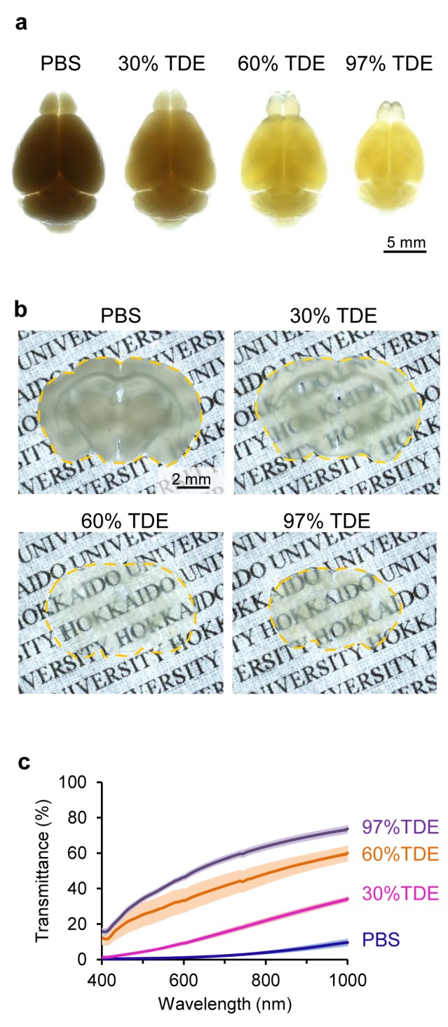
Figure 1
Fixed brain tissue clearing with TDE solutions.
(a, b) Fixed adult mouse brains after treatment with different concentrations of TDE solutions. Whole brains (a) and brain slices (400 u00b5m in thickness) (b) were immersed in each TDE solution for 2...
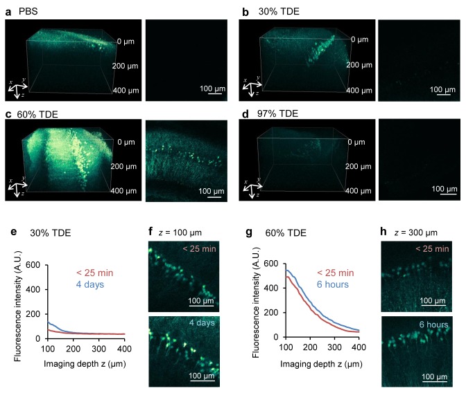
Figure 2
Enhancement of penetration depth on confocal microscopy.
(au2013d) Images of YFP-expressing neurons in the hippocampal slices of thy1-YFP-H mouse in PBS (a) and after 2 h of immersion in 30% TDE (b), 60% TDE (c), and 97% TDE (d) solution. Left, three-dimens...
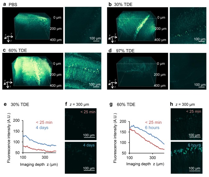
Figure 3
Enhancement of the penetration depth on two-photon microscopy.
(au2013d) Images of YFP-expressing neurons in the hippocampal slices of thy1-YFP-H mouse in PBS (a) and after 2 h of immersion in 30% TDE (b), 60% TDE (c), 97% TDE (d) solution. Left, three-dimensiona...
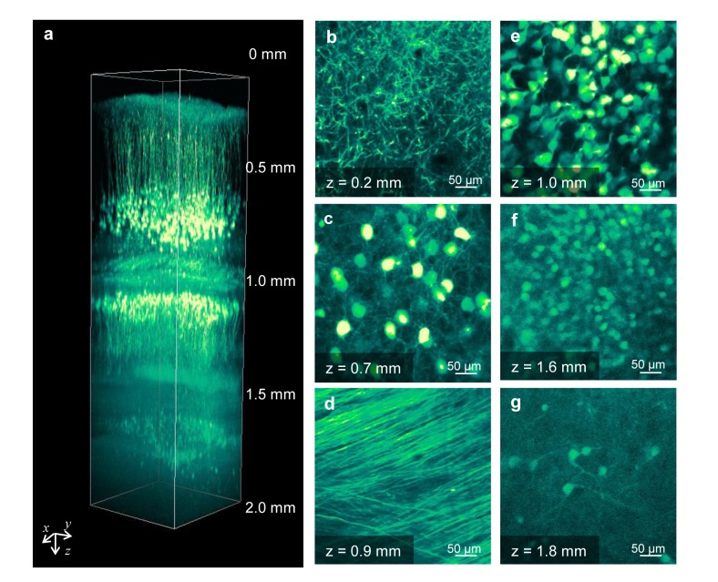
Figure 4
Two-photon deep imaging of a fixed whole brain immersed in 60% TDE.
(a) Three-dimensional reconstructed image of a whole mouse brain after 2 days of immersion in 60% TDE. (b-g) xy images at different depths from the cerebral cortex to the lower portion of the hippocam...
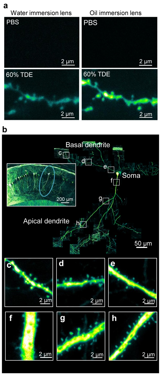
Figure 5
Images of dendritic spines along a single hippocampal neuron.
(a) Combination of TDE treatment and water/oil-immersion objective lens with a high NA for imaging dendritic spine shapes in deep (100 u00b5m) regions in a fixed brain slice. (b) Connected images of d...
Figure images are served from the NIH/NLM PubMed Central Open Access Subset or Europe PMC; copyright remains with the publishers and authors.
💬 Discussion
0 commentsNo comments yet. Be the first to start a discussion!
Leave a Comment