Abstract
Six percent of Americans, including 3 million high schoolers, use e-cigarettes, which contain potentially toxic substances, volatile organic compounds, and metals. We present the first human study on the effects of e-cigarette exposure in the oral cavity. By interrogating both immunoinflammatory responses and microbial functional dynamics, we discovered pathogen overrepresentation, higher virulence signatures, and a brisk proinflammatory signal in clinically healthy e-cigarette users, equivalent to patients with severe periodontitis. Using RNA sequencing and confocal and electron microscopy to validate these findings, we demonstrate that the carbon-rich glycol/glycerol vehicle is an important catalyst in transforming biofilm architecture within 24 hours of exposure. Last, a machine-learning classifier trained on the metagenomic signatures of e-cigarettes identified as e-cigarette users both those individuals who used e-cigarettes to quit smoking, and those who use both e-cigarettes and cigarettes. The present study questions the safety of e-cigarettes and the harm reduction narrative promoted by advertising campaigns.
🔬 Techniques
✨ Fluorophores
🧪 Sample Preparation
🔬 Cell Lines
🏭 Microscope Brands
🧪 Reagent Suppliers
💻 Software Details
💻 Code & Software
💾 Data Repositories
🏛️ Research Organizations (ROR)
Affiliated research institutions:
📋 Methods
Subject and site selection We obtained approval for this study from the Office of Responsible Research Practices at The Ohio State University [IRB (Institutional Review Board) protocol number 2014H0062 and e-IBC protocol number 2015R00000005], and the study was conducted in accordance with approved guidelines. We recruited 123 systemically [ASA I (American Society of Anaesthesiologists Physical Status Classification I)] and periodontally healthy individuals [attachment loss ≤ 1; less than three sites with 4 mm of probe depths (PD); bleeding index (BOP) ≤ 20%] following informed consent and clinical and radiographic examination to each of five groups: (i) smokers (25), (ii) nonsmokers (25), (iii) e-cigarette users (20), (iv) former smokers currently using e-cigarettes (25), and (v) concomitant cigarette and e-cigarette users (28). Current smokers were those who had at least a five pack-year history and had no prior history of e-cigarette use. Never smokers were those who had smoked less than 100 cigarettes in their lifetime and none in the past year, and e-cigarette users were those who used e-cigarettes daily for at least 3 months, with at least one cartridge per day or 1 ml of liquid per day. Former smokers were those who had quit smoking for at least 1 year. Exclusion criteria for all groups included controlled or uncontrolled diabetes, HIV infection, use of immunosuppressant medications, bisphosphonates, or steroids, antibiotic therapy or oral prophylactic procedures within the preceding 3 months, and fewer than 20 teeth in the dentition. Sample size was estimated on the basis of the probability of least an 80% chance of detecting clades of bacterial genes that differed in abundance by more than 1%.
Show full methods section
Subject and site selection We obtained approval for this study from the Office of Responsible Research Practices at The Ohio State University [IRB (Institutional Review Board) protocol number 2014H0062 and e-IBC protocol number 2015R00000005], and the study was conducted in accordance with approved guidelines. We recruited 123 systemically [ASA I (American Society of Anaesthesiologists Physical Status Classification I)] and periodontally healthy individuals [attachment loss ≤ 1; less than three sites with 4 mm of probe depths (PD); bleeding index (BOP) ≤ 20%] following informed consent and clinical and radiographic examination to each of five groups: (i) smokers (25), (ii) nonsmokers (25), (iii) e-cigarette users (20), (iv) former smokers currently using e-cigarettes (25), and (v) concomitant cigarette and e-cigarette users (28). Current smokers were those who had at least a five pack-year history and had no prior history of e-cigarette use. Never smokers were those who had smoked less than 100 cigarettes in their lifetime and none in the past year, and e-cigarette users were those who used e-cigarettes daily for at least 3 months, with at least one cartridge per day or 1 ml of liquid per day. Former smokers were those who had quit smoking for at least 1 year. Exclusion criteria for all groups included controlled or uncontrolled diabetes, HIV infection, use of immunosuppressant medications, bisphosphonates, or steroids, antibiotic therapy or oral prophylactic procedures within the preceding 3 months, and fewer than 20 teeth in the dentition. Sample size was estimated on the basis of the probability of least an 80% chance of detecting clades of bacterial genes that differed in abundance by more than 1%.
Sample collection
We collected and pooled subgingival plaque samples from 15 sites on six maxillary and mandibular anterior teeth using sterile endodontic paper points (Caulk-Dentsply, Milford, DE, USA). We also collected gingival crevicular fluid (GCF) by placing filter paper strips (Periopaper, Oraflow, Plainview, NY, USA) into the same sites. The fluid volume was measured with a calibrated device (Periotron 8000, Oraflow, Plainview, NY, USA). DNA isolation, metagenomic sequencing, and analysis We isolated bacterial DNA from paper points, using a Qiagen DNA MiniAmp kit (Qiagen, Valencia, CA, USA) after a 90-min incubation in lysozyme (2 mg/ml) (Thermo Fisher Scientific) and then quantified the DNA using a Qubit fluorometer. Library generation was completed. We used an Illumina TruSeq kit according to the manufacturer’s instructions for library generation, quantified and pooled the libraries and clustered them on the Illumina HiSeq 4000 (Illumina Inc., San Diego, CA) for 150–base pair (bp) paired-end sequencing. Sequences are deposited in the Sequence Read Archives under the project IDs PRJNA548383, PRJNA544061, and PRJNA508385. Trimmed and filtered sequences were uploaded to the MG-RAST metagenomics analysis pipeline (version 3.3.6) ( 13 ) (Argonne National Laboratory) for quality processing and basic functional analysis. The MG-RAST API and the custom Python library we have developed to access it and analyze/visualize results were used throughout the analysis process to download relevant data and pipeline results (available for download at http://github.com/smdabdoub/PyMGRAST ). We used Nonpareil ( 14 ) to estimate coverage per sample. Comparisons of functional potential between groups were made in the context of the KEGG (Kyoto Encyclopedia of Genes and Genomes) ( 15 ), and the SEED subsystem ( 16 ) ontological hierarchies and statistical analysis of differential functional potential were performed using the DESeq2 package for R ( 17 ). We used Kraken v1.1 ( 18 ) to determine the phylogenetic profile of each subject with a database constructed from a list of complete genomes from the Human Oral Microbiome Database, as of 19 September 2017. We computed alpha (within-group) and beta (between-group) diversity using PhyloToAST v1.4 and QIIME v1.9, respectively. The Shannon diversity index and Abundance Coverage Estimator (ACE) were used as estimators of species diversity and richness, and Bray-Curtis and Jaccard metrics were used to estimate beta diversity. We used principal coordinate analysis (PCoA) for dimensionality reduction and interrogated the significance of group-wise clustering using a permutational multivariate analysis of variance (MANOVA) (adonis function, vegan package for R). PCoA plots were generated with PhyloToAST (PCoA.py). We inducted CSS (cumulative sum scaling)–normalized species-level operational taxonomic unit (sOTU) counts into linear discriminant analysis (LDA) using scikit-learn v0.18.0 ( 19 ). Plots were visualized using PhyloToAST, and MANOVA/Wilks’ lambda was used to test for the significance of LDA clustering. We used SparCC ( 20 ) to test for differences in co-occurrence patterns between microbial communities from different ecosystems. Gephi ( 21 ) v0.9.1 was used to visualize the resultant networks. We used a machine learning algorithm (randomForest package in R) to test the ability of genes to discriminate between groups. Two-thirds of the dataset was used to train the algorithm, which was tested on the remaining data. This was iterated 10 times, and the mean “importance” was computed for each marker gene. The robustness of the classifier was evaluated using ROC (receiver operating characteristic) curves (ROCR package in R). Quality control All samples were sequenced in two runs; and to minimize batch effects, samples were randomly assigned to each run. Replicate sequencing was carried out for two samples in each batch, and the replicates showed good reliability across the five batches, with coefficient of variability (SD/mean) ranging from 0.26 to 1.3% for alpha diversity of taxonomy and 3.4 to 6.3% for predominant functions (carbohydrate metabolism, respiration, and virulence, disease, and defense).
Cytokine assay
We eluted GCF from the paper strips by adding 200 μl of phosphate-buffered saline and vortexing for 15 min. Levels of interferon-γ, interleukin-2 (IL-2), IL-4, IL-6, IL-8, IL-10, granulocyte-macrophage colony-stimulating factor (GM-CSF), and tumor necrosis factor–α (TNF-α) in GCF were determined using the Bio-Plex Pro Human Cytokine 8-plex assay (Bio-Rad Corporation, CA), according to the manufacturer’s instructions. In vitro validation studies Artificial saliva was made following the Marshall Group Research protocol for artificial saliva. SHI medium was prepared according to the protocol described by Tian et al . ( 22 ). The developed biofilms were developed using the modifications ( 23 ) from the protocol established by Guggenheim et al . ( 24 ). Briefly, sterilized, sintered hydroxyapatite (HA) disks (Clarkson Chromatography Products, South Williamsport, PA) were incubated in artificial saliva for 24 hours to establish a pellicle coat, following which multispecies commensal primary biofilms were generated by seeding six pioneer species [ Streptococcus oralis (ATCC 35037), S . sanguis (10556), S. mitis (49456), Actinomyces naeslundii (12104), Neisseria mucosa (25997), and Veillonella parvula (17745)] and incubating under aerobic conditions in a 1:1(v/v) mixture of SHI media and artificial saliva. Pathogen-rich biofilms were created by further seeding the commensal biofilms with an intermediate bridging colonizer [ Fusobacterium nucleatum (10953), secondary biofilm] followed 24 hours later by Porphyromonas gingivalis (33277), Filifactor alocis (35896), Selenomonas sputigena (35185), S. noxia (43541), Campylobacter gracilis (33236), Prevotella intermedia (25611), Parvimonas micra (33270), and Tannerella forsythia (43037) and incubating under anaerobic conditions for a further 24 hours (tertiary biofilms). Research grade cigarettes (IR4F) with a filter were obtained from the Kentucky Tobacco Research and Development Center (Lexington, KY). We prepared cigarette smoke extract (CSE) by bubbling smoke from two cigarettes into 5 ml of artificial saliva. To replicate our e-cigarette cohort’s usage, we used a moderate-sized e-cigarette vape pen, vaped by pressing “on” for 5 s and “off” for 30 s in between and repeated for 15 to 20 min. We prepared electronic cigarette vapor (ECV) by vaping two cartridges of nonflavored e-cigarette containing either 6 or 0 ml of nicotine through clean Pasteur pipettes into 5 ml of artificial saliva, until the whole cartridge was exhausted. CSE and ECV were prepared immediately before use. To maintain the consistency of CSE and ECV between experiments, an optical density of 0.65 at 600 nm represented 100% ( 25 ). CSE and ECV were diluted to 1%, and the biofilms were conditioned with these solutions for 24 hours. We isolated total RNA from the biofilms using the mirVana miRNA isolation kit (Applied Biosystems). Ribosomal RNA was depleted, and mRNA was enriched by modified capture hybridization approach (MICROBExpress mRNA enrichment kit, Thermo Fisher Scientific). Enriched mRNA served as a template for the polyadenylation reaction and complementary DNA synthesis. Microbial libraries were clustered on the Illumina HiSeq 4000 platform, and 150-bp paired-end sequencing was performed. The Illumina base-calling pipeline was used to process the raw fluorescence images and call sequences. Raw reads with >10% unknown nucleotides or with >50% low-quality nucleotides (quality value,
📊 Figures
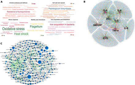
Fig. 1
Phylogenetic, functional, and immune characteristics of the subgingival microbiome of e-cigarette users.
( A ) Word map of the predominant microbial functions identified in e-cigarette users. The functions are colored by relative abundances (as indicated by the color map scale within each word map). ( B ...
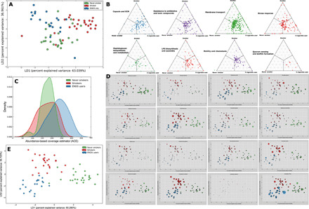
Fig. 2
Differences in microbial community structure and function between electronic cigarette users, smokers, and nonsmokers.
LDA of relative abundances of functional genes in periodontally and systemically healthy nonsmokers (green), smokers (red), and ENDS users (blue) is shown in ( A ). The microbial profiles of subjects ...
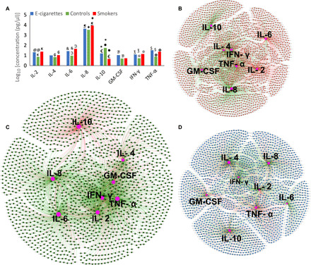
Fig. 3
Inflammatory burden imposed by the e-cigaretteu2013influenced microbiome.
Levels of selected immune mediators in periodontally and systemically healthy nonsmokers (green), smokers (red), and e-cigarette users (blue) is shown in ( A ). The y axis represents log 10 -transform...
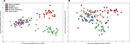
Fig. 4
The synergistic effects of smoking and e-cigarettes on the microbiome.
LDA of Bray-Curtis dissimilarity indices based on relative abundances of species level OTUs ( A ) and functional genes ( B ) in periodontally and systemically healthy individuals who exclusively use E...
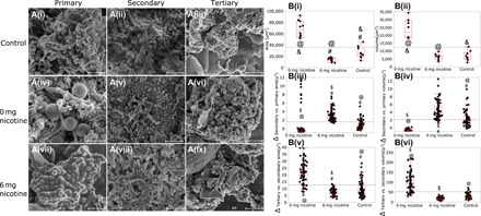
Fig. 5
Effect of e-cigarettes on sculpting the biofilm landscape.
( A ) SEM images of primary biofilms consisting of S. oralis , S. sanguis , S. mitis , A. naeslundii , N. mucosa , and V. parvula , secondary biofilms [following addition of an intermediate colonizer ...
Figure images are served from the NIH/NLM PubMed Central Open Access Subset or Europe PMC; copyright remains with the publishers and authors.
💬 Discussion
0 commentsNo comments yet. Be the first to start a discussion!
Leave a Comment