Abstract
Abnormalities of the endolysosomal and autophagy systems are found in Alzheimer's disease, but it is not clear whether defects in these systems are a cause or consequence of degenerative processes in the disease. In human neuronal models of monogenic Alzheimer's disease, APP and PSEN1 mutations disrupt lysosome function and autophagy, leading to impaired lysosomal proteolysis and defective autophagosome clearance. Processing of APP by γ-secretase is central to the pathogenic changes in the lysosome-autophagy system caused by PSEN1 and APP mutations: reducing production of C-terminal APP by inhibition of BACE1 rescued these phenotypes in both APP and PSEN1 mutant neurons, whereas inhibition of γ-secretase induced lysosomal and autophagic pathology in healthy neurons. Defects in lysosomes and autophagy due to PSEN1 mutations are rescued by CRISPR-knockout of APP. These data demonstrate a key role for proteolysis of the C-terminal of APP by γ-secretase in neuronal dysfunction in monogenic Alzheimer's disease.
🔬 Techniques
🔭 Microscopes
✨ Fluorophores
🧪 Sample Preparation
🔬 Cell Lines
🏭 Microscope Brands
🧪 Reagent Suppliers
💻 Software Details
🏷️ Research Resource Identifiers (RRIDs)
Verified research resources used in this paper:
🏛️ Research Organizations (ROR)
Affiliated research institutions:
📋 Methods
Key Resources Table REAGENT or RESOURCE SOURCE IDENTIFIER Antibodies
Mouse monoclonal anti-APP BioLegend Cat#802801; RRID: AB_2564648 Mouse anti-β-Amyloid, 1-16 BioLegend Cat#803001; RRID: AB_2564653 Rabbit polyclonal anti-Tubulin β-3 BioLegend Cat#802001; RRID: AB_2564645 Mouse monoclonal anti- β-actin Sigma Cat#A228; RRID: AB_476697 Rabbit polyclonal anti-LAMP1 abcam Cat#;Ab62562 RRID: AB_2134489 Rabbit polyclonal anti-LC3B Sigma Cat#L7543; RRID: AB_796155 Chicken polyclonal anti-MAP2 abcam Cat#Ab5392; RRID: AB_2138153 Experimental Models: Cell Lines Human: Non-demented control iPSC lines Israel et al., 2012 N/A Human: SFC840 iPSC lines StemBANCC N/A Human: AD3.1 iPSC lines StemBANCC N/A Human: PSEN1 Y115C iPSC lines Moore et al., 2015 N/A Human: PSEN1 M146I iPSC lines Moore et al., 2015 N/A Human: PSEN1 Intron4 iPSC lines Moore et al., 2015 N/A Human: APP V717I iPSC lines Moore et al., 2015 N/A Human: APP duplication iPSC lines Israel et al., 2012 N/A Human: Ts21 iPSC lines Park et al., 2008 N/A Oligonucleotides sgRNA-sense (S): ATCCAGAACTGGTGCAAGCGGGG This paper N/A sgRNA-antisense (AS): TTGGTTGGCTTCTACCACATTGG This paper N/A Software and Algorithms ImageJ https://imagej.net/Welcome RRID: SCR_003070 GraphPad Prism https://www.graphpad.com/ RRID: SCR_002798 Contact for Reagent and Resource Sharing Further information and requests for resources and reagents should be directed to and will be fulfilled by the Lead Contact, Rick Livesey ( r.livesey@ucl.ac.uk ).
Experimental Model and Subject Details Human iPSC lines
Non-demented control iPSC lines: Non-Demented-Control (NDC) ( Israel et al., 2012 ), SFC840 (StemBANCC), and AD3.1 (StemBANCC). All AD lines were previously reported and characterized: PSEN1 Y115C, M146I, intron 4, and APP V717I iPSCs ( Moore et al., 2015 ); APP duplication ( Israel et al., 2012 , Moore et al., 2015 ) and Ts21 iPSCs ( Park et al., 2008 , Moore et al., 2015 , Shi et al., 2012b ). This research was carried out in accordance with the UK Code of Practice for the Use of Human Stem Cell Lines.
Show full methods section
Key Resources Table REAGENT or RESOURCE SOURCE IDENTIFIER Antibodies
Mouse monoclonal anti-APP BioLegend Cat#802801; RRID: AB_2564648 Mouse anti-β-Amyloid, 1-16 BioLegend Cat#803001; RRID: AB_2564653 Rabbit polyclonal anti-Tubulin β-3 BioLegend Cat#802001; RRID: AB_2564645 Mouse monoclonal anti- β-actin Sigma Cat#A228; RRID: AB_476697 Rabbit polyclonal anti-LAMP1 abcam Cat#;Ab62562 RRID: AB_2134489 Rabbit polyclonal anti-LC3B Sigma Cat#L7543; RRID: AB_796155 Chicken polyclonal anti-MAP2 abcam Cat#Ab5392; RRID: AB_2138153 Experimental Models: Cell Lines Human: Non-demented control iPSC lines Israel et al., 2012 N/A Human: SFC840 iPSC lines StemBANCC N/A Human: AD3.1 iPSC lines StemBANCC N/A Human: PSEN1 Y115C iPSC lines Moore et al., 2015 N/A Human: PSEN1 M146I iPSC lines Moore et al., 2015 N/A Human: PSEN1 Intron4 iPSC lines Moore et al., 2015 N/A Human: APP V717I iPSC lines Moore et al., 2015 N/A Human: APP duplication iPSC lines Israel et al., 2012 N/A Human: Ts21 iPSC lines Park et al., 2008 N/A Oligonucleotides sgRNA-sense (S): ATCCAGAACTGGTGCAAGCGGGG This paper N/A sgRNA-antisense (AS): TTGGTTGGCTTCTACCACATTGG This paper N/A Software and Algorithms ImageJ https://imagej.net/Welcome RRID: SCR_003070 GraphPad Prism https://www.graphpad.com/ RRID: SCR_002798 Contact for Reagent and Resource Sharing Further information and requests for resources and reagents should be directed to and will be fulfilled by the Lead Contact, Rick Livesey ( r.livesey@ucl.ac.uk ).
Experimental Model and Subject Details Human iPSC lines
Non-demented control iPSC lines: Non-Demented-Control (NDC) ( Israel et al., 2012 ), SFC840 (StemBANCC), and AD3.1 (StemBANCC). All AD lines were previously reported and characterized: PSEN1 Y115C, M146I, intron 4, and APP V717I iPSCs ( Moore et al., 2015 ); APP duplication ( Israel et al., 2012 , Moore et al., 2015 ) and Ts21 iPSCs ( Park et al., 2008 , Moore et al., 2015 , Shi et al., 2012b ). This research was carried out in accordance with the UK Code of Practice for the Use of Human Stem Cell Lines.
Method Details Directed differentiation to human cortical neuron culture
Directed differentiation of iPSCs to cerebral cortex was carried out as described ( Moore et al., 2015 ). Briefly, dissociated iPSCs were plated on GelTrex-coated 6-well plates and neural induction were initiated by changing into culture medium that supports neuronal differentiation and neurogenesis, a 1:1 mixture of N2- and B27-containing media (supplemented with dorsomorphin and SB431542 to inhibit TGF β signaling during neural induction). Neuroepithelial cells were harvested with dispase and replated in laminin-coated plates with FGF2-containing media. FGF2 was withdrew for 4 days to promote differentiation, passaged with accutase, and maintained for up to 120 days with a medium change every other day. To establish identity and quality of cortical neuronal inductions, gene expression profiling was performed on a custom gene expression panel. RNA was isolated from induced cortical neurons, 85 – 90 days after induction, using an RNA extraction kit (QIAGEN), according to the manufacturer’s instructions. Expression levels of mRNAs enriched in deep and upper layer cortical neurons were assessed on the Nanostring nCounter platform ( Figure S1 ). For drug treatments, all compounds were dissolved in DMSO at the concentrations specified, and DMSO was used as vehicle control in all experiments. Compounds were added every 48 hr during treatment period: β-secretase inhibitor OM99-2 (Calbiochem); and γ-secretase inhibitor, DAPT (Sigma). Protein analysis Extracellular Aβ 1-42, Aβ 1-40, and Aβ 1-38 were measured in conditioned media using multiplexed MesoScale Discovery assays on a Quickplex SQ120 instrument (MesoScale Discovery). All statistical analysis was performed between the entire set of controls samples and all samples of each genotype, using Student’s t test with the Bonferroni correction for multiple testing.
Immunoblotting
For immunoblotting, whole cell lysate protein was extracted with RIPA buffer (Sigma). Samples were separated on a 4%–12% SDS-PAGE and transferred to PVDF membranes. Proteins were detected (Li-Cor Odyssey system) by incubation with specific primary antibodies and appropriate secondary antibodies. Antibodies used in this study are listed in Key Resources Table .
Live cell imaging and confocal microscopy
For live cell imaging, Rab5A-GFP (Catalog no. C10586 ), LAMP1-RFP (Catalog no. C10597 ), LC3-GFP (Catalog no. P36235 ) and p62-RFP (Catalog no. P36241 ) were expressed in day 80-85 neurons using commercial viral vectors (ThermoFisher Scientific). Neurons were transferred into live-imaging solution (Invitrogen) 16 hours after infection in order to improve imaging of fluorescent proteins. Cells were viable and appeared morphologically normal in imaging medium for at least 24 hours. As an alternative to expression of LAMP1-RFP, neuronal lysosomes were visualized by incubation of 100 nM LysoTracker Red DND-99 (Molecular Probes) with or without the addition of Pepstatin A, BODIPY FL Conjugate (Invitrogen) in fresh medium for 30 minutes at 37°C. Neurons were washed and then replaced with fresh culture medium before imaging. For imaging axonal transport of lysosomes, neurons were cultured in microfluidic devices (XONA microfluidics) mounted onto glass coverslips coated with Geltrex (Life Technologies). Time-lapse images were acquired using a Zeiss SP5 confocal microscope. Cultures were maintained at 37°C in a CO 2 environment chamber. Time-lapse images were acquired in a single focal plane at 2 s intervals for 60 stacks. Data were collected in the axon, up to approximately 900 microns away from the cell body (assuming the cell body is located at the opening of the micro-channel). For the analysis of vesicle size of fluorescently labeled particles, the image was first pre-processed to reduce noise and the coordinates were calculated using differences of Gaussian, and then single particle was tracked with TrackMate plugin ( Tinevez et al., 2017 ). ImageJ was used for generation of kymograph and analysis of particle motility as described previously ( Hung and Coleman, 2016 ). For immunostaining, cells were fixed in 4% paraformaldehyde (PFA) in PBS followed by permeabilization with Triton X-100 (Sigma). Fixed cells were blocked with 50% normal donkey serum (Sigma) in PBS, probed with primary antibodies diluted in blocking solution (antibodies listed in Key Resources Table ) and detected with goat anti-mouse, anti-chicken or anti-rabbit secondary antibody coupled to Alexa Fluor 488 or 594. Confocal images were acquired using a Zeiss SP5 confocal microscope.
CRISPR-mediated gene knockout
The following sites were used for designing the optimal sgRNA pairs for nickase targeting and prediction of off-target sites: CRISPR Design ( http://zlab.bio/guide-design-resources ) and WTSI Genome Editing ( https://www.sanger.ac.uk/htgt/wge/ ). The All-in-One nickase vector plasmid (gift from Prof S Jackson, Cambridge) ( Chiang et al., 2016 ). Pairs of complementary DNA oligos were purchased in standard desalted format from IDT: sgRNA-sense (S): 5′ ATCCAGAACTGGTGCAAGCGGGG 3′ sgRNA-antisense (AS): 5′ TTGGTTGGCTTCTACCACATTGG 3′ Both sense and antisense sgRNA oligos were cloned into the All-in-One nickase vector and the correct sequence was verified by Sanger sequencing. Plasmids were transfected into iPSCs by electroporation using the Neon Transfection System according to the manufacturer’s instructions (Life Technologies). Cells were trypsinised 48 hours after transfection, washed with Essential 8 medium (Thermo Fisher), and sorted based on EGFP signal into Geltrex (Life Technologies)-coated 10 cm 2 dish (Thermo Scientific) at a low density and individual colonies were picked manually for clonal expansion.
Quantification and Statistical Analysis
Unless otherwise specified, statistics analysis was performed using GraphPad Prism (Version 7). Unpaired Student’s t test was used to compare differences between two groups, assuming the data were normally distributed. One-way ANOVA with a post hoc Tukey test was used to analysis differences between more than two groups (i.e., genotypes). For precise p value calculation, a multiple t test was performed after ANOVA calculations. Significance threshold was defined as adjusted p value < 0.05. Error bars in all figures represent SEM. The number of biological replicates (n) is listed in the legend of each figure.
Experimental Model and Subject Details Human iPSC lines
Non-demented control iPSC lines: Non-Demented-Control (NDC) ( Israel et al., 2012 ), SFC840 (StemBANCC), and AD3.1 (StemBANCC). All AD lines were previously reported and characterized: PSEN1 Y115C, M146I, intron 4, and APP V717I iPSCs ( Moore et al., 2015 ); APP duplication ( Israel et al., 2012 , Moore et al., 2015 ) and Ts21 iPSCs ( Park et al., 2008 , Moore et al., 2015 , Shi et al., 2012b ). This research was carried out in accordance with the UK Code of Practice for the Use of Human Stem Cell Lines.
Method Details Directed differentiation to human cortical neuron culture
Directed differentiation of iPSCs to cerebral cortex was carried out as described ( Moore et al., 2015 ). Briefly, dissociated iPSCs were plated on GelTrex-coated 6-well plates and neural induction were initiated by changing into culture medium that supports neuronal differentiation and neurogenesis, a 1:1 mixture of N2- and B27-containing media (supplemented with dorsomorphin and SB431542 to inhibit TGF β signaling during neural induction). Neuroepithelial cells were harvested with dispase and replated in laminin-coated plates with FGF2-containing media. FGF2 was withdrew for 4 days to promote differentiation, passaged with accutase, and maintained for up to 120 days with a medium change every other day. To establish identity and quality of cortical neuronal inductions, gene expression profiling was performed on a custom gene expression panel. RNA was isolated from induced cortical neurons, 85 – 90 days after induction, using an RNA extraction kit (QIAGEN), according to the manufacturer’s instructions. Expression levels of mRNAs enriched in deep and upper layer cortical neurons were assessed on the Nanostring nCounter platform ( Figure S1 ). For drug treatments, all compounds were dissolved in DMSO at the concentrations specified, and DMSO was used as vehicle control in all experiments. Compounds were added every 48 hr during treatment period: β-secretase inhibitor OM99-2 (Calbiochem); and γ-secretase inhibitor, DAPT (Sigma). Protein analysis Extracellular Aβ 1-42, Aβ 1-40, and Aβ 1-38 were measured in conditioned media using multiplexed MesoScale Discovery assays on a Quickplex SQ120 instrument (MesoScale Discovery). All statistical analysis was performed between the entire set of controls samples and all samples of each genotype, using Student’s t test with the Bonferroni correction for multiple testing.
Immunoblotting
For immunoblotting, whole cell lysate protein was extracted with RIPA buffer (Sigma). Samples were separated on a 4%–12% SDS-PAGE and transferred to PVDF membranes. Proteins were detected (Li-Cor Odyssey system) by incubation with specific primary antibodies and appropriate secondary antibodies. Antibodies used in this study are listed in Key Resources Table .
Live cell imaging and confocal microscopy
For live cell imaging, Rab5A-GFP (Catalog no. C10586 ), LAMP1-RFP (Catalog no. C10597 ), LC3-GFP (Catalog no. P36235 ) and p62-RFP (Catalog no. P36241 ) were expressed in day 80-85 neurons using commercial viral vectors (ThermoFisher Scientific). Neurons were transferred into live-imaging solution (Invitrogen) 16 hours after infection in order to improve imaging of fluorescent proteins. Cells were viable and appeared morphologically normal in imaging medium for at least 24 hours. As an alternative to expression of LAMP1-RFP, neuronal lysosomes were visualized by incubation of 100 nM LysoTracker Red DND-99 (Molecular Probes) with or without the addition of Pepstatin A, BODIPY FL Conjugate (Invitrogen) in fresh medium for 30 minutes at 37°C. Neurons were washed and then replaced with fresh culture medium before imaging. For imaging axonal transport of lysosomes, neurons were cultured in microfluidic devices (XONA microfluidics) mounted onto glass coverslips coated with Geltrex (Life Technologies). Time-lapse images were acquired using a Zeiss SP5 confocal microscope. Cultures were maintained at 37°C in a CO 2 environment chamber. Time-lapse images were acquired in a single focal plane at 2 s intervals for 60 stacks. Data were collected in the axon, up to approximately 900 microns away from the cell body (assuming the cell body is located at the opening of the micro-channel). For the analysis of vesicle size of fluorescently labeled particles, the image was first pre-processed to reduce noise and the coordinates were calculated using differences of Gaussian, and then single particle was tracked with TrackMate plugin ( Tinevez et al., 2017 ). ImageJ was used for generation of kymograph and analysis of particle motility as described previously ( Hung and Coleman, 2016 ). For immunostaining, cells were fixed in 4% paraformaldehyde (PFA) in PBS followed by permeabilization with Triton X-100 (Sigma). Fixed cells were blocked with 50% normal donkey serum (Sigma) in PBS, probed with primary antibodies diluted in blocking solution (antibodies listed in Key Resources Table ) and detected with goat anti-mouse, anti-chicken or anti-rabbit secondary antibody coupled to Alexa Fluor 488 or 594. Confocal images were acquired using a Zeiss SP5 confocal microscope.
CRISPR-mediated gene knockout
The following sites were used for designing the optimal sgRNA pairs for nickase targeting and prediction of off-target sites: CRISPR Design ( http://zlab.bio/guide-design-resources ) and WTSI Genome Editing ( https://www.sanger.ac.uk/htgt/wge/ ). The All-in-One nickase vector plasmid (gift from Prof S Jackson, Cambridge) ( Chiang et al., 2016 ). Pairs of complementary DNA oligos were purchased in standard desalted format from IDT: sgRNA-sense (S): 5′ ATCCAGAACTGGTGCAAGCGGGG 3′ sgRNA-antisense (AS): 5′ TTGGTTGGCTTCTACCACATTGG 3′ Both sense and antisense sgRNA oligos were cloned into the All-in-One nickase vector and the correct sequence was verified by Sanger sequencing. Plasmids were transfected into iPSCs by electroporation using the Neon Transfection System according to the manufacturer’s instructions (Life Technologies). Cells were trypsinised 48 hours after transfection, washed with Essential 8 medium (Thermo Fisher), and sorted based on EGFP signal into Geltrex (Life Technologies)-coated 10 cm 2 dish (Thermo Scientific) at a low density and individual colonies were picked manually for clonal expansion.
Supplemental Information Document S1. Figures S1–S5 Document S2. Article plus Supplemental Information
📊 Figures
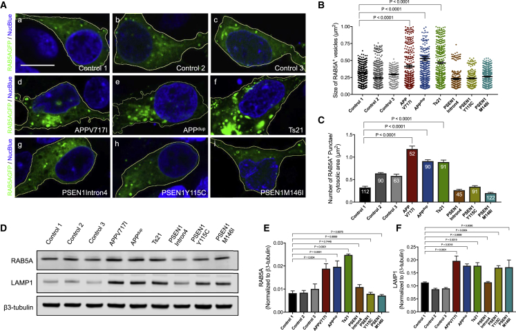
Figureu00a01
Alzheimeru2019s Disease Mutations in APP , but Not PSEN1 , Result in Early Endosome Abnormalities in Human Cortical Neurons (A) Representative images of neurons generated from three independent non-de...
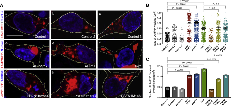
Figureu00a02
Accumulation of Enlarged Late Endosomes and Lysosomes in Human Neurons with PSEN1 or APP Mutations (A) Representative images of neurons expressing a LAMP1-RFP fusion protein (red; blue, nuclei labeled...
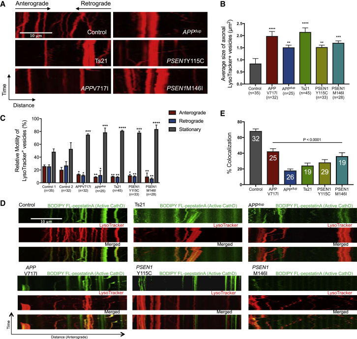
Figureu00a03
Impaired Axonal Lysosome Transport and Proteolysis Deficits in Human APP and PSEN1 Mutant Neurons (A) Representative kymographs showing transport of lysosomes labeled with LysoTracker Red DND 99. Scal...
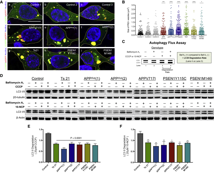
Figureu00a04
Defective Degradation of Autophagosomes in Human APP and PSEN1 Mutant Neurons (A) Increased size of autophagosomes in human cortical excitatory neurons with APP and PSEN1 mutations, as detected by liv...
Figureu00a05
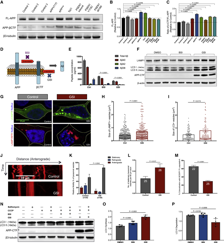
Gamma-Secretase Inhibition Disrupts Function of the Lysosomal-Autophagic System in Human Neurons (Au2013C) APP-u03b2-CTF levels are significantly increased in human cortical excitatory neurons with AP...
Figureu00a06
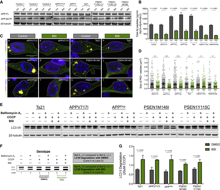
Dysfunction in the Lysosome-Autophagy System in Familial AD Neurons Is Reversed by u03b2-Secretase Inhibition (A) BSI reduces APP-u03b2CTF level in all genotypes. Representative western blots of FL-AP...
Figureu00a07
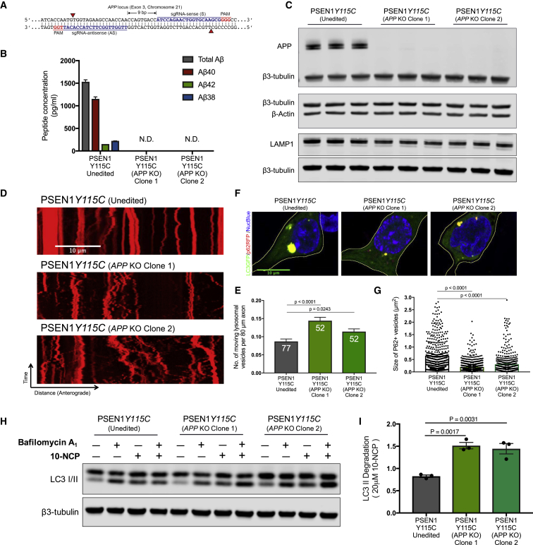
Autophagy Defects in PSEN1 Mutant Neurons Are Rescued by CRISPR Knockout of APP (A) Double nickase strategy with sense (S) and antisense (AS) single guide RNAs (sgRNAs) separated by 9u00a0bp at exon 3...
Figure images are served from the NIH/NLM PubMed Central Open Access Subset or Europe PMC; copyright remains with the publishers and authors.
💬 Discussion
0 commentsNo comments yet. Be the first to start a discussion!
Leave a Comment