Abstract
The goal of this study was to develop near-infrared (NIR) resonant gold-gold sulfide nanoparticles (GGS-NPs) as dual contrast and therapeutic agents for cancer management via multiphoton microscopy followed by higher intensity photoablation. We demonstrate that GGS-NPs exposed to a pulsed, NIR laser exhibit two-photon induced photoluminescence that can be utilized to visualize cancerous cells in vitro. When conjugated with anti-HER2 antibodies, these nanoparticles specifically bind SK-BR-3 breast carcinoma cells that over-express the HER2 receptor, enabling the cells to be imaged via multiphoton microscopy with an incident laser power of 1 mW. Higher excitation power (50 mW) could be employed to induce thermal damage to the cancerous cells, producing extensive membrane blebbing within seconds leading to cell death. GGS-NPs are ideal multifunctional agents for cancer management because they offer the ability to pinpoint precise treatment sites and perform subsequent thermal ablation in a single setting.
🔬 Techniques
🔭 Microscopes
💻 Software
✨ Fluorophores
🧪 Sample Preparation
🔬 Cell Lines
🏭 Microscope Brands
🧪 Reagent Suppliers
🔴 Lasers
💻 Software Details
🏛️ Research Organizations (ROR)
Affiliated research institutions:
📋 Methods
GGS-NP synthesis and functionalization GGS-NPs were synthesized using a variation of the procedures described by Averitt et al 35 and Schwartzberg et al. 30 Solutions of HAuCl 4 (2 mM, Alfa Aesar, Ward Hill, MA) and Na 2 S 2 O 3 (1 mM, Sigma, Saint Louis, MO) were prepared in milli-Q water, aged two days at room temperature, and mixed in small quantities at volumetric ratios ranging from 1:1 to 1:2 (HAuCl 4 :Na 2 S 2 O 3 ). The ratio that produced nanoparticles resonant near 800 nm as determined with a UV-visible spectrophotometer (Cary 50, Varian, Walnut Creek, CA) was used to synthesize a large batch of nanoparticles for in vitro experiments. GGS-NPs were visualized with transmission electron microscopy and the diameter of at least 50 nanoparticles per sample was measured with ImageJ software (NIH, Bethesda, MD). Calculation of the mean diameter revealed a slight batch-to-batch variability in average particle size, which ranged from 26 nm to 37 nm. In the initial reaction solutions we also observed ∼5 nm diameter colloidal gold and 50–100 nm flat triangular nanoparticles; however, most of these particles were removed from solution by a multi-step centrifugation process so the final product used in experiments consisted of a majority of GGS-NPs. Dynamic light scattering was also incorporated to assess nanoparticle size and analysis of multiple batches with a ZetaSizer NanoZS (Malvern Instruments, Worcestershire, UK) revealed an average hydrodynamic diameter of 42.2 nm, in good agreement with the transmission electron microscopy (TEM) results. GGS-NPs were conjugated to either anti-HER2 (NeoMarkers, Freemont, CA) or nonspecific anti-IgG (Sigma) antibodies using 2,000 Da orthopyridyl-disulfide-poly(ethylene glycol)-N-hydroxysuccinimide (OPSS-PEG-NHS, Creative PEGWorks, Winston Salem, NC) as a linker. PEG-antibody conjugates were prepared by reacting one part 125 μM OPSSPEG-NHS with 9 parts 1 mg/mL antibody at 4°C overnight. This reaction produces a stable amide bond between primary amines on the antibody and carboxyl groups on the PEG chain that are exposed when the NHS terminus is cleaved in water. The particles were suspended in milli-Q water and exposed to PEG-antibody conjugates for 1 hour at 4°C at a 100:1 volumetric ratio. Following antibody coupling, GGS-NPs were reacted with a solution of mPEG-SH (5 mM, 5,000 Da, Laysan Bio, Inc., Arab, AL) for a minimum of 4 hours at 4°C (1:200 volumetric ratio) to passivate any exposed gold surface area. GGS-NPs coated with mPEG-SH only (no antibody) were also synthesized for use as a negative control. Self-assembly of PEG-antibody and mPEG-SH onto the nanoparticle surface is possible due to dative interactions between sulfur and gold. Following antibody and/or PEG modification, GGS-NPs were centrifuged to remove unbound molecules, aspirated, and suspended in phosphate buffered saline (PBS) at an optical density of 2.0 (∼4.2 × 10 11 particles/ml) unless otherwise noted.
Show full methods section
GGS-NP synthesis and functionalization GGS-NPs were synthesized using a variation of the procedures described by Averitt et al 35 and Schwartzberg et al. 30 Solutions of HAuCl 4 (2 mM, Alfa Aesar, Ward Hill, MA) and Na 2 S 2 O 3 (1 mM, Sigma, Saint Louis, MO) were prepared in milli-Q water, aged two days at room temperature, and mixed in small quantities at volumetric ratios ranging from 1:1 to 1:2 (HAuCl 4 :Na 2 S 2 O 3 ). The ratio that produced nanoparticles resonant near 800 nm as determined with a UV-visible spectrophotometer (Cary 50, Varian, Walnut Creek, CA) was used to synthesize a large batch of nanoparticles for in vitro experiments. GGS-NPs were visualized with transmission electron microscopy and the diameter of at least 50 nanoparticles per sample was measured with ImageJ software (NIH, Bethesda, MD). Calculation of the mean diameter revealed a slight batch-to-batch variability in average particle size, which ranged from 26 nm to 37 nm. In the initial reaction solutions we also observed ∼5 nm diameter colloidal gold and 50–100 nm flat triangular nanoparticles; however, most of these particles were removed from solution by a multi-step centrifugation process so the final product used in experiments consisted of a majority of GGS-NPs. Dynamic light scattering was also incorporated to assess nanoparticle size and analysis of multiple batches with a ZetaSizer NanoZS (Malvern Instruments, Worcestershire, UK) revealed an average hydrodynamic diameter of 42.2 nm, in good agreement with the transmission electron microscopy (TEM) results. GGS-NPs were conjugated to either anti-HER2 (NeoMarkers, Freemont, CA) or nonspecific anti-IgG (Sigma) antibodies using 2,000 Da orthopyridyl-disulfide-poly(ethylene glycol)-N-hydroxysuccinimide (OPSS-PEG-NHS, Creative PEGWorks, Winston Salem, NC) as a linker. PEG-antibody conjugates were prepared by reacting one part 125 μM OPSSPEG-NHS with 9 parts 1 mg/mL antibody at 4°C overnight. This reaction produces a stable amide bond between primary amines on the antibody and carboxyl groups on the PEG chain that are exposed when the NHS terminus is cleaved in water. The particles were suspended in milli-Q water and exposed to PEG-antibody conjugates for 1 hour at 4°C at a 100:1 volumetric ratio. Following antibody coupling, GGS-NPs were reacted with a solution of mPEG-SH (5 mM, 5,000 Da, Laysan Bio, Inc., Arab, AL) for a minimum of 4 hours at 4°C (1:200 volumetric ratio) to passivate any exposed gold surface area. GGS-NPs coated with mPEG-SH only (no antibody) were also synthesized for use as a negative control. Self-assembly of PEG-antibody and mPEG-SH onto the nanoparticle surface is possible due to dative interactions between sulfur and gold. Following antibody and/or PEG modification, GGS-NPs were centrifuged to remove unbound molecules, aspirated, and suspended in phosphate buffered saline (PBS) at an optical density of 2.0 (∼4.2 × 10 11 particles/ml) unless otherwise noted.
Quantification of antibody bound to nanoparticles
To verify that antibody and mPEG-SH successfully bound the GGS-NP surface we monitored changes in hydrodynamic diameter, which should increase upon addition of biomolecules. Using a Malvern ZetaSizer NanoZS, four nanoparticle formulations were studied: (1) Bare GGS-NPs, (2) GGS-NPs coated with mPEG-SH, (3) GGS-NPs coated with mPEG-SH and anti-IgG antibodies, and (4) GGS-NPs coated with mPEG-SH and anti-HER2 antibodies. The mean hydrodynamic diameter of each formulation was calculated as an average from three sets of nanoparticles, with individual sets also being tested in triplicate. Bare nanoparticles demonstrated a mean hydrodynamic diameter of 42.2 nm, which increased to 58.0 nm upon addition of mPEG-SH. A further increase occurred when either antibody formulation was included, with diameter of 69.8 nm for anti-IgG coated nanoparticles and 63.4 nm for anti-HER2 coated nanoparticles. These results suggested the antibody and mPEG-SH were able to self-assemble on the nanoparticle surface using the disulfide or thiol terminus, respectively. To quantify the amount of antibody present on the nanoparticles, targeted (coated with mPEG-SH and antibody) and control (only mPEG-SH coated) nanoparticles were incubated with 10 μg/ml horseradish peroxidase (HRP)-conjugated goat anti-mouse IgG (for quantification of mouse anti-human HER2) or HRP-conjugated rabbit anti-goat IgG (for quantification of goat anti-mouse IgG) (both HRP antibodies from Sigma). Nonspecific reaction sites were blocked with a 3% solution of bovine serum albumin (BSA, Sigma) in PBS. To remove unbound HRP-labeled antibodies, the nanoparticles were centrifuged twice at 1500 g for eight minutes and suspended in 3% BSA. The HRP bound to GGS-NPs was developed with 3,3′,5,5′-tetramethylbenzidine dihydrochloride (Sigma) for 15 minutes and the reaction was stopped by addition of 2M sulfuric acid. The developed HRP was compared to a standard curve of the appropriate HRP-conjugated anti-IgG by determining the absorbance at 450 nm with a spectrophotometer. The total amount of peroxidase-labeled anti-IgG present in solution was divided by the total number of GGS-NPs in solution to determine the number of antibodies per nanoparticle. The number of nanoparticles was calculated from the Beer-Lambert law with the extinction coefficient of GGS-NPs derived from Mie theory as described by Averitt et al. 35 Cell culture SK-BR-3 breast carcinoma cells (American Type Culture Collection, Manassas, VA), which over-express the HER2 receptor, 34 were cultured in McCoy’s 5A growth medium (Sigma) containing 10% fetal bovine serum and 1% penicillin-streptomycin at 37°C in a 5% CO 2 environment. For experiments, cells transferred to 15 mL conical tubes (6 × 10 5 cells/tube) were centrifuged (115 g, 3 minutes) to form pellets that were subsequently suspended in 1 mL of one of the four following solutions: (1) nanoparticles coated with anti-HER2 and mPEG-SH, (2) nanoparticles coated with anti-IgG and mPEG-SH, (3) nanoparticles coated with only mPEG-SH, or (4) PBS. The nanoparticle solutions consisted of approximately 4.2 × 10 11 GGS-NPs. Cells were incubated in these suspensions for 30 minutes at 37°C in a hybridization chamber (VWR International, West Chester, PA) with constant rotation at 7 rpm. Following incubation, the samples were centrifuged (115 g, 3 minutes), aspirated, and diluted in PBS to remove any particles not bound to the cells. This rinsing procedure was repeated thrice followed by resuspension in growth media (1 mL). The cells were cultured on chambered coverglass overnight before experiments were performed. For studies of the effect of thermal therapy on cell membrane structure, the cells were labeled with 5 μM DiI (Molecular Probes, Eugene, OR) for 10 minutes prior to incubation with the nanoparticles as described above. Multiphoton microscopy and photothermal therapy A Zeiss Laser Scanning Microscope (LSM) 510 META (Carl Zeiss, Inc., Thornwood, NJ) equipped with a femtosecondpulsed Ti:sapphire laser source (Chameleon, Coherent, Inc., Santa Clara, CA) was used to perform multiphoton microscopy and photothermal therapy experiments. The wavelength of the output laser beam was tuned to match the peak extinction of the GGS-NPs and operated with a pulse width of 140 fs and repetition rate of 90 MHz. A short-pass dichroic mirror was used to reflect incident NIR light onto the sample through a 20× objective (numerical aperture (NA) = 0.75) or a 63× objective (NA = 1.4) and to collect photoluminescence. Background signal was reduced with an infrared-blocking filter and the META detector was used to collect TPL from the GGS-NPs between 451–644 nm. For imaging nanoparticles on cells, incident laser power was 1 mW with a pixel dwell time of 12.8 μsec and the laser beam was raster-scanned across a 450 μm × 450 μm area. Calculating laser intensity by dividing power by the area of the Airy disc, this corresponds to a fluence of 0.96 J/cm 2 . To perform photothermal ablation, the samples were repositioned and laser power was increased to 50 mW (48.1 J/cm 2 ) while maintaining the same dwell time. Samples were treated with a single pass of the laser. One hour after laser treatment cell viability was assessed by labeling cells with Calcein AM (1 μM, Molecular Probes, Eugene, OR), a live cell stain, and ethidium homodimer-1 (4 μM, EthD-1, Molecular Probes), a dead cell stain. Fluorescence microscopy was performed with an inverted Zeiss Axiovert 135 phase contrast microscope (Carl Zeiss).
📊 Figures
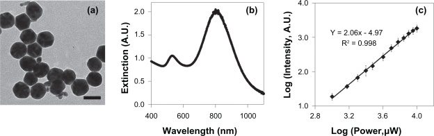
Figure 1
a) TEM of GGS-NPs. Scale bar = 40 nm. b) Extinction spectrum of the GGS-NPs. c) GGS-NPs displayed a quadratic dependence of luminescence intensity on excitation power when exposed to an 800 nm pulsed ...
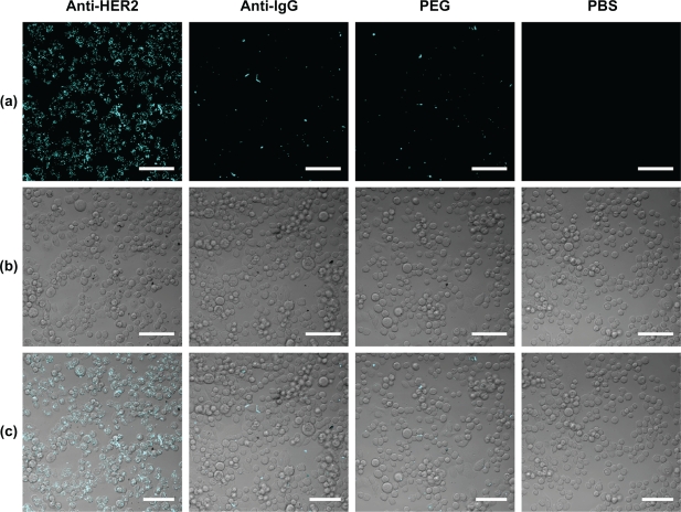
Figure 2
a) Two-photon induced photoluminescence images of SK-BR-3 cells exposed to 1 mW with the pulsed laser tuned to 800 nm. b) Brightfield images of SK-BR-3 cells in the same field-of-view as the luminesce...
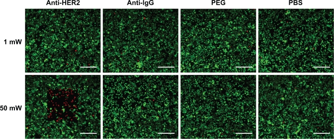
Figure 3
Calcein AM staining indicated that cancerous cells remained viable (evidenced by green fluorescent signal) when exposed to 1 mW laser power, regardless of nanoparticle presence. At 50 mW laser output ...
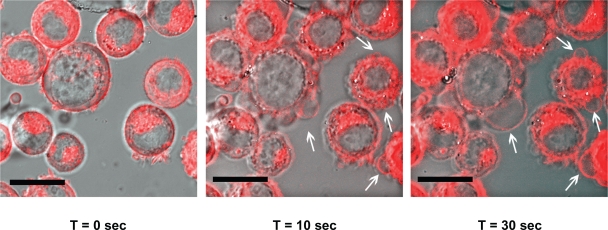
Figure 4
Time-lapse photography of SK-BR-3 cells exposed to anti-HER2 functionalized GGS-NPs and 50 mW laser power. The fluorescent red DiI membrane stain indicates regions of membrane blebbing generated by lo...
Figure images are served from the NIH/NLM PubMed Central Open Access Subset or Europe PMC; copyright remains with the publishers and authors.
💬 Discussion
0 commentsNo comments yet. Be the first to start a discussion!
Leave a Comment