Abstract
AbstractParkinson’s disease is a slowly progressive neurodegenerative disease characterised by dysfunction and death of selectively vulnerable midbrain dopaminergic neurons and the development of human in vitro cellular models of the disease is a major challenge in Parkinson’s disease research. We constructed an automated cell culture platform optimised for long-term maintenance and monitoring of different cells in three dimensional microfluidic cell culture devices. The system can be flexibly adapted to various experimental protocols and features time-lapse imaging microscopy for quality control and electrophysiology monitoring to assess cellular activity. Using this system, we continuously monitored the differentiation of Parkinson’s disease patient derived human neuroepithelial stem cells into midbrain specific dopaminergic neurons. Calcium imaging confirmed the electrophysiological activity of differentiated neurons and immunostaining confirmed the efficiency of the differentiation protocol. This system is the first example of an automated Organ-on-a-Chip culture and has the potential to enable a versatile array of in vitro experiments for patient-specific disease modelling.
🔬 Techniques
🔭 Microscopes
✨ Fluorophores
🧪 Sample Preparation
🔬 Cell Lines
🏭 Microscope Brands
🧪 Reagent Suppliers
💻 Software Details
🏛️ Research Organizations (ROR)
Affiliated research institutions:
📋 Methods
System
Construction and Design The Pelican is composed of a sterile automation enclosure that abuts a sterile manual enclosure on one side and an incubator on another. The automation enclosure contains a set of devices that may physically communicate via a four-axis gantry robot within a customised housing support. The manual enclosure is a cell culture hood, adapted for restricted communication of material with the automation enclosure. The automation enclosure currently includes a de-lidder, eight-fold and 96fold parallel dispenser, three-axis fourfold liquid handling robot (pipettor) with disposable tips, confocal microscope, and camera. The assembly (Fig. 7a,b ), all of the components are described in the Supplementary Experimental Procedures. Figure 7 CAD drawing of automated workstation. ( a ) Top view inside the Pelican without housing. ( b ) Front view of the Pelican with housing. Yellow (imaging station), light blue (liquid handling station), green (level of stainless steel work surface) and orange (waste containers). Perforated surface = work surface. (1) Storage incubator. (2) Liquid-handler. (3) Hotels (manual working bench). (4) Position of microscopy station. (5) Position of liquid dispenser. (6) Automated work bench. Coordinates shown here are consistent with the remainder of the manuscript. See also Figs S1 and S2 .
Cell culture
All work with human iPSCs and thereof derived cells has been approved by the Ethics Review Panel (ERP) of the University Luxembourg as well as by the Luxembourgish Comité National d’Ethique de Recherche (CNER). The CNER reference number is 201305/04. All experiments were performed in accordance with relevant guidelines and regulations. We confirm that written informed consent was obtained, by our cell line suppliers, for the establishment of stem cell lines, from all donors.
Show full methods section
System
Construction and Design The Pelican is composed of a sterile automation enclosure that abuts a sterile manual enclosure on one side and an incubator on another. The automation enclosure contains a set of devices that may physically communicate via a four-axis gantry robot within a customised housing support. The manual enclosure is a cell culture hood, adapted for restricted communication of material with the automation enclosure. The automation enclosure currently includes a de-lidder, eight-fold and 96fold parallel dispenser, three-axis fourfold liquid handling robot (pipettor) with disposable tips, confocal microscope, and camera. The assembly (Fig. 7a,b ), all of the components are described in the Supplementary Experimental Procedures. Figure 7 CAD drawing of automated workstation. ( a ) Top view inside the Pelican without housing. ( b ) Front view of the Pelican with housing. Yellow (imaging station), light blue (liquid handling station), green (level of stainless steel work surface) and orange (waste containers). Perforated surface = work surface. (1) Storage incubator. (2) Liquid-handler. (3) Hotels (manual working bench). (4) Position of microscopy station. (5) Position of liquid dispenser. (6) Automated work bench. Coordinates shown here are consistent with the remainder of the manuscript. See also Figs S1 and S2 .
Cell culture
All work with human iPSCs and thereof derived cells has been approved by the Ethics Review Panel (ERP) of the University Luxembourg as well as by the Luxembourgish Comité National d’Ethique de Recherche (CNER). The CNER reference number is 201305/04. All experiments were performed in accordance with relevant guidelines and regulations. We confirm that written informed consent was obtained, by our cell line suppliers, for the establishment of stem cell lines, from all donors.
Human neuroepithelial stem cell culture
In order to demonstrate the capability of the automated system to maintain and monitor many types of cells including PD specific cell lines, we used a human neuroepithelial stem cell line from a healthy donor (hNESC K7) and a human neuroepithelial stem cell line derived from a patient carrying the Parkinson’s disease related mutation p.I368N in PINK1 (40066C5N). These cells were maintained and differentiated into midbrain-specific dopaminergic neurons within an OrganoPlate, by automating an existing macroscopic cell culture protocol 20 , that we previously adapted for microfluidic cell culture 35 . In brief, to culture hNESCs in an OrganoPlate, they were harvested from wells of a 6 well plate. The harvested hNESCs were then re-suspended on Matrigel (catalogue number 354277, lot number 3318549, Discovery Labware, Inc., Two Oak Park, Bedford, MA, USA). 0.7 μL of this Matrigel-cell mix was loaded in assigned chips of the OrganoPlate at a density of 0.03 million cells/μL. After seeding the cells, the plate was loaded into position B3 in the hotel of the Pelican. Afterwards, the plate was moved by the robotic arm to the storage incubator, at 37 °C and 5% CO2.
Dopaminergic neuronal differentiation
The culture medium preparation “N2B27 medium” consisted of mixed equal amounts of Neurobasal medium (invitrogen/life technologies) and DMEM/F12 medium (invitrogen/life technologies) supplemented with 1% penicillin/streptomycin (life technologies), 2 mM L-glutamine (life technologies), 0.5 X B27 supplement without Vitamin A (life technologies) and 0.5 X N2 supplement (life technologies). The medium to maintain the hNESC in culture “maintenance medium” consisted of N2B27 medium with 0.5 μM PMA (Enzo life sciences), 3 μM CHIR (Axon Medchem) and 150 μM Ascorbic Acid (Sigma Aldrich). The differentiation medium formulation to induce the differentiation of hNESCs towards midbrain dopaminergic neurons “differentiation medium with PMA” consisted of N2B27 medium with 200 μM ascorbic acid, 0.01 ng/μL BDNF (Peprotech), 0.01 ng/μL GDNF (Peprotech), 0.001 ng/μL TGFβ3 (Peprotech), 2.5 μM dbcAMP (Sigma Aldrich) and 1 μM PMA. The function of PMA in this medium preparation was to stimulate the sonic hedghog (SHH) pathway in the cultured hNESCs. Differentiation medium with PMA was changed, every 2 days during the first 6 days of culture in the differentiation process. For the maturation of differentiated neurons, PMA was no longer added to the differentiation medium “differentiation medium without PMA” from day 7 onwards, which was changed every 2 days during 3 weeks. To monitor cellular morphology during differentiation, bright field images were acquired automatically in the Pelican using the microscopy station.
Calcium imaging assay
A calcium imaging assay was done on 15 representative chips of the OrganoPlate at day 24 of differentiation. At room temperature, 50 μL of 5 μM cell permeant Fluo-4 AM (Life technologies) in neurobasal medium (Invitrogen/Life technologies) was manually added to the medium inlet well and 20 μ L to the medium outlet well of selected chips of the OrganoPlate. Then, the plate was incubated for 30 min at 37 °C and 5% CO2. The plate was then placed in an onstage incubator within the microscope. Calcium time-series of spontaneously firing hNESC-derived neurons were then automatically acquired. Images were sampled at a rate of 1 Hz for approximately 5 min, stored as image stacks and analysed using custom Matlab (version 2016b; MathWorks Inc.) scripts. Regions of interest corresponding to individual cells were automatically segmented with an established technique 37 and fluorescence traces were generated for each segmented cell and presented as relative changes in fluorescence intensity ∆ F/F .
Immunofluorescence staining assay
Immunostaining for the dopaminergic neuronal markers class 3 beta tubulin (TUB β III) and tyrosine hydroxylase (TH), the penultimate enzyme in the biosynthesis of dopamine 20 , 49 , 50 , was performed on representative chips at day 24 of differentiation. Differentiated cells were fixed with 4% paraformaldehyde (PFA) in 1 × phosphate-buffered saline (PBS) for 15 min, by manually adding 70 μL in medium well inlet and 30 μL in medium well outlet followed by permeabilisation with 0.05% Triton-X 100 in 1 × PBS (3 min on ice), and blocking with 10% fetal calf serum (FCS) in 1 × PBS (1 h). After washing with 1 × PBS, the primary antibodies mouse anti-TUBβIII (1:2000, Covance) and rabbit anti-TH (1:2000, Santa cruz biotechnology), were incubated for 90 min at room temperature. After washing with 1 × PBS, the secondary antibodies Alexa Fluor 488 Goat Anti-Mouse and Alexa Fluor 568 Goat Anti-Rabbit together with a stain DNA (Hoechst 33342, Invitrogen), were incubated for 2 hours at room temperature. After washing with 1 × PBS and water, confocal images of representative culture chambers were acquired using a confocal microscope (Zeiss LSM 710). Automating the differentiation of human neuroepithelial stem cell into dopaminergic neurons LACS Config Editor was used to develop automated pipelines for the differentiation of hNESCs into dopaminergic neurons and time-lapse imaging microscopy. The automated pipelines were drafted according to the SiLA communication protocol and command format as previously described 9 , 42 , 51 . In brief, SiLA uses a Simple Object Access Protocol (SOAP) and a Web Service Description Language (WSDL) documentation, both of which are based on XML. A full library of commands for each device is downloaded once and stored in LACS as a configuration document of the Pelican. LACS Config Editor was used to incorporate the automated pipelines in this configuration file used herein to automate the differentiation of hNESCs into dopaminergic neurons. On the workbench, gel-embedded hNESCs were manually loaded into the culture lanes of a 2-lane OrganoPlate as described above. Then, the plate was put inside the Pelican through Hotel B, and placed inside the storage incubator by the robotic arm. The dispenser was fitted with a 5 μL cassette from Biotek and used as a dispensing medium for the media change.
Statistical Analysis
Three representative chips (n = 3) were selected to illustrate the results of this study, in which the statistical analyses were performed by determination of the mean value and the standard deviation of the proportion of dopaminergic neurons within the overall neuronal population.
Electronic supplementary material Supplementary Video 1 Supplementary Video 2 Supplementary Video 3 Supplementary Material
Electronic supplementary material Supplementary information accompanies this paper at 10.1038/s41598-018-34828-3.
📊 Figures
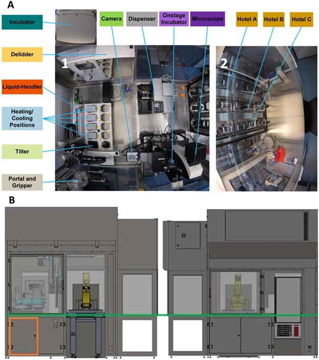
Figure 1
Pelican automated cell culture observatory. ( A ) Top view inside the Pelican automation workstation without housing: (1) Wide angle lens image of the automated enclosure. (2) Wide angle lens image of...
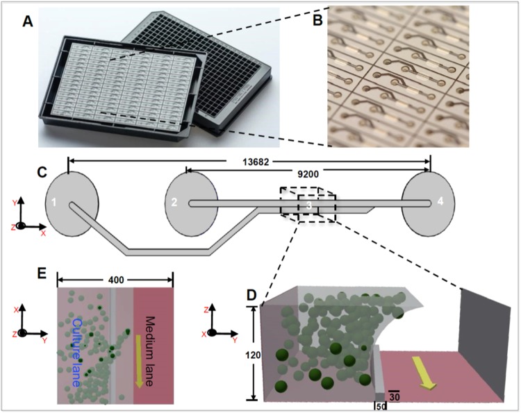
Figure 2
Microfluidic cell culture device: OrganoPlate. ( A ) Photograph of underside (left) and upper views (right) of an OrganoPlate. ( B ) bottom plate of OrganoPlate with selective chips. ( C ) A schematic...
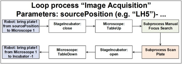
Figure 3
Semi-automated image acquisition pipeline. See Supplementary Fig. S5 for more details.
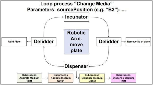
Figure 4
Automated media change pipeline. See Supplementary Fig. S4 for more details.
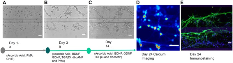
Figure 5
Dopaminergic neuronal differentiation. Bright field images of hNESCs with media components at ( A ) 1 day ( B ) 4 days and ( C ) 21 days after seeding. ( D ) Calcium imaging frame of a firing event ta...
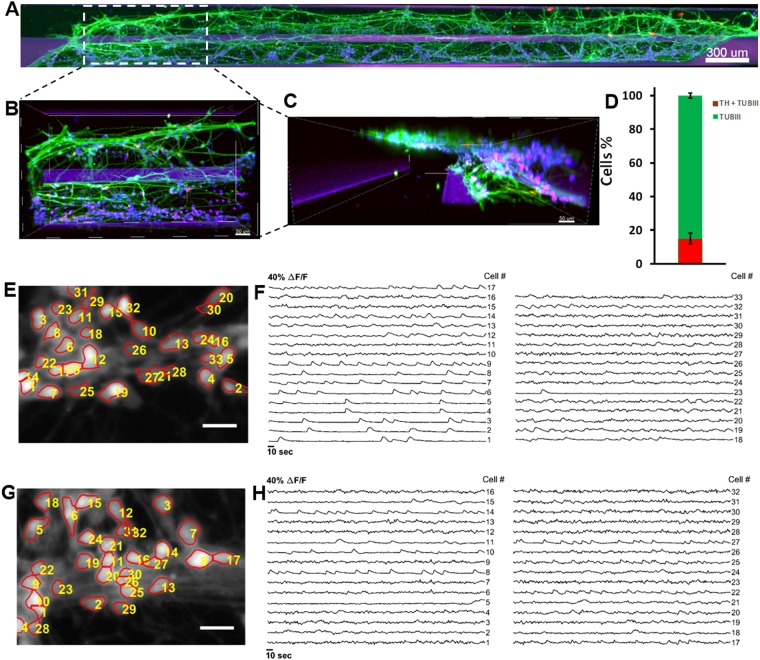
Figure 6
Immunostaining and automated cell segmentation of calcium time-series hNESC differentiated in chips of the OrganoPlate inside the Pelican. ( A ) Top view of an entire chip in the OrganoPlate showing d...
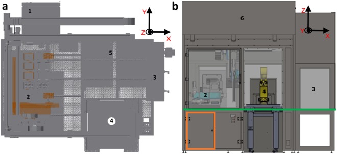
Figure 7
CAD drawing of automated workstation. ( a ) Top view inside the Pelican without housing. ( b ) Front view of the Pelican with housing. Yellow (imaging station), light blue (liquid handling station), g...
Figure images are served from the NIH/NLM PubMed Central Open Access Subset or Europe PMC; copyright remains with the publishers and authors.
💬 Discussion
0 commentsNo comments yet. Be the first to start a discussion!
Leave a Comment