Abstract
Autophagy is a membrane-mediated degradation process, which is governed by sequential functions of Atg proteins. Although Atg proteins are highly conserved in eukaryotes, protozoa possess only a partial set of Atg proteins. Nonetheless, almost all protozoa have the complete factors belonging to the Atg8 conjugation system, namely, Atg3, Atg4, Atg7, and Atg8. Here, we report the biochemical properties and subcellular localization of the Atg8 protein of the human malaria parasite Plasmodium falciparum (PfAtg8). PfAtg8 is expressed during intra-erythrocytic development and associates with membranes likely as a lipid-conjugated form. Fluorescence microscopy and immunoelectron microscopy show that PfAtg8 localizes to the apicoplast, a four membrane-bound non-photosynthetic plastid. Autophagosome-like structures are not observed in the erythrocytic stages. These data suggest that, although Plasmodium parasites have lost most Atg proteins during evolution, they use the Atg8 conjugation system for the unique organelle, the apicoplast.
🔬 Techniques
🧬 Organisms
✨ Fluorophores
🧪 Sample Preparation
🔬 Cell Lines
🏭 Microscope Brands
🧪 Reagent Suppliers
🏛️ Research Organizations (ROR)
Affiliated research institutions:
📋 Methods
Parasite culture
P. falciparum strain FCR3 was cultured in human B + erythrocytes as described [61] . In some subcellular localization experiments, the 3D7 parasite strain transfected with pSSPF2/GFP-ACP was used; the transfectant was cultured in the standard culture medium supplemented with 5 nM WR99210 [33] . Where indicated, chloroquine (Sigma-Aldrich) was added to the culture medium. For synchronizing the culture, the red blood cells infected by the late stage schizont were recovered from asynchronous culture by 60% Percoll (GE healthcare) density centrifugation at 2000× g for 20 min. After 4 h the culture was treated with 5% D-sorbitol [62] , yielding parasites tightly synchronized in the early ring stage (0–4 h after parasite invasion of the erythrocyte).
Cloning of PfAtg8 cDNA and generation of anti-PfAtg8 antibodies
RNA extraction and cDNA synthesis were carried out as described previously [63] , [64] . GST-fused PfAtg8 recombinant protein was generated using a wheat germ cell-free system [65] . Two independent anti-PfAtg8 antisera (#1 and #2) were raised in two New Zealand white rabbits and the antibodies were purified using GST-PfAtg8 recombinant protein. Animal experimental protocols were approved by the Institutional Animal Care and Use Committee of Tokyo Medical and Dental University (No. 0110115A).
Immunoblotting
Parasites were collected from erythrocytes by treatment with 0.15% saponin (Sigma) in phosphate-buffered saline (PBS) with Complete Protease Inhibitor cocktail (Roche Applied Science), washed three times in PBS and lysed in sample buffer. Parasite extracts were loaded onto 13.5% SDS gel and transferred to a PVDF membrane. Blots were blocked with 5% skim milk in Tris-buffered saline with 0.01% Tween 20 (TBST) and stained with primary antibodies overnight at 4°C. The following primary antibodies were used: rabbit anti-PfAtg8 and mouse monoclonal anti-PfHSP70 antibodies (1∶100) [66] , [67] . After washing with TBST, blots were stained with HRP-conjugated secondary antibodies and visualized with SuperSignal West Pico Chemiluminescent substrate (Thermo Fisher Scientific). Subcellular fractionation Asynchronous parasites were harvested as described above. Parasite pellets were disrupted by three cycles of freezing/thawing in MSE buffer (225 mM mannitol, 75 mM sucrose, 0.1 mM EDTA, and 3 mM Tris-HCl [pH 7.4]). Cell debris and intact erythrocytes were removed by centrifugation at 800× g for 5 min. The supernatant was spun at 13,000× g for 15 min to separate the LSP, and the supernatant was centrifuged again at 100,000× g for 60 min to generate the high-speed pellet (HSP) and high-speed supernatant (HSS). The LSP and HSP were resuspended in the same buffer. To analyze solubility, each sample was incubated with 2 M urea or 2% Triton X-100 on ice for 1 h, and then centrifuged at 100,000× g for 1 h. The samples were precipitated with ice-cold acetone, resuspended in SDS-PAGE sample buffer, and analyzed by SDS-PAGE.
Show full methods section
Parasite culture
P. falciparum strain FCR3 was cultured in human B + erythrocytes as described [61] . In some subcellular localization experiments, the 3D7 parasite strain transfected with pSSPF2/GFP-ACP was used; the transfectant was cultured in the standard culture medium supplemented with 5 nM WR99210 [33] . Where indicated, chloroquine (Sigma-Aldrich) was added to the culture medium. For synchronizing the culture, the red blood cells infected by the late stage schizont were recovered from asynchronous culture by 60% Percoll (GE healthcare) density centrifugation at 2000× g for 20 min. After 4 h the culture was treated with 5% D-sorbitol [62] , yielding parasites tightly synchronized in the early ring stage (0–4 h after parasite invasion of the erythrocyte).
Cloning of PfAtg8 cDNA and generation of anti-PfAtg8 antibodies
RNA extraction and cDNA synthesis were carried out as described previously [63] , [64] . GST-fused PfAtg8 recombinant protein was generated using a wheat germ cell-free system [65] . Two independent anti-PfAtg8 antisera (#1 and #2) were raised in two New Zealand white rabbits and the antibodies were purified using GST-PfAtg8 recombinant protein. Animal experimental protocols were approved by the Institutional Animal Care and Use Committee of Tokyo Medical and Dental University (No. 0110115A).
Immunoblotting
Parasites were collected from erythrocytes by treatment with 0.15% saponin (Sigma) in phosphate-buffered saline (PBS) with Complete Protease Inhibitor cocktail (Roche Applied Science), washed three times in PBS and lysed in sample buffer. Parasite extracts were loaded onto 13.5% SDS gel and transferred to a PVDF membrane. Blots were blocked with 5% skim milk in Tris-buffered saline with 0.01% Tween 20 (TBST) and stained with primary antibodies overnight at 4°C. The following primary antibodies were used: rabbit anti-PfAtg8 and mouse monoclonal anti-PfHSP70 antibodies (1∶100) [66] , [67] . After washing with TBST, blots were stained with HRP-conjugated secondary antibodies and visualized with SuperSignal West Pico Chemiluminescent substrate (Thermo Fisher Scientific). Subcellular fractionation Asynchronous parasites were harvested as described above. Parasite pellets were disrupted by three cycles of freezing/thawing in MSE buffer (225 mM mannitol, 75 mM sucrose, 0.1 mM EDTA, and 3 mM Tris-HCl [pH 7.4]). Cell debris and intact erythrocytes were removed by centrifugation at 800× g for 5 min. The supernatant was spun at 13,000× g for 15 min to separate the LSP, and the supernatant was centrifuged again at 100,000× g for 60 min to generate the high-speed pellet (HSP) and high-speed supernatant (HSS). The LSP and HSP were resuspended in the same buffer. To analyze solubility, each sample was incubated with 2 M urea or 2% Triton X-100 on ice for 1 h, and then centrifuged at 100,000× g for 1 h. The samples were precipitated with ice-cold acetone, resuspended in SDS-PAGE sample buffer, and analyzed by SDS-PAGE.
Expression of PfAtg8 in mammalian cells
PfAtg8 cDNA was inserted into a pCI-neo mammalian expression plasmid (Promega) and transfected into HEK293T cells [68] using Lipofectamine 2000 reagent (Invitrogen). Total cell lysates were subjected to SDS-PAGE and immunoblot analysis.
Immunofluorescence microscopy
Parasite thin blood smears were fixed with 4% paraformaldehyde/PBS for 10 min and samples were permealized with 0.1% Triton X-100/PBS for 15 min. After blocking with 3% bovine serum albumin/PBS for 1 h, samples were incubated with primary and secondary antibodies for 2 h and 1 h, respectively. The smears were mounted with Prolong Gold (Invitrogen). All reactions were carried out at room temperature. Samples were observed with a confocal laser microscope (FV1000D IX81, Olympus) using a 60x PlanApoN oil immersion lens (1.42 NA; Olympus). The following primary antibodies were used: purified rabbit anti-PfAtg8 (1∶200 for #1, and 1∶100 for #2) antibody, mouse anti-apical membrane antigen (AMA)1 (1∶500) [67] , anti-rhoptry-associated protein 1 (RAP1, 1∶200) [67] , anti-rhoptry neck protein 2 (RON2, 1∶200) [69] , and anti-ring-infected erythrocyte surface antigen (RESA) (23/9, 1∶200) [70] antibodies, rabbit anti- PfHU (organellar histone-like protein) antibody [34] , and rat anti-GFP antibody (Nacalai Tesque). For visualizing the mitochondrion, parasites were preincubated for 30 min with complete culture medium containing 100 nM MitoTracker Red CMXRos (Molecular Probes).
Immunoelectron microscopy
Mature schizont stage parasites were enriched from synchronous culture using MACS 25LD columns (MiltenyiBiotec) as previously described [71] . For immunoelectron microscopy of P. falciparum , the previously described pre-embedding silver enhancement immunogold method [72] was used with slight modifications. The parasitized erythrocytes were fixed in 4% paraformaldehyde and 0.0075% glutaraldehyde dissolved in 0.1 M sodium phosphate buffer (PB) (pH 7.4) for 2 h and then washed three times with PB. Then the cells were permeabilized in liquid nitrogen and incubated in a blocking buffer containing 0.005% saponin, 10% goat serum, 0.1% cold water fish gelatin, and 10% bovine serum albumin for 30 min, and reacted with rabbit anti-PfAtg8 (#1) or rat monoclonal anti-GFP (IgG2a, Nacalai Tesque #04404-84) in blocking buffer at 4°C overnight. Next the cells were washed in PB containing 0.005% saponin and incubated with goat anti-rabbit IgG or anti-rat IgG conjugated with colloidal gold (1.4-nm diameter, Nanogold, Nanoprobes) in blocking buffer for 2 h at room temperature. Cells were washed five times with PB containing 0.005% saponin for 10 min, washed with PB for 5 min, and fixed with 1% glutaraldehyde for 10 min. After washing, the gold partiles were intensified using a silver enhancement kit (HQ silver, Nanoprobes) for 6 min at 20°C in the dark. After washing in distilled water, the cells were post-fixed with 0.03% OsO 4 for 15 min at 4°C. After washing with PB, cells were resuspended in 2% gelatin (Sigma) and pelleted again. Microcentrifuge tubes were plunged into ice-cold water to quickly solidify the gelatin with the cells. The tip of the tube was cut open and the cell pellets were retrieved into 15% ethanol, and cut into 1-mm 3 blocks. The blocks were suspended and dehydrated with a graded series of ethanol concentrations, and embedded in epoxy resin. Ultrathin sections were doubly stained with uranyl acetate and lead citrate and observed using a Hitachi H7100 electron microscope.
Supporting Information Figure S1 Sequence alignment of Atg5 homologs. Alignment of the sequences of S. cerevisiae Atg5, H. sapiens Atg5 and P. falciparum Atg5. Asterisk (*) shows the position of the Lys residue that receives Atg12 conjugation in yeast and human. This Lys is conserved in PfAtg5. (TIF) Click here for additional data file. Figure S2 Sequence alignment of Atg12 homologs. Alignment of the sequences of S. cerevisiae Atg12, H. sapiens Atg12 and P. falciparum Atg12. Asterisk (*) shows the C-terminal Gly residue essential for conjugation with Atg5 in yeast and human. PfAtg12 lacks this Gly residue. (TIF) Click here for additional data file. Figure S3 Sequence alignment of Atg18 homologs. Alignment of the sequences of S. cerevisiae Atg18, H. sapiens WIPI1 and P. falciparum Atg18. Asterisk (*) shows the motif required for PtdIns 3-phosphate binding. (TIF) Click here for additional data file.
📊 Figures
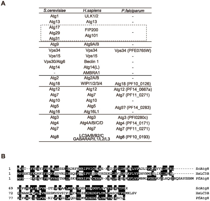
Figure 1
Atg protein sets are only partially conserved in P. falciparum .
(A) List of Atg proteins in S. cerevisiae , Homo sapiens and P. falciparum . u2013, no ortholog found. It has been suggested that the mammalian FIP200u2013Atg101 complex and the yeast Atg17u201329u201...
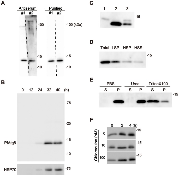
Figure 2
PfAtg8 is associated with membranes.
(A) Specificity of the two independently generated anti-PfAtg8 antibodies (#1 and #2). Crude antisera and purified antibodies were used for immunoblotting of lysates of asynchronized P. falciparum par...
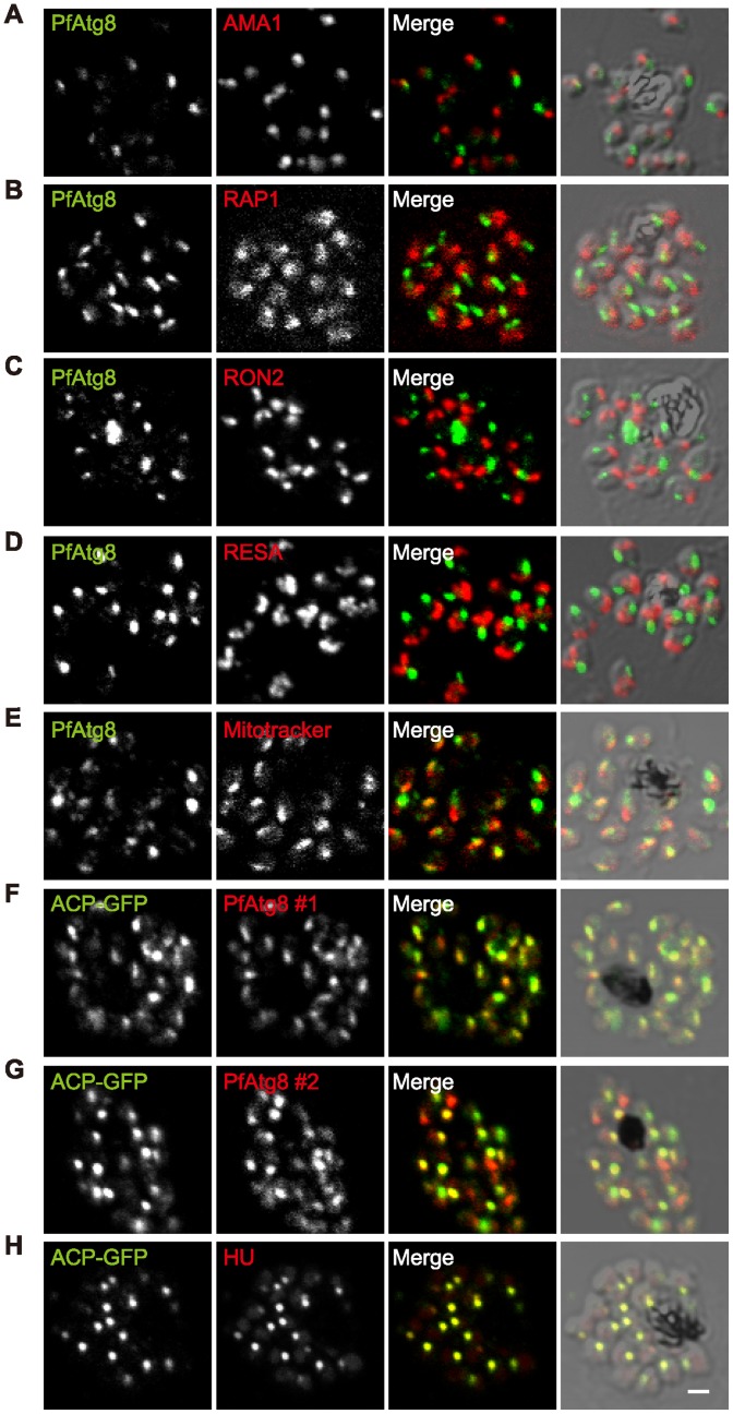
Figure 3
PfAtg8 localizes to the apicoplast.
P. falciparum FCR3 (Au2013E) and P. falciparum 3D7 transfected with ACP-GFP (Fu2013H) were stained with the indicated organelle markers and visualized by confocal microscopy (because ACP-GFP was not u...
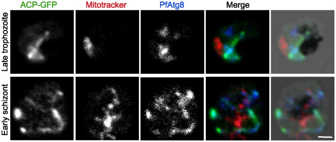
Figure 4
PfAtg8 localizes to tubular and branched apicoplasts.
P. falciparum transfectant expressing ACP-GFP at late trophozoite and early schizont stages was stained with anti-GFP and anti-PfAtg8 antibodies and MitoTrackerRed CMXRos, and visualized by confocal m...
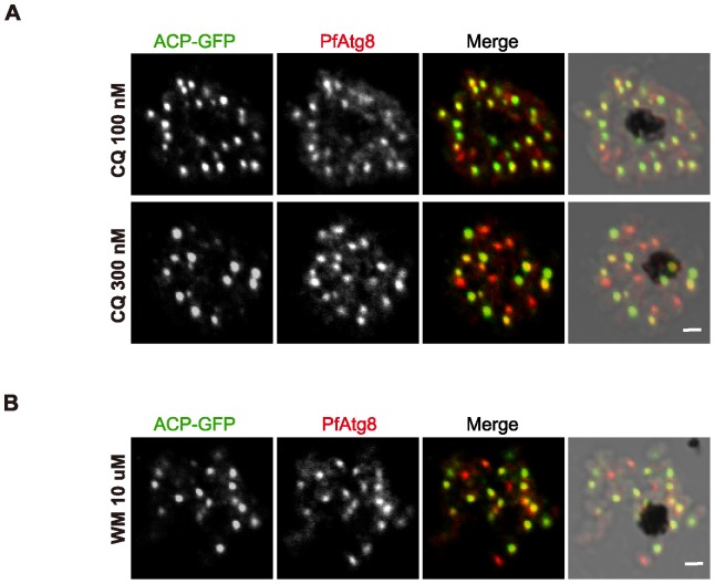
Figure 5
PfAtg8 localization is not affected by chloroquine or wortmannin treatment.
P. falciparum transfectant expressing ACP-GFP was treated with chloroquine (100 or 300 nM) (A), or wortmannin (10 u03bcM) (B) for 2 h. Scale bar, 1 u03bcm.
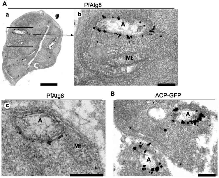
Figure 6
PfAtg8 is associated with the apicoplast membrane.
(A) P. falciparum FCR3 parasites at the schizont stage were analyzed by immunoelectron microscopy (immunogold and silver enhancement method) with an antibody against PfAtg8 (#1). (a) A schizont in an ...
Figure images are served from the NIH/NLM PubMed Central Open Access Subset or Europe PMC; copyright remains with the publishers and authors.
💬 Discussion
0 commentsNo comments yet. Be the first to start a discussion!
Leave a Comment