Abstract
Deep learning algorithms are powerful tools to analyse, restore and transform bioimaging data, increasingly used in life sciences research. These approaches now outperform most other algorithms for a broad range of image analysis tasks. In particular, one of the promises of deep learning is the possibility to provide parameter-free, one-click data analysis achieving expert-level performances in a fraction of the time previously required. However, as with most new and upcoming technologies, the potential for inappropriate use is raising concerns among the biomedical research community. This perspective aims to provide a short overview of key concepts that we believe are important for researchers to consider when using deep learning for their microscopy studies. These comments are based on our own experience gained while optimising various deep learning tools for bioimage analysis and discussions with colleagues from both the developer and user community. In particular, we focus on describing how results obtained using deep learning can be validated and discuss what should, in our views, be considered when choosing a suitable tool. We also suggest what aspects of a deep learning analysis would need to be reported in publications to describe the use of such tools to guarantee that the work can be reproduced. We hope this perspective will foster further discussion between developers, image analysis specialists, users and journal editors to define adequate guidelines and ensure that this transformative technology is used appropriately.
🔬 Techniques
💻 Software Details
🏛️ Research Organizations (ROR)
Affiliated research institutions:
📊 Figures
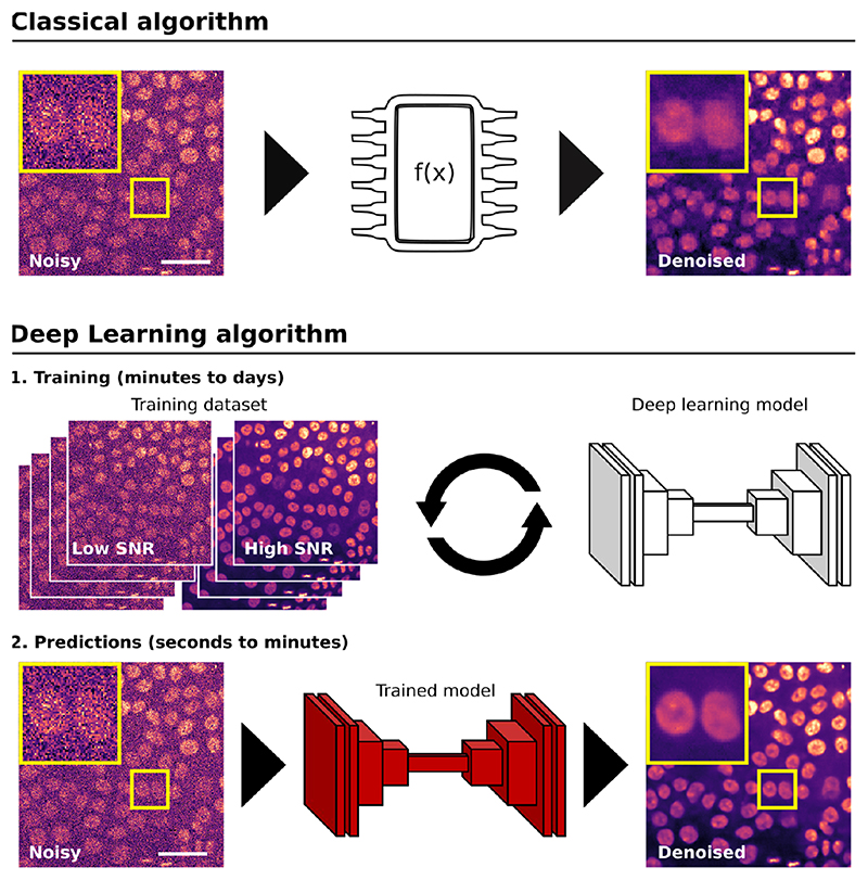
Figure 1
Using classical or DL algorithms to analyse microscopy images.
This figure illustrates the critical steps required when using classical or DL-based algorithms to analyse microscopy images, using denoising as an example. When using a classical algorithm, the resea...
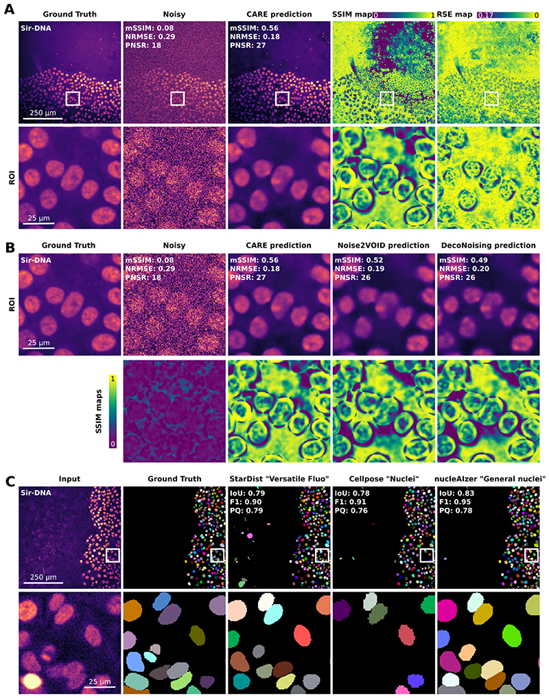
Figure 2
Using quality metrics to assess the performance of DL models.
Figure illustrating that comparing DL-based predictions to ground truth images is a powerful strategy to assess a DL model performance. ( A , B ) Noisy images of breast cancer cells labelled with SiR-...
Figure images are served from the NIH/NLM PubMed Central Open Access Subset or Europe PMC; copyright remains with the publishers and authors.
💬 Discussion
0 commentsNo comments yet. Be the first to start a discussion!
Leave a Comment