Abstract
Coral animals harbor diverse microorganisms in their tissues, including archaea, bacteria, viruses, and zooxanthellae. The extent to which coral-bacterial associations are specific and the mechanisms for their maintenance across generations in the environment are unknown. The high diversity of bacteria in adult coral colonies has made it challenging to identify species-specific patterns. Localization of bacteria in gametes and larvae of corals presents an opportunity for determining when bacterial-coral associations are initiated and whether they are dynamic throughout early development. This study focuses on the early onset of bacterial associations in the mass spawning corals Montastraea annularis, M. franksi, M. faveolata, Acropora palmata, A. cervicornis, Diploria strigosa, and A. humilis. The presence of bacteria and timing of bacterial colonization was evaluated in gametes, swimming planulae, and newly settled polyps by fluorescence in situ hybridization (FISH) using general eubacterial probes and laser-scanning confocal microscopy. The coral species investigated in this study do not appear to transmit bacteria via their gametes, and bacteria are not detectable in or on the corals until after settlement and metamorphosis. This study suggests that mass-spawning corals do not acquire, or are not colonized by, detectable numbers of bacteria until after larval settlement and development of the juvenile polyp. This timing lays the groundwork for developing and testing new hypotheses regarding general regulatory mechanisms that control bacterial colonization and infection of corals, and how interactions among bacteria and juvenile polyps influence the structure of bacterial assemblages in corals.
🔬 Techniques
🔭 Microscopes
✨ Fluorophores
🧪 Sample Preparation
🏭 Microscope Brands
🏛️ Research Organizations (ROR)
Affiliated research institutions:
📋 Methods
Belize Fisheries Department provided permits and facilitated the research on coral larvae at Carrie Bow Cay, Belize. Coral gametes in the Florida Keys were collected under permit FKNMS-2006-025.
Gamete collection
A summary of the dates and locations of spawning events from which gametes were collected for this study is presented in Table 1 . Gametes were collected from Looe Key Buoy #21 during an A. palmata spawning event in August 2007 and a M. faveolata spawning event in September 2007. Diploria strigosa gametes were collected from colonies at Grecian Rocks reef in Key Largo during a spawning event in September 2007. Gametes were obtained from spawning colonies with non-invasive nylon mesh collection tents attached to polypropylene collection jars, such as the apparatus shown in Figure 1 . 10.1371/journal.pone.0010898.g001 Figure 1 Spawning tent used to collect gametes from coral colonies. Photo: Erich Bartels. 10.1371/journal.pone.0010898.t001 Table 1 A list of the locations and timing of all gamete collections described in this study. Species Spawning Year Collection Location Acropora cervicornis August 2005 Carrie Bow Cay, Belize 16°48.18′N, 88°04.93′W Montastraea franksi September 2006 Carrie Bow Cay, Belize 16°48.18′N, 88°04.93′W Montastraea annularis September 2006 Carrie Bow Cay, Belize 16°48.18′N, 88°04.93′W Montastraea faveolata September 2006 Carrie Bow Cay, Belize 16°48.18′N, 88°04.93′W Acropora palmata September 2006 Carrie Bow Cay, Belize 16°48.18′N, 88°04.93′W Acropora palmata August 2007 Looe Key, Florida Keys, US 24°32.75′ N, 81°24.35′ W Montastraea faveolata September 2007 Looe Key, Florida Keys, US 24°32.75′ N, 81°24.35′ W Diploria strigosa September 2007 Grecian Rocks, Key Largo, FL 25°06.91′N, 080°18.20′W Acropora humilis August 2007 Pago Bay, Guam 13°42.67′N, 144°79.86′E In the Belize 2005 and 2006 collections, gametes were collected from A. palmata colonies with non-invasive nylon mesh collection tents and polypropylene collection jars, similar to the design used for gamete collection in the Florida Keys. Small colonies of A. cervicornis , M. franksi, M. faveolata , and M. annularis were collected from the reefs surrounding Carrie Bow Cay, Belize and were placed on submerged racks where they were maintained for several days. Each night, the colonies were placed in 5-gallon buckets full of freshly collected reef water for several hours, and upon gamete release, gametes were collected from the buckets via glass Pasteur pipets. Every night after gamete collection was complete, the colonies were returned to the submerged racks. After gamete collection, coral colonies were reattached on their reef using Splash Zone underwater epoxy (Z-Spar). Fertilization was achieved by mixing gametes from separate colonies in buckets of reef water, and the resulting embryos developed into swimming larvae in flow-through reef water (Belize), or fresh, filtered reef water (0.45 µm filtered Looe Key reef water) that was changed twice per day (Florida Keys). Samples were fixed for microscopy (see below) at sequential time points including gametes (t = 0 h) and larvae at subsequent 24 h intervals until settlement. In the 2007 M. faveolata collection from Florida, some larvae were reared in the above conditions until 96 h post-gamete release. At 96 h, swimming larvae were moved to 500 ml beakers containing 0.45 µm filtered reef water and untreated glass microscopy slides. A small percentage of the larvae settled and metamorphosed on the microscopy slides, and those settled individuals were fixed for microscopy (as described below) after attachment. Larvae were classified as metamorphosed if they had transformed into a juvenile polyp. A few days after metamorphosis, carbonate skeleton deposition was observed. In the 2007 Guam collection, Acropora humilis gametes were collected from laboratory colonies. Colonies (15–20 cm diameter in size) were collected one week before the spawning event from the fore reef slope (2–5 m depth) in Pago Bay, Guam. Colonies were maintained in 72 L tanks (1 per tank) with unfiltered flow-through seawater. For gamete collection colonies were kept without running seawater and aeration only. Egg and sperm bundles from different colonies were mixed upon release, and developing larvae were maintained in 13 L of filtered seawater (200 µm), which was changed twice daily. After 8 days, larvae were pipetted into wax-coated 15 ml petri dishes, each of which contained a glass microscopy slide. One to three small pieces of the crustose coralline alga (CCA) Hydrolithon sp. were placed on the glass slides to facilitate larval settlement because only occasional settlement was observed in preliminary experiments without CCA added to the petri dishes. Typically, 3–5 larvae settled on the glass slide, which produced a sufficient number of settled polyps on the glass slides for FISH experiments at distinct time intervals. Fixation of Larvae for FISH Gametes and swimming larvae were rinsed three times in sterile filtered seawater (0.22 µm), fixed in paraformaldehyde (4% in buffer: 20 mM K 2 HPO 4 , 0.5 M NaCl, pH 7.4) overnight at 4°C, and transferred to 70% ethanol for long-term storage at −20°C. To fix polyps that settled on glass slides, the slides were dipped three times in separate 50 ml polypropylene tubes of sterile filtered seawater (0.22 µm). The slides were then submerged in 4% paraformaldehyde in 50 ml polypropylene tubes and fixed overnight at 4°C. The 4% paraformaldehyde was discarded, and the tube was filled with 70% ethanol for long-term storage at −20°C.
Show full methods section
Belize Fisheries Department provided permits and facilitated the research on coral larvae at Carrie Bow Cay, Belize. Coral gametes in the Florida Keys were collected under permit FKNMS-2006-025.
Gamete collection
A summary of the dates and locations of spawning events from which gametes were collected for this study is presented in Table 1 . Gametes were collected from Looe Key Buoy #21 during an A. palmata spawning event in August 2007 and a M. faveolata spawning event in September 2007. Diploria strigosa gametes were collected from colonies at Grecian Rocks reef in Key Largo during a spawning event in September 2007. Gametes were obtained from spawning colonies with non-invasive nylon mesh collection tents attached to polypropylene collection jars, such as the apparatus shown in Figure 1 . 10.1371/journal.pone.0010898.g001 Figure 1 Spawning tent used to collect gametes from coral colonies. Photo: Erich Bartels. 10.1371/journal.pone.0010898.t001 Table 1 A list of the locations and timing of all gamete collections described in this study. Species Spawning Year Collection Location Acropora cervicornis August 2005 Carrie Bow Cay, Belize 16°48.18′N, 88°04.93′W Montastraea franksi September 2006 Carrie Bow Cay, Belize 16°48.18′N, 88°04.93′W Montastraea annularis September 2006 Carrie Bow Cay, Belize 16°48.18′N, 88°04.93′W Montastraea faveolata September 2006 Carrie Bow Cay, Belize 16°48.18′N, 88°04.93′W Acropora palmata September 2006 Carrie Bow Cay, Belize 16°48.18′N, 88°04.93′W Acropora palmata August 2007 Looe Key, Florida Keys, US 24°32.75′ N, 81°24.35′ W Montastraea faveolata September 2007 Looe Key, Florida Keys, US 24°32.75′ N, 81°24.35′ W Diploria strigosa September 2007 Grecian Rocks, Key Largo, FL 25°06.91′N, 080°18.20′W Acropora humilis August 2007 Pago Bay, Guam 13°42.67′N, 144°79.86′E In the Belize 2005 and 2006 collections, gametes were collected from A. palmata colonies with non-invasive nylon mesh collection tents and polypropylene collection jars, similar to the design used for gamete collection in the Florida Keys. Small colonies of A. cervicornis , M. franksi, M. faveolata , and M. annularis were collected from the reefs surrounding Carrie Bow Cay, Belize and were placed on submerged racks where they were maintained for several days. Each night, the colonies were placed in 5-gallon buckets full of freshly collected reef water for several hours, and upon gamete release, gametes were collected from the buckets via glass Pasteur pipets. Every night after gamete collection was complete, the colonies were returned to the submerged racks. After gamete collection, coral colonies were reattached on their reef using Splash Zone underwater epoxy (Z-Spar). Fertilization was achieved by mixing gametes from separate colonies in buckets of reef water, and the resulting embryos developed into swimming larvae in flow-through reef water (Belize), or fresh, filtered reef water (0.45 µm filtered Looe Key reef water) that was changed twice per day (Florida Keys). Samples were fixed for microscopy (see below) at sequential time points including gametes (t = 0 h) and larvae at subsequent 24 h intervals until settlement. In the 2007 M. faveolata collection from Florida, some larvae were reared in the above conditions until 96 h post-gamete release. At 96 h, swimming larvae were moved to 500 ml beakers containing 0.45 µm filtered reef water and untreated glass microscopy slides. A small percentage of the larvae settled and metamorphosed on the microscopy slides, and those settled individuals were fixed for microscopy (as described below) after attachment. Larvae were classified as metamorphosed if they had transformed into a juvenile polyp. A few days after metamorphosis, carbonate skeleton deposition was observed. In the 2007 Guam collection, Acropora humilis gametes were collected from laboratory colonies. Colonies (15–20 cm diameter in size) were collected one week before the spawning event from the fore reef slope (2–5 m depth) in Pago Bay, Guam. Colonies were maintained in 72 L tanks (1 per tank) with unfiltered flow-through seawater. For gamete collection colonies were kept without running seawater and aeration only. Egg and sperm bundles from different colonies were mixed upon release, and developing larvae were maintained in 13 L of filtered seawater (200 µm), which was changed twice daily. After 8 days, larvae were pipetted into wax-coated 15 ml petri dishes, each of which contained a glass microscopy slide. One to three small pieces of the crustose coralline alga (CCA) Hydrolithon sp. were placed on the glass slides to facilitate larval settlement because only occasional settlement was observed in preliminary experiments without CCA added to the petri dishes. Typically, 3–5 larvae settled on the glass slide, which produced a sufficient number of settled polyps on the glass slides for FISH experiments at distinct time intervals. Fixation of Larvae for FISH Gametes and swimming larvae were rinsed three times in sterile filtered seawater (0.22 µm), fixed in paraformaldehyde (4% in buffer: 20 mM K 2 HPO 4 , 0.5 M NaCl, pH 7.4) overnight at 4°C, and transferred to 70% ethanol for long-term storage at −20°C. To fix polyps that settled on glass slides, the slides were dipped three times in separate 50 ml polypropylene tubes of sterile filtered seawater (0.22 µm). The slides were then submerged in 4% paraformaldehyde in 50 ml polypropylene tubes and fixed overnight at 4°C. The 4% paraformaldehyde was discarded, and the tube was filled with 70% ethanol for long-term storage at −20°C.
FISH and Microscopy Analysis
FISH was performed on fixed whole gametes or larvae in microfuge tubes, or settled polyps on microscopy slides with hybridization buffer (0.9 M NaCl, 20 mM Tris-HCl [pH 7.4], 0.01% sodium dodecyl sulfate) containing 35% percent formamide. Samples were probed with a suite of general eubacterial probes, added in equimolar amounts: EUB338I ( 5′-GCTGCCTCCCGTAGGAGT-3′ ); EUB338II ( 5′-GCA GCC ACC CGT AGG TGT-3′ ), and EUB338III ( 5′-GCT GCC ACC CGT AGG TGT-3′ ) [38] . Negative control samples were probed with the negative control probe, NONEUB ( 5′-ACT CCT ACG GGA GGC AGC-3′ ) [38] . Probes were ordered as CY3-end labeled oligonucleotides (Integrated DNA Technologies, Coralville, IA). For each set of FISH reactions, a sample in which the bacterial content and location was known was probed with the EUB338 probe suite as a positive control for probe and reagent quality. All probes were added to hybridization buffer at a final concentration of 5 ng/µl. After 2 h hybridization at 46°C, the hybridization buffer was removed from the samples. Samples were incubated in wash buffer (0.7 M NaCl, 20 mM Tris-HCl [pH 7.4], 50 mM EDTA, 0.01% sodium dodecyl sulfate) for 20 minutes at 48°C. The samples were rinsed with Milli-Q water and mounted in VectaShield ( Vector Labs , Burlingame, CA). Slides were visualized on an LSM510 laser scanning confocal microscope (Zeiss, Jena, Germany). Approximately 50–100 individuals of each stage were imaged, depending on sample availability.
📊 Figures
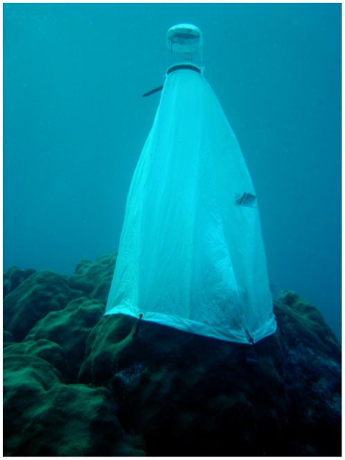
Figure 1
Spawning tent used to collect gametes from coral colonies.
Photo: Erich Bartels.
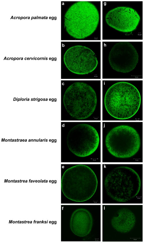
Figure 2
General eubacterial FISH in gametes of six Caribbean coral species.
FISH visualization of the CY3-labeled general eubacterial probe suite (EUB338I/EUB338II/EUB338III) on eggs from the six species of Caribbean scleractinian corals, Montastraea annularis, M. franksi, M....
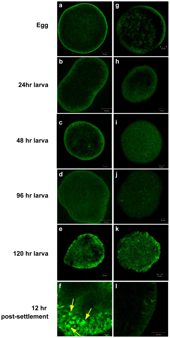
Figure 3
General eubacterial FISH in early development of Montastraea faveolata .
FISH visualization of the CY3-labeled general eubacterial probe suite (EUB338I/EUB338II/EUB338III) on Montastraea faveolata eggs, larvae, and settled juvenile polyps. Panels au2013f, EUB338; panels gu...
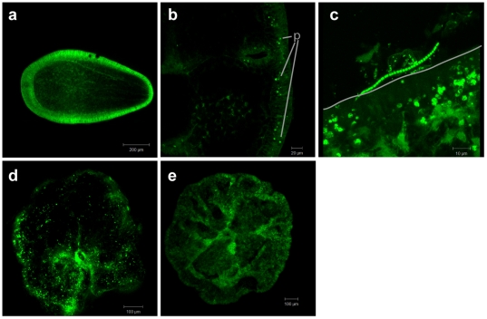
Figure 4
Bacteria in juvenile Acropora humilis polyps.
FISH visualization of signal from the CY3-labeled general eubacterial probe suite (EUB338I/EUB338II/EUB338III) on early stages of Acropora humilis . Panel a, a cross-section of a 129 h planula larva p...
Figure images are served from the NIH/NLM PubMed Central Open Access Subset or Europe PMC; copyright remains with the publishers and authors.
💬 Discussion
0 commentsNo comments yet. Be the first to start a discussion!
Leave a Comment