Abstract
Epidemiological evidence suggests that cadmium (Cd) exposure causes pulmonary damage such as emphysema and lung cancer. However, relatively little is known about the mechanisms involved in Cd pulmonary toxicity. In the present study, the effects of Cd exposure on human fetal lung fibroblasts (MRC-5 cells) were evaluated by determination of lipid peroxidation, intra-cellular production of reactive oxygen species (ROS), and changes of mitochondrial membrane potential. A time- and dose-dependent increase of both lactate dehydrogenase leakage and malondialdehyde formation was observed in Cd-treated cells. A close correlation between these two events suggests that lipid peroxidation may be one of the main pathways causing its cytotoxicity. It was also noted that Cd-induced cell injury and lipid peroxidation were inhibited by catalase and superoxide dismutase, two antioxidant enzymes. By using the fluorescent probe 2',7'-dichlorofluorescin diacetate, a significant increase of ROS production in Cd-treated MRC-5 cells was detected. The inhibition of dichlorofluorescein fluorescence by catalase, not superoxide dismutase, suggests that hydrogen peroxide is the main ROS involved. Moreover, the significant dose-dependent changes of mitochondrial membrane potential in Cd-treated MRC-5 cells, demonstrated by increased fluorescence of rhodamine 123 examined using a laser-scanning confocal microscope, also indicate the involvement of mitochondrial damage in Cd cytotoxicity. These findings provide in vitro evidence that Cd causes oxidative cellular damage in human fetal lung fibroblasts, which may be closely associated with the pulmonary toxicity of Cd.
🔬 Techniques
✨ Fluorophores
🧪 Sample Preparation
🔬 Cell Lines
🏭 Microscope Brands
🧪 Reagent Suppliers
🏛️ Research Organizations (ROR)
Affiliated research institutions:
📋 Methods
Cels and chemicals. The human fetal lung fibroblast cell line (MRC-5 cells) was from American Type Culture Collection (ATCC; Rockville, MD). Cadmium chloride (CdCl2) and thiobarbituric acid (TBA) were purchased from Merck (Darmstadt, Germany); minimum essential medium (MEM) was from Gibco (Buffalo, NY); and fetal bovine serum (FBS) was from Cytosystems (Castle Hill, Australia). Rhodamine 123 (Rh-123), sodium dodecyl sulphate (SDS), penicillin, streptomycin, catalase (CAT), and superoxide dismutase (SOD) were from Sigma Chemical Co. (St. Louis, MO). 2',7',-Dichlorofluorescin diacetate (DCFH-DA) was purchased from Molecular Probes, Eugene, OR.
Cell culture and treatments. MRC-5 cells were cultured in complete MEM (10% FBS, 100 units/ml penicillin, 100 mg/ml streptomycin, pH 7.4) at 37?C in 95% 02 and 5% CO2. Cells in logarithmic growth phases (approximately 90% confluence) were used for various experiments.
In the dose-response study, various
Address correspondence to C.N. Ong, Department of Community, Occupational, and Family Medicine, National University of Singapore, Singapore 119074.
This research project was supported in part by the China Medical Board, New York, USA and the Program on Environmental and Occupational Health, Centre for Environmental and Occupational Health, National University of Singapore. The authors would like to thank H.Y. Ong, B.L. Lee, A.L. New, and M. Chan for their technical support.
Received 3 December 1996; accepted 1 April 1997.
Volume 105, Number 7, July 1997 * Environmental Health Perspectives
Show full methods section
Cels and chemicals. The human fetal lung fibroblast cell line (MRC-5 cells) was from American Type Culture Collection (ATCC; Rockville, MD). Cadmium chloride (CdCl2) and thiobarbituric acid (TBA) were purchased from Merck (Darmstadt, Germany); minimum essential medium (MEM) was from Gibco (Buffalo, NY); and fetal bovine serum (FBS) was from Cytosystems (Castle Hill, Australia). Rhodamine 123 (Rh-123), sodium dodecyl sulphate (SDS), penicillin, streptomycin, catalase (CAT), and superoxide dismutase (SOD) were from Sigma Chemical Co. (St. Louis, MO). 2',7',-Dichlorofluorescin diacetate (DCFH-DA) was purchased from Molecular Probes, Eugene, OR.
Cell culture and treatments. MRC-5 cells were cultured in complete MEM (10% FBS, 100 units/ml penicillin, 100 mg/ml streptomycin, pH 7.4) at 37?C in 95% 02 and 5% CO2. Cells in logarithmic growth phases (approximately 90% confluence) were used for various experiments.
In the dose-response study, various
Address correspondence to C.N. Ong, Department of Community, Occupational, and Family Medicine, National University of Singapore, Singapore 119074.
This research project was supported in part by the China Medical Board, New York, USA and the Program on Environmental and Occupational Health, Centre for Environmental and Occupational Health, National University of Singapore. The authors would like to thank H.Y. Ong, B.L. Lee, A.L. New, and M. Chan for their technical support.
Received 3 December 1996; accepted 1 April 1997.
Volume 105, Number 7, July 1997 * Environmental Health Perspectives
712
Articles - Cd-induced oxidative cellular damage in MRC-5 cells
concentrations of CdCI2 dissolved in FBS- free MEM were incubated with MRC-5 cells for 16 hr. In the time-course study, cells were treated with 35 pM CdCl2 up to 20 hr. The inhibitory effects of CAT (1000 U/ml) and SOD (500 U/ml) on Cd- induced cell damage were tested after they were cultured together with 35 pM CdCl2 for 16 hr.
Determination of lactate dehydrogenase leakage. Lactate dehydrogenase (LDH) activity was determined using an Abbott VP Biochemical Analyzer with the test kit (Abbott Laboratories, Chicago, IL), as described by Shen et al. (15). At the end of the experiment, an aliquot of medium (0.2 ml) was taken out for measuring extracellu- lar LDH activity. The total LDH activity was determined after cells were disrupted thoroughly using sonication. The percent- age of LDH leakage was then calculated to reflect the cytotoxicity of CdCl2.
Measurement of lipid peroxidation. Malondialdehyde (MDA), an end product of lipid peroxidation, was measured to esti- mate the extent of lipid peroxidation in MRC-5 cells. MDA concentration in cell homogenate was determined using a TBA method as described by Uchiyama and Mihara (16), with modifications. Briefly, at the end of the experiment, cells were col- lected using a cell scraper and washed with PBS. Cell homogenate (0.5 ml in PBS with 1% SDS) was mixed with 3 ml 1% phos- phoric acid and 1 ml 0.67% TBA and heated in boiling water for 60 min. After cooling, 1.5 ml n-butanol was added. After centrifugation, the absorbance of the butanol phase was read at 535 nm and 520 nm. The difference between 535 nm and 520 nm was used to calculate the MDA concentration, which was expressed as nanomoles per milligram protein.
Detection of ROSformation and effects of antioxidant enzymes. Cd-induced ROS formation in MRC-5 cells was detected by using a fluorescent probe, 2',7'-diclorofluo- rescin diacetate (DCFH-DA), as described by Shen et al. (17). DCFH-DA diffuses through the cell membrane readily and is enzymatically hydrolyzed by intracellular esterases to nonfluorescent diclorofluorescin (DCFH), which is then rapidly oxidized to highly fluorescent diclorofluorescein (DCF) in the presence of ROS. The DCF fluores- cence intensity is believed to be parallel to the amount of ROS formed intracellularly (18). The stock DCFH-DA (2 mM) was prepared in absolute ethanol and kept at -70'C in the dark. Cells collected from cul- ture flasks using a cell scraper were washed twice with PBS prior to the analysis. Each fluorescence cuvette contained 0.6 x 10⁵
cells in 3 ml PBS. CdCl2 was added to the
cells simultaneously with DCFH-DA (final concentration 2 FM) and incubated at 37?C up to 4 hr. The fluorescence intensity was monitored using a Perkin-Elmer spectrofluo- rometer LS-5B (Perkin Elmer, Beaconsfield, U.K.) with excitation wavelength at 485 nm and emission wavelength at 530 nm.
The inhibitory effects of CAT (1,000 U/ml) and SOD (500 U/ml) on ROS pro- duction were evaluated by the following approach: both enzymes were first pre- incubated with MRC-5 cells in culture flasks for 6 hr; cells were then collected and washed with PBS for the fluorescence test as described above.
Determination of mitochondrial mem- brane potentiaL MMP in intact
MRC-5 cells was determined using Rh-123, a fluo- rescent dye. Mitochondria are stained by Rh-123 because of the high negative electri- cal potential across the mitochondrial mem- brane, and the diffusion of Rh-123 is direct- ly proportional to the degree of MMP (14). MRC-5 cells were cultured in MEM in cov- erglass chambers. Before analysis, the cul- tured cells were washed once with HEPES- containing Hanks' balanced salt buffer (HBSS; 1.26 mM CaCl2, 5.36 mM KCl, 0.44 mM KH2PO4, 0.49 mM MgCl2 6H20, 0.41 mM MgSO47H2O, 0.137 M NaCl, 0.34 mM Na2HPO4 7H20, 20 mM HEPES). Cells were then incubated with different concentrations of CdCl2 (0, 8.75, 17.5, and 35 pM) for 1 hr, followed by incubation with 6 pg/inl Rh-123 for 30 min. After the removal of CdCl2 and Rh- 123 with HBSS, cells were evaluated imme- diately using a laser-scanning inverted con- focal microscope (Carl Zeiss LSM 410, Jena, Germany). Rh-123 was excited using 488 nm laser line with a laser power of 10%. The emission signal was observed with a combination of a 510 nm dichroic mirror and a 515-516 nm cut-off filter. A heat platform was fitted to the microscope and set at 37?C throughout the analysis. The quantification of the Rh-123 fluores- cence intensity in different groups was per- formed.
Statistical analysis. Data are presented as mean ? standard deviation (SD) and ana- lyzed using one-way analysis of variance (ANOVA) with Scheffe's test or Student's t- test. A p-value of
📊 Figures
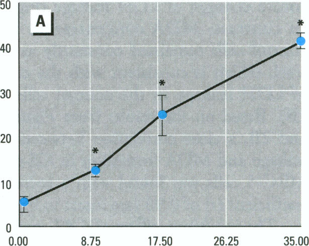
Figure 1. A
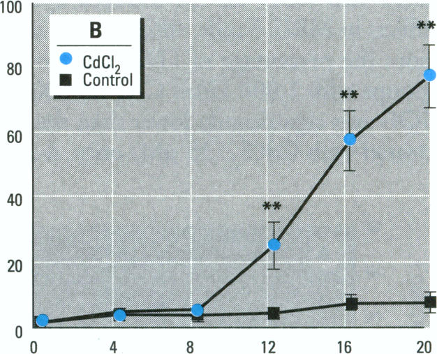
Figure 1. B
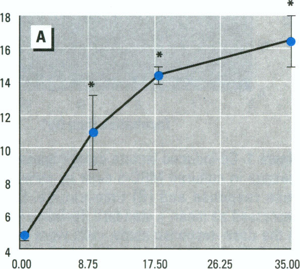
Figure 2. A
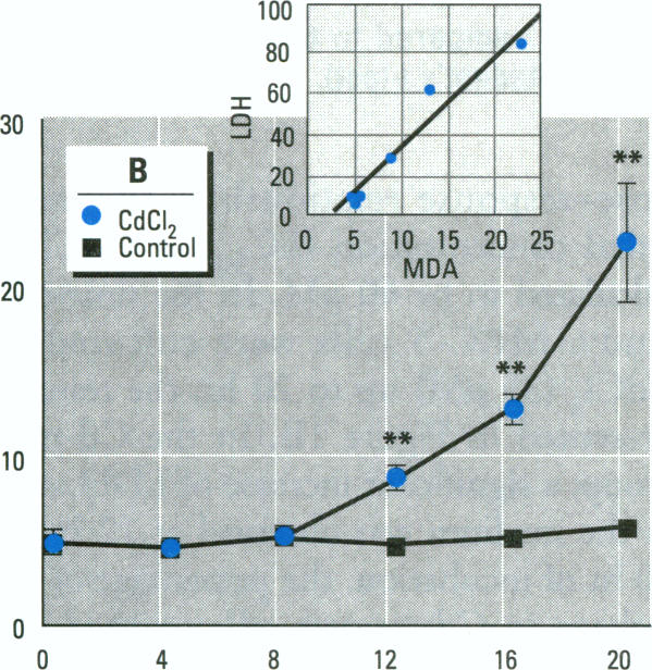
Figure 2. B
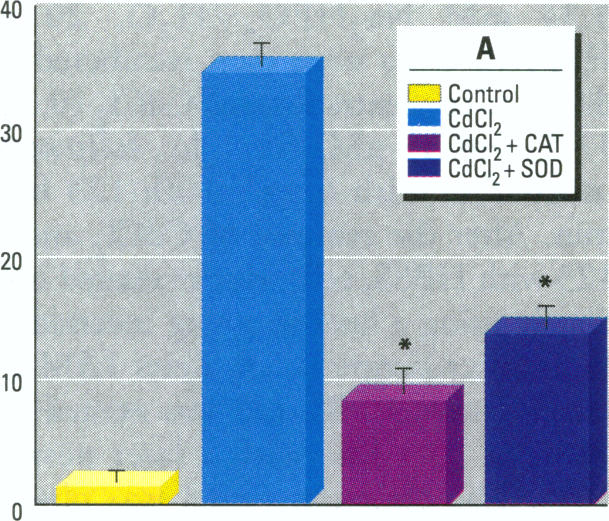
Figure 3. A
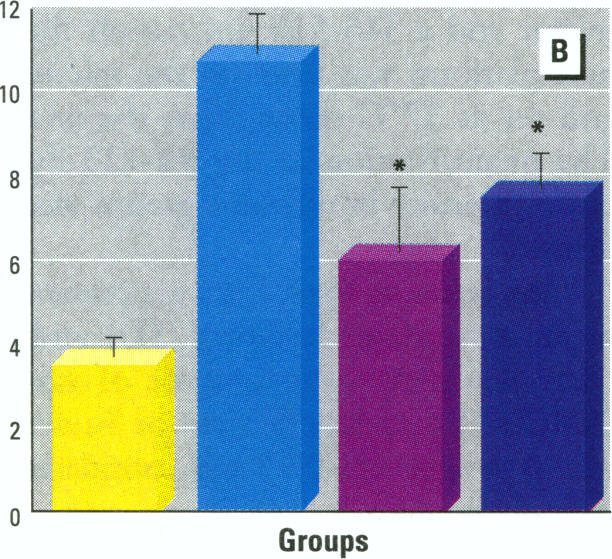
Figure 3. B
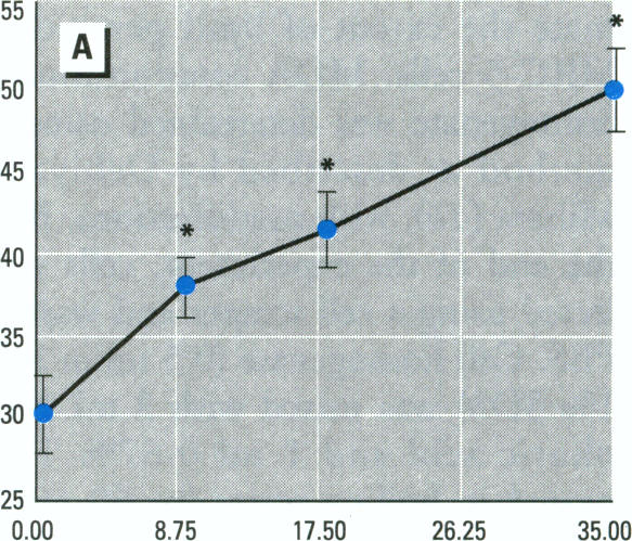
Figure 4. A
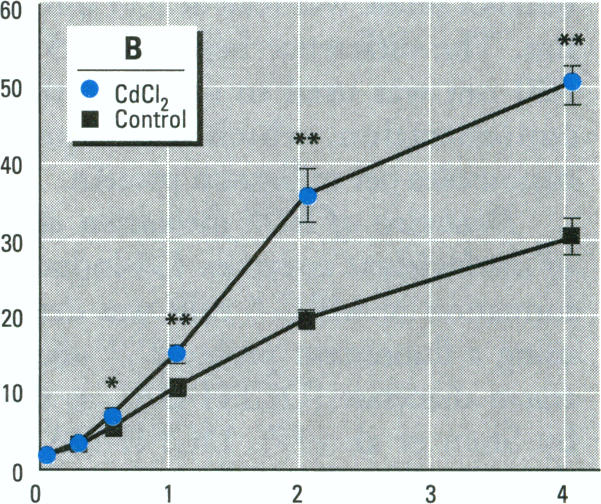
Figure 4. B
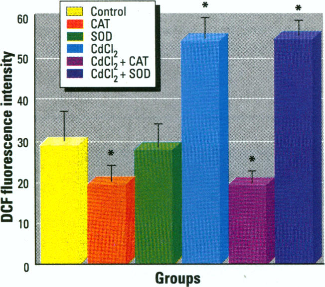
Figure 5.
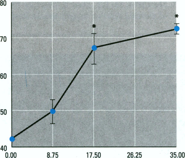
Figure 6.
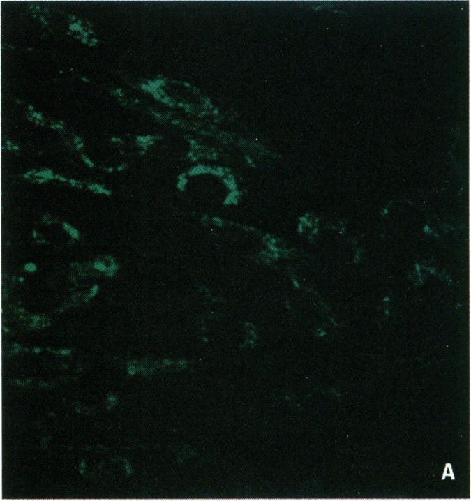
Figure 7. A
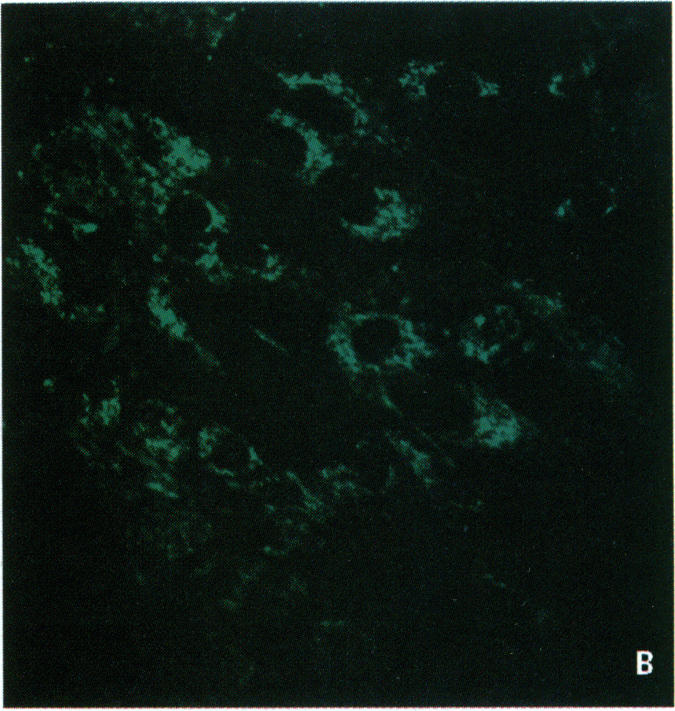
Figure 7. B
Figure images are served from the NIH/NLM PubMed Central Open Access Subset or Europe PMC; copyright remains with the publishers and authors.
💬 Discussion
0 commentsNo comments yet. Be the first to start a discussion!
Leave a Comment