Abstract
Cancer heterogeneity increasingly requires ultrasensitive techniques that allow early diagnosis for personalized treatment. In addition, they should preferably be non-invasive tools that do not damage surrounding tissues or contribute to body toxicity. In this context, liquid biopsy of biological samples such as urine, blood, or saliva represents an ideal approximation of what is happening in real time in the affected tissues. Plasmonic nanoparticles are emerging as an alternative or complement to current diagnostic techniques, being able to detect and quantify novel biomarkers such as specific peptides and proteins, microRNA, circulating tumor DNA and cells, and exosomes. Here, we review the latest ideas focusing on the use of plasmonic nanoparticles in coded and label-free surface-enhanced Raman scattering (SERS) spectroscopy. Moreover, surface plasmon resonance (SPR) spectroscopy, colorimetric assays, dynamic light scattering (DLS) spectroscopy, mass spectrometry or total internal reflection fluorescence (TIRF) microscopy among others are briefly examined in order to highlight the potential and versatility of plasmonics.
🔬 Techniques
✨ Fluorophores
🧪 Sample Preparation
🔬 Cell Lines
🏛️ Research Organizations (ROR)
Affiliated research institutions:
📋 Methods
2. SERS Methods Surface-enhanced Raman scattering (SERS) is a recognized spectroscopic technique with ultrasensitive spatial resolution, which outperforms classical techniques such fluorescence: it provides narrow bands of 0.1 nm in comparison with 20–80 nm bandwidth for fluorescence, being suitable for multiplexing detection in the same sample allowing the identification of several targets in simple assays. Obtaining a high volume of valuable data in parallel saves time and costs to clinicians [ 29 ]. Another advantage over fluorescence, is that plasmonic labels are not vulnerable of photobleaching, and the limit of detection (LOD) achieved with SERS is around three orders of magnitude smaller than LOD obtained with fluorescence [ 30 , 31 ]. SERS combines Raman spectroscopy, which provides specific structural information based on molecules’ vibration modes, with the capacity of signal-amplification of plasmonic nanoparticles. The plasmonic effect enhances Raman scattering intensity through the electromagnetic mechanism (EM), derived eminently from the plasmonic nanostructure and the excitation light, with additional contribution from the chemical effect (CT), due to the charge-transfer between the nanostructure and the molecule. EM can be explained as the contribution of the enhancement of the incident field at a certain point on the surface where the analyte is positioned and the enhancement of the re-emitted Stokes scattering by the molecule. CT mechanism occurs through the formation of a new analyte-metal surface complex, where the electronic properties of the attached molecule are distorted enabling new transitions within the complex. Modified polarizability of the analyte leads to an enhancement of the Raman cross-section. Both mechanisms occur together adding their effects [ 25 ]. Since each molecule has a unique Raman spectrum, it has become a fingerprint that can be used for the characterization of even single tumor cells. Furthermore, it is a non-destructive technique that can be applied to a little amount of solid and liquid samples requiring minimum preparation. Among the SERS nanosensors developed in the last decades to diagnose cancer, some approaches used bare plasmonic nanoparticles whereas most of them use a Raman probe, whose spectral variations in shift or intensity will be monitored. Table 1 summarizes the latest advances in both approximations. 2.1. Label-Free SERS The nonspecificity and high background habitually difficult the assignment of Raman bands making this approach less popular. On the other hand, direct measurements are advantageous for their fast, cost-effective and easy-to-use performance. Furthermore, the lack of accuracy can be overcome through amplification processes of the target, suitable in genetic material detection because of the several techniques for replicate the nucleic acid biomarkers that exist [ 44 ]. Specifically, for nucleic acid sequences, direct adsorption onto plasmonic materials through the negatively charged phosphate groups enables direct label-free readout strategies saving time-consuming labeling [ 45 , 46 ]. Koo et al. designed a label-free SERS diagnostic technology for differentiating between high- and low-risk prostate cancer (PC) ( Figure 3 A) [ 10 ]. T2:ERG, PCA3, and KLK2 miRNA (PC specific targets) from urine samples are amplified to enhance SERS intensity, through an isothermal process, which results in the stabilization of native miRNA in double-stranded DNA. After mixing the targets with silver nanoparticles (AgNPs) of 40 nm in size, SERS spectra were collected. Differences among Raman patterns of three targets spectra were identified and clustered into three groups through principal component analysis (PCA). With this statistical method, the high number of variables is reduced to a small set of dominant dimensions to further stablish a risk stratification scoring system by analyzing a training cohort ( n = 80, 20 healthy patients, 60 PC samples divided into low- and high-risk). This methodology provides results in 90 min, shortening alternative methods, which habitually take more than 4 h. Lin et al. developed a similar label-free SERS-based dispositive to detect blood circulating DNA (cDNA) of nasopharyngeal cancer (NPC) [ 34 ]. In this approach, the whole cDNA is considered a biomarker, saving even more time compared to alternative methods that isolate determined targets. It consists in extracting cDNA from NPC patients ( n = 120) and control subjects ( n = 120), to further mix with AgNPs of 35 nm in size. Although as-synthesized AgNPs are stabilized by chloride anions, DNA divalent phosphate backbones displace mentioned monovalent anions. After 3h of incubation, one drop of MgSO 4 is added in order to aggregate the sample causing hot spots to increase the SERS enhancement. Differences between SERS spectra of control and NPC group were obtained after multivariate algorithm analysis based on PCA combined with linear discriminant analysis (LDA) in order to identify the significant variables, achieving diagnostic sensitivity of 83.3% and specificity of 82.5% among NPC and control group. Following the analogous strategy, Carmicheal et al. [ 33 ] designed a label-free substrate for early diagnose of pancreatic cancer based on the identification of exosomes extracted from serum of healthy donors ( n = 10) and pancreatic cancer (PaC) patients ( n = 10). Plasmonic material consists of gold slides, and positively charged AuNPs of 10 nm are deposited onto it to caption negatively charged exosomes from C18/HPAF, MiaPaCa, and HPDE pancreatic cancer cells. Collection of SERS spectra was then submitted to PCA algorithm, providing good results in terms of sensitivity and specificity. Previous studies developed by Park et al. focused also their efforts on discerning among exosomes originated from healthy and lung cancer cells by SERS-PCA [ 47 ]. Stremersch et al. differentiated exosomes from melanoma and healthy red blood cells by developing SERS-partial least squares discriminant analysis [ 48 ]. Moving to tissue samples as biomarker, Girish et al. fabricated a catheter to classify and stablish grades (healthy, premalignant, and malignant) of oral cancer by testing oral squamous cell carcinoma, verrucous carcinoma, and premalignant leukoplakia ( Figure 3 B) [ 32 ]. The sensor is made by a TiO 2 leaf-like nanostructure equipped with AgNPs of 30 nm in size, which serves to extract cancerous tissue from mouth and directly make the SERS measurements. From averaged spectra from the four different groups, discriminant function scores were obtained. PCA-DA cross-validation was carried out with an accuracy of 97.24%. It is worth noting that this patient exam can be developed in around 30 min, outperforming conventional histopathology process time based on invasive biopsy, which includes surgery, cryogenization, and biochemistry techniques. Previous studies of label-free SERS in cancer tissues are reported in literature [ 49 , 50 ]. 2.2. Encoded-SERS A refined SERS-based strategy consists in synthesizing hybrid nanostructures composed by the plasmonic nanoparticle functionalized with a molecule with high Raman cross-section (named reporter or probe); then, plasmonic nanoparticle can be protected with an out layer of polymer or silica. These SERS-encoded particles (SEPs), can be functionalized with specific antibodies or proteins to provide selectivity. This powerful strategy can be applied to the detection from smaller biomarkers until tissue exemplars. For example, Lee et al. ( Figure 4 A), developed a SERS-based sensor for quantitative detection of exosomal miRNA in serum samples of breast cancer patients [ 39 ]. Hybrid structure consists in a head-flocked gold nanopillar substrate functionalized with a specific locked nucleic acid (LNA) probes for capturing miR-21, miR-222, and miR-200c added in known concentrations to human serum. Through a sandwich strategy, LNA probes functionalized with Cy3 as Raman reporter are hybridized to further monitoring SERS peak intensity of Cy3 at 1150 cm −1 . The closeness of gold nanoparticles creates self-assembled hotspots achieving LOD of 1 attomolar. The strategy proposed by Li et al. to detect miR-107 as biomarker for prostate cancer in urine samples consists in functionalizing both AuNPs and Au/Ag alloy nanocuboids with probes half-complementary to the whole miR-107 sequence [ 36 ]. In this way, the presence of the biomarker promotes the selective and spontaneous self-assembly of the Au spheres onto the Au/Ag nanocuboids with the specific interparticle gap (=2.3 nm) optimizing the SERS signal intensity. Indeed, inherent peroxidase-like activity of AuNPs (nanozymes) [ 51 , 52 ], generate catalytic cascades that oxidize TMB (3,3’,5,5’-Tetramethylbenzidine), which has a strong and recognizable SERS spectrum. Correlating TMBox SERS signal with the amount of miR-107, LOD in the range of femtomolar is achieved. These two mentioned works are innovative because they profit the assembly of plasmonic nanoparticles to create hot spots, obtaining really competitive LODs. However, other approaches are based on the frequency shift of the monitored peak. These approximations habitually present difficulties in terms of reproducibility, because the shifts are usually in the order of fewer than 1 cm –1 . Zhu et al. [ 43 ] developed an Ag multiplex plasmonic substrate where Raman reports [4-mercaptobenzoic acid (MBA), 5,5‘-Dithiobis(succinimidyl-2-nitrobenzoate (DSNB) and 6-thioguanine (6TG)] were attached and functionalized against miRNA (miR-26a-5p and miR-223) and α-fetoprotein (AFP) for discriminate primary liver cancer. In the presence of the target binding, vibrational frequencies of Raman reporters are shifted. By inferring the shift in a previous calibration curve, quantification of these biomarkers can be established. Zhang et al. proposed another diagnostic approach based on frequency shift for detecting circulating tumor DNA of lung cancer in serum samples [ 38 ]. To overcome the low concentration of ctDNA (pico-to-femtomolar), a first step of enzymatic amplification of the target mediated by RNase-II was done. Amplification combined with SERS allows the detection of one single-base pair mutation on KARS G12 gene. In this case, monitoring of the Raman shift of the molecule reporter DSNB 1334 cm −1 peak correlates with ctDNA achieving a LOD of 1.2 x 10 −16 M. The improvement provided by Wang et al. consists in including magnetic properties along with the plasmonic ones, facilitating the process of separation of the analyte from the whole sample. They developed a method based on an exosomal capturing probe based on a gold shell magnetic nanostructure with antibodies against surface protein CD63 and a detecting probe consisting in AuNPs functionalized with aptamers/Raman probes (Aptamer H2/DTNB, aptamer CEA/MMC, and aptamer PSMA/2NAT), for breast cancer, colorectal cancer, and prostate cancer, respectively [ 41 ]. After adding a known concentration of capturing and detection probes to blood samples, the immunocomplex takes place, and magnetic beads are collected with a magnet. By measuring the decrease in intensity of the respective Raman reporter signal in the supernatant, quantitative analysis can be done. Moreover, Bai et al. use magnetic properties for simultaneous detection. In order to detect specific proteins for lung cancer (AFP, carcinoembryonic antigen (CEA), and ferritin (FER)) from serum, they designed a plasmonic structure codified with three different tags labeled to respective antibodies [ 35 ]. This structure will be linked through a immunosandwich assay to a magnetic bead, leading to a core-satellite structure made with both plasmonic and magnetic structures. This approximation permits a simultaneous detection with LOD of 0.15, 20, and 4 pg/mL for AFP, CEA, and FER, respectively. Finally, the novelty provided by Lee et al. [ 39 ] consists in a dispositive able to detect two kinds of biomarkers simultaneously: CTCs and DNA. They developed a dual-function nanodevice for capturing nasopharyngeal CTCs and detecting Epstein–Barr virus (EBV) DNA from plasma samples with a LOD of 10 −13 M [ 37 ]. For this aim, a 3D structure of Si nanowires forming microscale pyramids coated with AgNPs of 200 nm was functionalized with Anti-EpCAM antibodies to capture the above-mentioned cells and at the same time, with a probe DNA specific for EBV. EBV target DNA will be attached to a second nanostructure consisting in AgNps functionalized with 4-MBA as Raman probe. The hybridization between both probe and target DNA will cause the capturing of the second nanostructure, giving rise to the clear spectra of 4-MBA. Other approaches were developed to detect CTCs and exosomes [ 53 ]. 2.3.
Show full methods section
2. SERS Methods Surface-enhanced Raman scattering (SERS) is a recognized spectroscopic technique with ultrasensitive spatial resolution, which outperforms classical techniques such fluorescence: it provides narrow bands of 0.1 nm in comparison with 20–80 nm bandwidth for fluorescence, being suitable for multiplexing detection in the same sample allowing the identification of several targets in simple assays. Obtaining a high volume of valuable data in parallel saves time and costs to clinicians [ 29 ]. Another advantage over fluorescence, is that plasmonic labels are not vulnerable of photobleaching, and the limit of detection (LOD) achieved with SERS is around three orders of magnitude smaller than LOD obtained with fluorescence [ 30 , 31 ]. SERS combines Raman spectroscopy, which provides specific structural information based on molecules’ vibration modes, with the capacity of signal-amplification of plasmonic nanoparticles. The plasmonic effect enhances Raman scattering intensity through the electromagnetic mechanism (EM), derived eminently from the plasmonic nanostructure and the excitation light, with additional contribution from the chemical effect (CT), due to the charge-transfer between the nanostructure and the molecule. EM can be explained as the contribution of the enhancement of the incident field at a certain point on the surface where the analyte is positioned and the enhancement of the re-emitted Stokes scattering by the molecule. CT mechanism occurs through the formation of a new analyte-metal surface complex, where the electronic properties of the attached molecule are distorted enabling new transitions within the complex. Modified polarizability of the analyte leads to an enhancement of the Raman cross-section. Both mechanisms occur together adding their effects [ 25 ]. Since each molecule has a unique Raman spectrum, it has become a fingerprint that can be used for the characterization of even single tumor cells. Furthermore, it is a non-destructive technique that can be applied to a little amount of solid and liquid samples requiring minimum preparation. Among the SERS nanosensors developed in the last decades to diagnose cancer, some approaches used bare plasmonic nanoparticles whereas most of them use a Raman probe, whose spectral variations in shift or intensity will be monitored. Table 1 summarizes the latest advances in both approximations. 2.1. Label-Free SERS The nonspecificity and high background habitually difficult the assignment of Raman bands making this approach less popular. On the other hand, direct measurements are advantageous for their fast, cost-effective and easy-to-use performance. Furthermore, the lack of accuracy can be overcome through amplification processes of the target, suitable in genetic material detection because of the several techniques for replicate the nucleic acid biomarkers that exist [ 44 ]. Specifically, for nucleic acid sequences, direct adsorption onto plasmonic materials through the negatively charged phosphate groups enables direct label-free readout strategies saving time-consuming labeling [ 45 , 46 ]. Koo et al. designed a label-free SERS diagnostic technology for differentiating between high- and low-risk prostate cancer (PC) ( Figure 3 A) [ 10 ]. T2:ERG, PCA3, and KLK2 miRNA (PC specific targets) from urine samples are amplified to enhance SERS intensity, through an isothermal process, which results in the stabilization of native miRNA in double-stranded DNA. After mixing the targets with silver nanoparticles (AgNPs) of 40 nm in size, SERS spectra were collected. Differences among Raman patterns of three targets spectra were identified and clustered into three groups through principal component analysis (PCA). With this statistical method, the high number of variables is reduced to a small set of dominant dimensions to further stablish a risk stratification scoring system by analyzing a training cohort ( n = 80, 20 healthy patients, 60 PC samples divided into low- and high-risk). This methodology provides results in 90 min, shortening alternative methods, which habitually take more than 4 h. Lin et al. developed a similar label-free SERS-based dispositive to detect blood circulating DNA (cDNA) of nasopharyngeal cancer (NPC) [ 34 ]. In this approach, the whole cDNA is considered a biomarker, saving even more time compared to alternative methods that isolate determined targets. It consists in extracting cDNA from NPC patients ( n = 120) and control subjects ( n = 120), to further mix with AgNPs of 35 nm in size. Although as-synthesized AgNPs are stabilized by chloride anions, DNA divalent phosphate backbones displace mentioned monovalent anions. After 3h of incubation, one drop of MgSO 4 is added in order to aggregate the sample causing hot spots to increase the SERS enhancement. Differences between SERS spectra of control and NPC group were obtained after multivariate algorithm analysis based on PCA combined with linear discriminant analysis (LDA) in order to identify the significant variables, achieving diagnostic sensitivity of 83.3% and specificity of 82.5% among NPC and control group. Following the analogous strategy, Carmicheal et al. [ 33 ] designed a label-free substrate for early diagnose of pancreatic cancer based on the identification of exosomes extracted from serum of healthy donors ( n = 10) and pancreatic cancer (PaC) patients ( n = 10). Plasmonic material consists of gold slides, and positively charged AuNPs of 10 nm are deposited onto it to caption negatively charged exosomes from C18/HPAF, MiaPaCa, and HPDE pancreatic cancer cells. Collection of SERS spectra was then submitted to PCA algorithm, providing good results in terms of sensitivity and specificity. Previous studies developed by Park et al. focused also their efforts on discerning among exosomes originated from healthy and lung cancer cells by SERS-PCA [ 47 ]. Stremersch et al. differentiated exosomes from melanoma and healthy red blood cells by developing SERS-partial least squares discriminant analysis [ 48 ]. Moving to tissue samples as biomarker, Girish et al. fabricated a catheter to classify and stablish grades (healthy, premalignant, and malignant) of oral cancer by testing oral squamous cell carcinoma, verrucous carcinoma, and premalignant leukoplakia ( Figure 3 B) [ 32 ]. The sensor is made by a TiO 2 leaf-like nanostructure equipped with AgNPs of 30 nm in size, which serves to extract cancerous tissue from mouth and directly make the SERS measurements. From averaged spectra from the four different groups, discriminant function scores were obtained. PCA-DA cross-validation was carried out with an accuracy of 97.24%. It is worth noting that this patient exam can be developed in around 30 min, outperforming conventional histopathology process time based on invasive biopsy, which includes surgery, cryogenization, and biochemistry techniques. Previous studies of label-free SERS in cancer tissues are reported in literature [ 49 , 50 ]. 2.2. Encoded-SERS A refined SERS-based strategy consists in synthesizing hybrid nanostructures composed by the plasmonic nanoparticle functionalized with a molecule with high Raman cross-section (named reporter or probe); then, plasmonic nanoparticle can be protected with an out layer of polymer or silica. These SERS-encoded particles (SEPs), can be functionalized with specific antibodies or proteins to provide selectivity. This powerful strategy can be applied to the detection from smaller biomarkers until tissue exemplars. For example, Lee et al. ( Figure 4 A), developed a SERS-based sensor for quantitative detection of exosomal miRNA in serum samples of breast cancer patients [ 39 ]. Hybrid structure consists in a head-flocked gold nanopillar substrate functionalized with a specific locked nucleic acid (LNA) probes for capturing miR-21, miR-222, and miR-200c added in known concentrations to human serum. Through a sandwich strategy, LNA probes functionalized with Cy3 as Raman reporter are hybridized to further monitoring SERS peak intensity of Cy3 at 1150 cm −1 . The closeness of gold nanoparticles creates self-assembled hotspots achieving LOD of 1 attomolar. The strategy proposed by Li et al. to detect miR-107 as biomarker for prostate cancer in urine samples consists in functionalizing both AuNPs and Au/Ag alloy nanocuboids with probes half-complementary to the whole miR-107 sequence [ 36 ]. In this way, the presence of the biomarker promotes the selective and spontaneous self-assembly of the Au spheres onto the Au/Ag nanocuboids with the specific interparticle gap (=2.3 nm) optimizing the SERS signal intensity. Indeed, inherent peroxidase-like activity of AuNPs (nanozymes) [ 51 , 52 ], generate catalytic cascades that oxidize TMB (3,3’,5,5’-Tetramethylbenzidine), which has a strong and recognizable SERS spectrum. Correlating TMBox SERS signal with the amount of miR-107, LOD in the range of femtomolar is achieved. These two mentioned works are innovative because they profit the assembly of plasmonic nanoparticles to create hot spots, obtaining really competitive LODs. However, other approaches are based on the frequency shift of the monitored peak. These approximations habitually present difficulties in terms of reproducibility, because the shifts are usually in the order of fewer than 1 cm –1 . Zhu et al. [ 43 ] developed an Ag multiplex plasmonic substrate where Raman reports [4-mercaptobenzoic acid (MBA), 5,5‘-Dithiobis(succinimidyl-2-nitrobenzoate (DSNB) and 6-thioguanine (6TG)] were attached and functionalized against miRNA (miR-26a-5p and miR-223) and α-fetoprotein (AFP) for discriminate primary liver cancer. In the presence of the target binding, vibrational frequencies of Raman reporters are shifted. By inferring the shift in a previous calibration curve, quantification of these biomarkers can be established. Zhang et al. proposed another diagnostic approach based on frequency shift for detecting circulating tumor DNA of lung cancer in serum samples [ 38 ]. To overcome the low concentration of ctDNA (pico-to-femtomolar), a first step of enzymatic amplification of the target mediated by RNase-II was done. Amplification combined with SERS allows the detection of one single-base pair mutation on KARS G12 gene. In this case, monitoring of the Raman shift of the molecule reporter DSNB 1334 cm −1 peak correlates with ctDNA achieving a LOD of 1.2 x 10 −16 M. The improvement provided by Wang et al. consists in including magnetic properties along with the plasmonic ones, facilitating the process of separation of the analyte from the whole sample. They developed a method based on an exosomal capturing probe based on a gold shell magnetic nanostructure with antibodies against surface protein CD63 and a detecting probe consisting in AuNPs functionalized with aptamers/Raman probes (Aptamer H2/DTNB, aptamer CEA/MMC, and aptamer PSMA/2NAT), for breast cancer, colorectal cancer, and prostate cancer, respectively [ 41 ]. After adding a known concentration of capturing and detection probes to blood samples, the immunocomplex takes place, and magnetic beads are collected with a magnet. By measuring the decrease in intensity of the respective Raman reporter signal in the supernatant, quantitative analysis can be done. Moreover, Bai et al. use magnetic properties for simultaneous detection. In order to detect specific proteins for lung cancer (AFP, carcinoembryonic antigen (CEA), and ferritin (FER)) from serum, they designed a plasmonic structure codified with three different tags labeled to respective antibodies [ 35 ]. This structure will be linked through a immunosandwich assay to a magnetic bead, leading to a core-satellite structure made with both plasmonic and magnetic structures. This approximation permits a simultaneous detection with LOD of 0.15, 20, and 4 pg/mL for AFP, CEA, and FER, respectively. Finally, the novelty provided by Lee et al. [ 39 ] consists in a dispositive able to detect two kinds of biomarkers simultaneously: CTCs and DNA. They developed a dual-function nanodevice for capturing nasopharyngeal CTCs and detecting Epstein–Barr virus (EBV) DNA from plasma samples with a LOD of 10 −13 M [ 37 ]. For this aim, a 3D structure of Si nanowires forming microscale pyramids coated with AgNPs of 200 nm was functionalized with Anti-EpCAM antibodies to capture the above-mentioned cells and at the same time, with a probe DNA specific for EBV. EBV target DNA will be attached to a second nanostructure consisting in AgNps functionalized with 4-MBA as Raman probe. The hybridization between both probe and target DNA will cause the capturing of the second nanostructure, giving rise to the clear spectra of 4-MBA. Other approaches were developed to detect CTCs and exosomes [ 53 ]. 2.3.
SERS Imaging
When we move to targets like CTCs and cancer tissues, SERS can be implemented as an optical imaging technique by monitoring specific molecules. The crucial advantage in SERS imaging is the high spatial resolution of the technique as it can achieve values of < 0.5 microns in the visible range, permitting the mapping of samples with high resolution. Another advantage is also the multiplexing analysis of several analytes in the same sample. In this regard, Nima et al. synthesized 4 SEPs hybrids consisting of Au@Ag nanorod, covered with four different Raman label/antibody against breast cancer as follows: 4-MBA/anti-EpCAM, p-nitrobenzoic acid/anti-IGF-I receptor β, p-aminobenzoic acid/anti-CD44, and 4-(methylsulfanyl)thiophenol/anti-cytokeratin. After incubation of SEPs with blood containing MCF-7 cancer cells, a SERS-map of a single cancer cell was registered, obtaining the clear signal of the four Raman probes onto the cell surface [ 54 ]. Li et al., followed the same approach to multicolor imaging breast cancer cells tissues but substituting the thiolated compounds habitually used as Raman labels for alkynes and nitriles ( Figure 4 C) [ 40 ]. They highlight the predisposition of thiols to oxidation plus the limitation of spectral overlap. By functionalizing AuNPs of 60 nm with Tag 1 (4-ethynyl-bipheny/ER), Tag 2 (phenylthiocyanate/EGFR), and Tag 3 (4-(phenylethynyl)aniline/PR) and incubating with the tissue samples, the profiling of ER, EGFR, and PR expression in breast cancer and normal tissue sections can be done. Multiplexing approaches were used also by Bodelón et al., who employed SEPs made with gold octahedra functionalized against EGFR, EpCAM, and CD44, to detect and image one single cell in vitro of human epithelial carcinoma A431, discriminating from non-tumoral murine fibroblast 3T3 2.2 [ 55 ]. Multiplexing detection was done also in solid samples of formalin-fixed human prostate tissue by Lutz et al. By conjugating composites of organic–inorganic nanoparticles (COINs) with specific antibodies against cytokeratin-18 and PSA, a controlled aggregation of silver nanoparticles was produced, which generate images with subcellular spatial resolution [ 56 ]. Following the same rationality [ 57 ], Wang et al. developed four SEPs against cell-surface biomarkers of human breast cancer (EGFR, HER2, CD44, and CD24). The plus point of this work is the enhancement of the method by immersing the tissue surfaces into a NP-staining solution submitted to high-frequency mechanical vibration in order to promote the convection and improvement of the plasmonic materials to the biomarker targets. This approach significantly improves the speed of the ratiometric SERS-imaging. Last, an encouraging application of SERS is as a tool image endoscopy during tumor resection in order to detect minimal lesions residually left during intraoperative surgery (responsible of metastasis origin in most cases). Here, Davis et al. proposed a multiplex imaging of bladder cancer tissue made in real-time whereas transurethral resection is giving place [ 42 ]. For that aim, AuNPs functionalized with antibodies against CA9 and CD47 protein biomarkers were administrated ex-vivo in bladder cancer patients after principal surgery, to further SERS-mapping the resection margins of the tumor area in order to classify the tissue as normal or tumor, offering an in situ guide for resections. This approximation overcomes limitations of current endoscopy techniques in detecting small tumor cells, which habitually give rise to postoperative exams, high rates of re-excision, and definitively worse prognosis [ 58 ].
📊 Figures
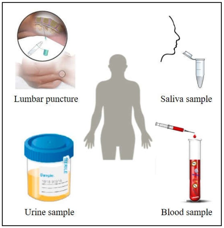
Figure 1
Body fluids as blood, saliva, urine or cerebrospinal fluid are excellent candidates to perform non-invasive cancer diagnosis based on plasmonic sensors. Adapted from [ 9 ] with permission. Copyright F...
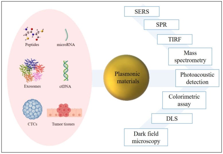
Figure 2
Plasmonic materials improve the detection of cancer biomarkers through several techniques.
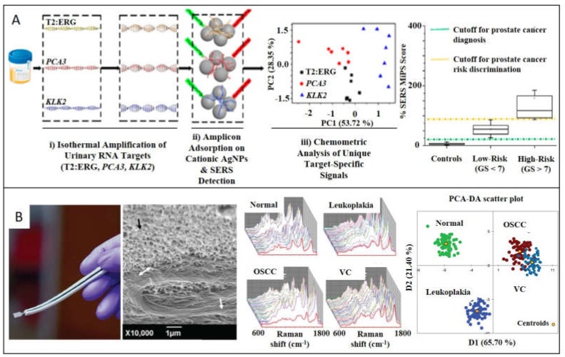
Figure 3
SERS detection of cancer biomarkers through label-free SERS nanoparticles. ( A ) Urine samples from patients with prostate cancer were collected to further isothermally amplify RNA targets (T2:ERG, PC...
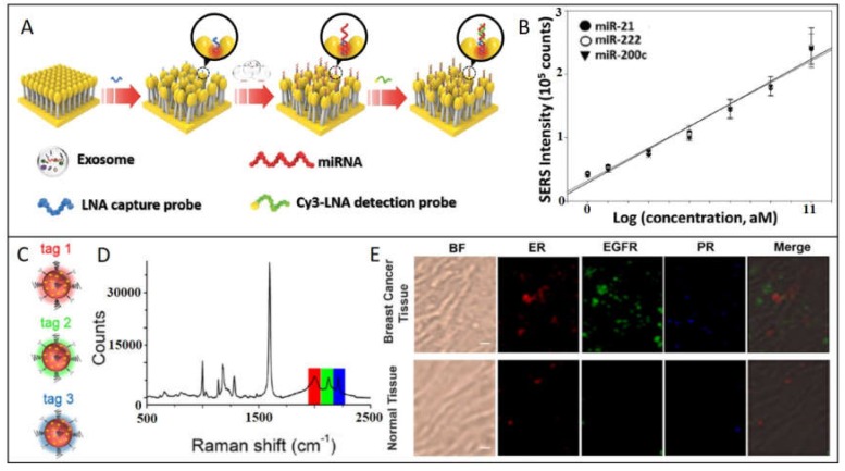
Figure 4
SERS detection of cancer biomarkers using SERS-encoded nanoparticles. ( A ) Schematic depiction of exosomal miRNA detection through the head-flocked gold nanopillars SERS sensor. locked nucleic acid (...
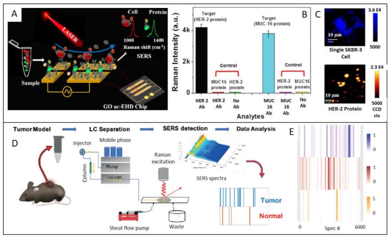
Figure 5
( A ) Schematic illustration of identification of circulating protein and cell on the GO functionalized ac-EHD chip. ( B ) Device sensitivity to capture HER 2 protein (black bar) and MUC-16 (light blu...
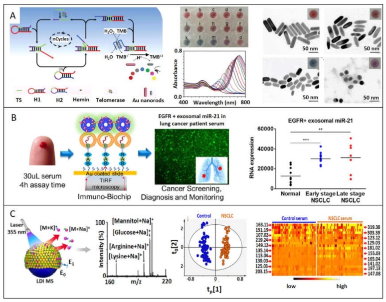
Figure 6
Plasmonic materials improve the detection of cancer biomarkers through several techniques: ( A ) Colorimetric assay is performed based on telomerase activity, proportional to the etching of Au nanorod...
Figure images are served from the NIH/NLM PubMed Central Open Access Subset or Europe PMC; copyright remains with the publishers and authors.
💬 Discussion
0 commentsNo comments yet. Be the first to start a discussion!
Leave a Comment