Abstract
BACKGROUND: During the coronavirus disease 2019 pandemic, several acral chilblain-like lesions were observed in young patients with suspected, but mostly unconfirmed, infection with severe acute respiratory syndrome coronavirus 2. The histopathologic aspect of these lesions is as yet poorly known. OBJECTIVE: To investigate the pathologic features of chilblain-like lesions. METHODS: Biopsies were obtained from 17 cases of chilblain-like lesions during the coronavirus disease 2019 pandemic in France and were studied by routine histologic examination, immunohistochemistry, and direct immunofluorescence. The patients had suspected but unconfirmed infection with severe acute respiratory syndrome coronavirus 2 (negative nasopharyngeal polymerase chain reaction and serologic test results). RESULTS: Chilblain-like lesions showed many features in common with those reported in idiopathic and autoimmune-related chilblains, including epidermal necrotic keratinocytes, dermal edema, perivascular and perieccrine sweat gland lymphocytic (predominantly CD3/CD4+) inflammation, and frequent vascular changes (endothelialitis, microthromboses, fibrin deposition, and immunoreactant deposits on vessels). CONCLUSIONS: Chilblain-like lesions show histopathologic features similar to those of idiopathic and autoimmune-related chilblains, with a high rate of vascular changes and direct immunofluorescence positivity. The role of severe acute respiratory syndrome coronavirus 2 in the development of these puzzling lesions remains to be elucidated.
🔬 Techniques
🧪 Sample Preparation
🔬 Cell Lines
💾 Data Repositories
🏛️ Research Organizations (ROR)
Affiliated research institutions:
📋 Methods
Biopsies were obtained from 17 cases of chilblain-like lesions during the coronavirus disease 2019 pandemic in France and were studied by routine histologic examination, immunohistochemistry, and direct immunofluorescence. The patients had suspected but unconfirmed infection with severe acute respiratory syndrome coronavirus 2 (negative nasopharyngeal polymerase chain reaction and serologic test results).
Patients and methods
This case series study included 17 patients (11 men and 6 women; mean age 32 years; range 15-63 years) who were referred to our dermatology department in April 2020 for cutaneous, red-violaceous, edematous, rarely necrotic, chilblain-like lesions localized on the toes (n = 9), the feet (heel and sole; n = 6), and fingers (n = 2). Among these patients, 5 mentioned recent nonspecific general symptoms preceding the onset of cutaneous lesions (cough, fever, or weakness) and 6 suspected a possible contamination from a family member (although only 2 of them had had positive test results for SARS-CoV-2). No clinical or biological evidence in favor of an underlying autoimmune disease was present (basic evaluation included routine blood tests and levels of inflammatory markers, D-dimers, antinuclear antibodies, complement, cryoglobulins, and antiphospholipid antibodies). A nasopharyngeal polymerase chain reaction test for SARS-CoV-2 and serologic testing for antibodies to this virus were performed concomitantly with the skin biopsies; all these tests had negative results. Two skin biopsies were obtained from chilblain-like lesions from each of these patients. Formalin-fixed, paraffin-embedded skin specimens were studied microscopically for various epidermal and dermal changes associated with idiopathic and autoimmune-related chilblains. To characterize cells participating in the inflammation, the sections were immunolabeled for CD3, CD4, CD8 (T, T-helper, and T-suppressor/cytotoxic cells), CD20 (B cells), CD79a (plasma cells), CD30 (activated T cells), CD68 (histiomonocytic cells), and CD303/BDCA-2 (plasmacytoid dendritic cells) with an immunoperoxidase technique and diaminobenzidine as chromogen. Snap-frozen biopsies were studied by direct immunofluorescence for the presence of immunoreactants (IgA, IgG, IgM, and C3).
Show full methods section
Biopsies were obtained from 17 cases of chilblain-like lesions during the coronavirus disease 2019 pandemic in France and were studied by routine histologic examination, immunohistochemistry, and direct immunofluorescence. The patients had suspected but unconfirmed infection with severe acute respiratory syndrome coronavirus 2 (negative nasopharyngeal polymerase chain reaction and serologic test results).
Patients and methods
This case series study included 17 patients (11 men and 6 women; mean age 32 years; range 15-63 years) who were referred to our dermatology department in April 2020 for cutaneous, red-violaceous, edematous, rarely necrotic, chilblain-like lesions localized on the toes (n = 9), the feet (heel and sole; n = 6), and fingers (n = 2). Among these patients, 5 mentioned recent nonspecific general symptoms preceding the onset of cutaneous lesions (cough, fever, or weakness) and 6 suspected a possible contamination from a family member (although only 2 of them had had positive test results for SARS-CoV-2). No clinical or biological evidence in favor of an underlying autoimmune disease was present (basic evaluation included routine blood tests and levels of inflammatory markers, D-dimers, antinuclear antibodies, complement, cryoglobulins, and antiphospholipid antibodies). A nasopharyngeal polymerase chain reaction test for SARS-CoV-2 and serologic testing for antibodies to this virus were performed concomitantly with the skin biopsies; all these tests had negative results. Two skin biopsies were obtained from chilblain-like lesions from each of these patients. Formalin-fixed, paraffin-embedded skin specimens were studied microscopically for various epidermal and dermal changes associated with idiopathic and autoimmune-related chilblains. To characterize cells participating in the inflammation, the sections were immunolabeled for CD3, CD4, CD8 (T, T-helper, and T-suppressor/cytotoxic cells), CD20 (B cells), CD79a (plasma cells), CD30 (activated T cells), CD68 (histiomonocytic cells), and CD303/BDCA-2 (plasmacytoid dendritic cells) with an immunoperoxidase technique and diaminobenzidine as chromogen. Snap-frozen biopsies were studied by direct immunofluorescence for the presence of immunoreactants (IgA, IgG, IgM, and C3).
📊 Figures
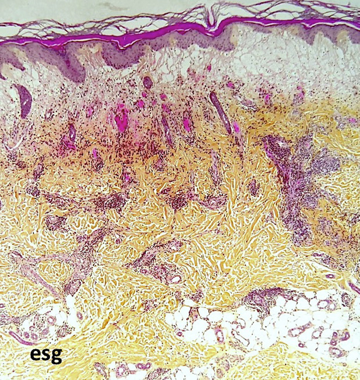
Fig 1
Chilblain-like lesion. Scanning magnification shows diffuse upper dermal edema and a dense dermal (perivascular and perieccrine sweat gland) inflammatory infiltrate. (Hematoxylin-eosin-saffron stain; ...
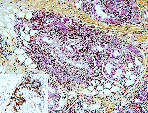
Fig 2
Chilblain-like lesion. Dense lymphocytic infiltrate around an eccrine sweat gland. Inset: perieccrine clusters of CD303 + /BDCA-2 plasmacytoid dendritic cells. (Hematoxylin-eosin-saffron stain; origin...
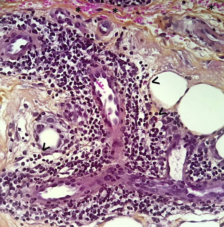
Fig 3
Chilblain-like lesion. Dense lymphocytic dermal perivascular infiltrate admixed with occasional eosinophils (arrowheads). Endothelial cell swelling and extravasated red blood cells (asterisks).
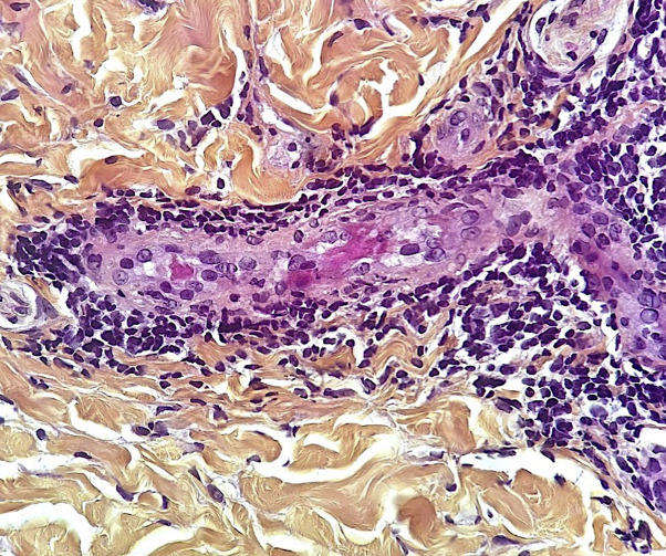
Fig 4
Chilblain-like lesion. Eosinophilic thrombi within the lumen of a dermal venule. (Hematoxylin-eosin-saffron stain; original magnification: u00d7400.)
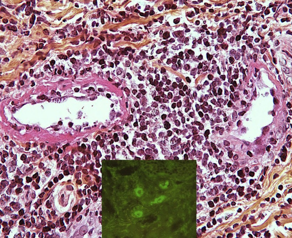
Fig 5
Chilblain-like lesion. Eosinophilic fibrin deposits on the wall of a dermal venule. Inset shows vascular deposits of immunoglobulin M. (Hematoxylin-eosin-saffron stain; original magnification: u00d740...
Figure images are served from the NIH/NLM PubMed Central Open Access Subset or Europe PMC; copyright remains with the publishers and authors.
💬 Discussion
0 commentsNo comments yet. Be the first to start a discussion!
Leave a Comment