Abstract
Cellular uptake pathway of nanoparticle (NP) is different from that of free drugs. Therefore, NP-mediated nanotherapeutics can be designed to overcome the adverse effects of free drugs. However, synthetic NPs are typically trapped in the endosome and have difficulty to reach the cytosol because of the characteristic endocytosis, where the endosomal membranes wrap-up the introduced NPs. In this study, the Spacer molecules linking the apoptotic anticancer drug and the gold NP (AuNP) are designed and cellular uptake procedure and drug deployment in the cancer cells are controlled. X-ray nanoscopy and two-photon microscopy are employed to observe the AuNPs in a cell in-situ without additional dye molecule or imaging agent introduction on an AuNP. We confirm that the effective design of the Spacer molecules importantly control the cellular interaction of the AuNPs. This technology can be generalized to broad biomedical applications utilizing nanotherapeutics-mediated diagnosis and new-concepted disease treatment technologies.
🔬 Techniques
🧪 Sample Preparation
🔬 Cell Lines
🏭 Microscope Brands
🧪 Reagent Suppliers
🔴 Lasers
🏛️ Research Organizations (ROR)
Affiliated research institutions:
📋 Methods
DOX-tethered surface-modified AuNPs The average core size obtained by transmission electron microscopy (TEM) is approximately 20 nm for all the AuNPs. The concentration of standard AuNP stock solution (×1) is controlled at 1 mM of Au ( Supporting Information ). The concentration of the standard AuNP solution is 2.4 × 10 12 AuNPs/mL. Then the number of DOX per unit AuNP is controlled as 1.15 × 10 3 DOX/AuNP. The carboxyl groups of the designed spacers are activated by N -hydroxysuccinimide and N , N ′-dicyclohexylcarbodiimide. The activated acid is then reacted with the amine group of DOX. All the reaction mixtures are dialyzed against Milli-Q water for purification. For respective AuNP, the spacers containing 0.05 mg DOX (M w : 543.52, 9.2 ×10 −8 mole DOX, 5.5×10 16 DOX molecules) are introduced to a 20 mL standard citrate-covered AuNP stock solution (1 mmol·L −1 of Au). Then, the number of DOX per unit AuNP is controlled as 1.15×10 3 DOX/AuNP. The completion of the reaction is confirmed by the absence of the thiol-containing DOX-conjugated spacers in the reaction solvent (confirmed by UV-vis spectrum and fluorescence, data is not shown). To the aforementioned DOX-conjugated AuNPs, negatively and positively charged thiol ligands are further introduced to modify the surface properties of the DOX-conjugated AuNPs: thioglycolic acid (SH-CH 2 COOH, designated as AuNP 1) and 6-thioguanine (SH-C 5 H 4 N 5 , designated as AuNP 2). The obtained AuNP solutions are purified by dialysis for more than 2 days. The detailed procedures are provided in Supporting Information .
Show full methods section
DOX-tethered surface-modified AuNPs The average core size obtained by transmission electron microscopy (TEM) is approximately 20 nm for all the AuNPs. The concentration of standard AuNP stock solution (×1) is controlled at 1 mM of Au ( Supporting Information ). The concentration of the standard AuNP solution is 2.4 × 10 12 AuNPs/mL. Then the number of DOX per unit AuNP is controlled as 1.15 × 10 3 DOX/AuNP. The carboxyl groups of the designed spacers are activated by N -hydroxysuccinimide and N , N ′-dicyclohexylcarbodiimide. The activated acid is then reacted with the amine group of DOX. All the reaction mixtures are dialyzed against Milli-Q water for purification. For respective AuNP, the spacers containing 0.05 mg DOX (M w : 543.52, 9.2 ×10 −8 mole DOX, 5.5×10 16 DOX molecules) are introduced to a 20 mL standard citrate-covered AuNP stock solution (1 mmol·L −1 of Au). Then, the number of DOX per unit AuNP is controlled as 1.15×10 3 DOX/AuNP. The completion of the reaction is confirmed by the absence of the thiol-containing DOX-conjugated spacers in the reaction solvent (confirmed by UV-vis spectrum and fluorescence, data is not shown). To the aforementioned DOX-conjugated AuNPs, negatively and positively charged thiol ligands are further introduced to modify the surface properties of the DOX-conjugated AuNPs: thioglycolic acid (SH-CH 2 COOH, designated as AuNP 1) and 6-thioguanine (SH-C 5 H 4 N 5 , designated as AuNP 2). The obtained AuNP solutions are purified by dialysis for more than 2 days. The detailed procedures are provided in Supporting Information .
Cell preparation
A549 cells are cultured in RPMI-1640 medium containing 10% fetal bovine serum and 1% penicillin-streptomycin. A low-serum growth supplement containing 20% fetal bovine serum and 1% penicillin-streptomycin is also added. The cells are cultured in a humidified atmosphere with 5% CO 2 at 37°C. A trypsin (0.25%)-EDTA solution is used to detach the cells from the culture flask. Detailed procedures are in Supporting Information . SEM−EDXS The cells are loaded onto a cover glass and then cultured for 24 h. Thereafter, the cells are treated with a series of surface-modified AuNPs for the designated time. The samples are fixed with 2.5% glutaraldehyde in Dulbecco's phosphate buffered saline for 20 min at room temperature, washed thrice with deionized water, and then air-dried. The samples are Ni-coated using a coater (Quorum Technology, SC7640 model). SEM images are captured using an XL30SFEG (Philips) SEM connected to an EDXS system at an acceleration voltage of 20 kV. SEM and EDXS measurements are performed using a XL30SFEG electron probe microanalyzer equipped with a Genesis System EDAX detector. The detailed procedures are described in Supporting Information . TPM An upright microscope (BX51; Olympus) is used to perform TPM. The system is equipped with a Ti:sapphire femtosecond laser (Chameleon; Coherent) with a tuning range of 680 nm to 1020 nm. To generate the two-photon effect, the laser source is operated at 80 MHz repetition rate and 140 fs pulse width. The beam first passes through a half-wave plate and then through a polarizer for power control. The beam is expanded twice by passing it through a pair of plano-convex lenses, and then the expanded and excited light are passed through a resonant x - y scanner (counter rotation scanner, GSI Lumonics) operating at 8 KHz scanning speed. Details are provided in Supporting Information .
Zebrafish preparation and cancer cell microinjection
Zebrafish is maintained in E3 embryo media (15 mM NaCl, 0.5 mM KCl, 1 mM MgSO 4 , 1 mM CaCl 2 , 0.15 mM KH 2 PO 4 , 0.05 mM Na 2 HPO 4 , 0.7 mM NaHCO 3 , 10–5% methylene blue; pH 7.4) containing 1-phenyl 2-thiourea (Sigma-Aldrich) to prevent pigmentation at 29°C. Red fluorescence labeled cancer cell pellet and matrigel (BD Biosciences) are mixed with 1/1 (vol/vol) at 4°C. To this call-gel mixture, doxorubicin or doxorubicin-tethered surface-modified AuNP solutions are mixed with 3/1 (vol/vol). This cell suspension mixture containing 1000 cells is loaded into a borosilicate glass needle and implanted into each zebrafish of 4 day-post-fertilization embryo through the yolk in a single injection by using an electronically regulated air-pressure microinjector (Eppendorf, FemtoJet express). After injection, zebrafish is washed once with E3 embryo media and examined the presence of fluorescent cells. Forty zebrafishes are selected and transferred to 24-well plate containing 1 mL of fresh E3 embryo media for photography. E3 embryo media is changed daily and maintained under normal fish husbandry conditions for 3 days. For the work with zebrafish, all the experiments were performed in accordance with relevant guidelines and regulations.
📊 Figures
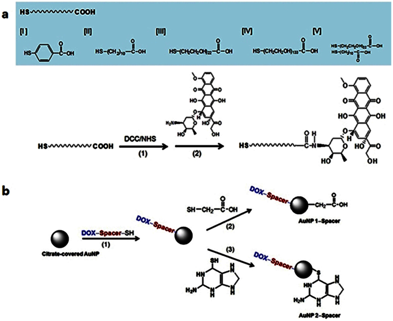
Figure 1
(a) Synthetic procedure of the Spacer molecules. (b) Synthetic procedure of the drug-conjugated and surface-modified AuNPs.
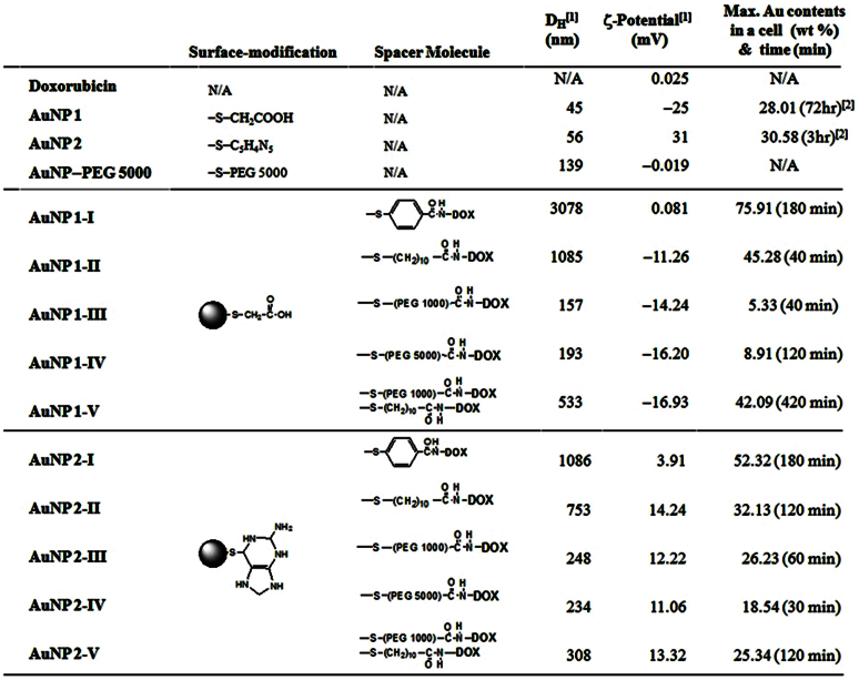
Figure 2
Hydrodynamic diameter (D H ) and zeta (u03b6)-potential of the AuNPs.
[1] Measured in de-ionized water at 25u00b0C. The concentration of the AuNP solution is controlled by 1u2005mM Au. [2] The maximum values for AuNP 1 and AuNP 2 are obtained with different time interva...
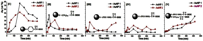
Figure 3
Time-dependent Au wt% in A549 cells treated with the AuNPs having designed Spacers I, II, II, IV and V, combinated with cationic (u2013CH 2 COOH, AuNP 1) and anionic (u2013C 5 H 4 N 5 , AuNP 2) surface-modification.
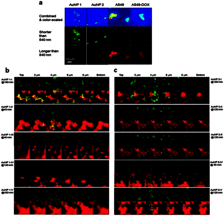
Figure 4
Depth profiles images of a cell obtained by two-photon microscopy (TPM).
From top to bottom of cell, 10u2005u03bcm depth is scanned. The size of each image is 50u2005u03bcm u00d7 50u2005u03bcm. (a) The images of anionic AuNP 1, cationic AuNP 2, A549 cell and DOX-treated A5...
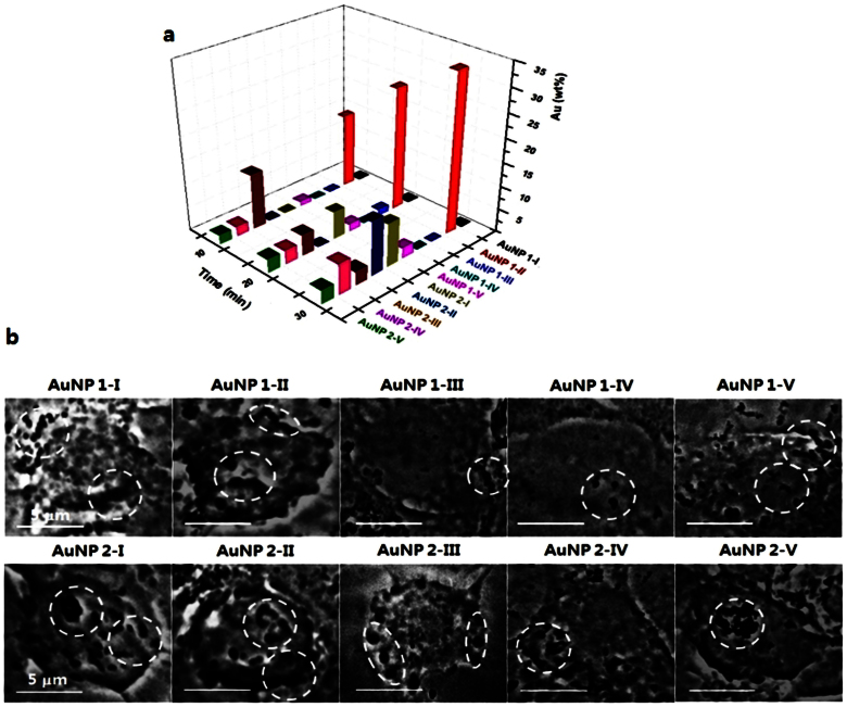
Figure 5
(a) Au wt% at 10, 20 and 30u2005min obtained by EDXS. (b) Zone-plate X-ray nanoscopy images obtained at 30u2005min. The scale-bar is 5u2005u03bcm.
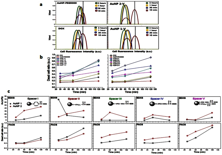
Figure 6
(a) Fluorescence-activated cell sorting (FACS) results of PEG 5000-conjugated AuNP, free DOX and AuNP 1-V and 2-V. The results are obtained at 10u2005min, 30u2005min, 1u2005hr and 2u2005hr. (b) Dead c...
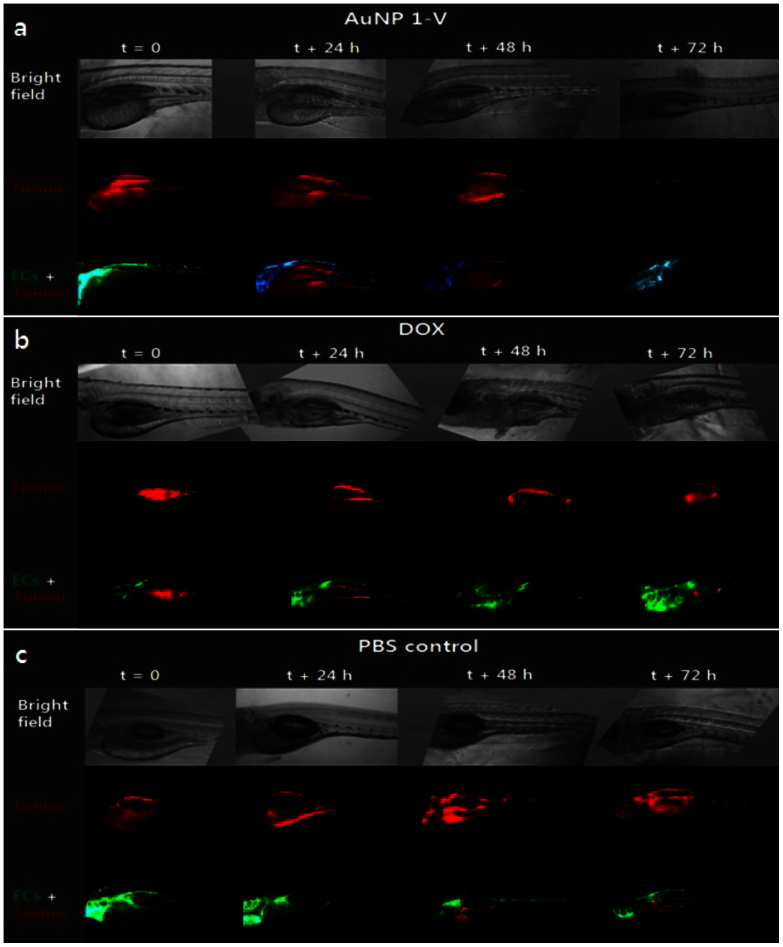
Figure 7
In-vivo tumor xenografts in zebrafish embryos treated with (a) AuNP 1-V, (b) free DOX and (c) PBS control.
Figure images are served from the NIH/NLM PubMed Central Open Access Subset or Europe PMC; copyright remains with the publishers and authors.
💬 Discussion
0 commentsNo comments yet. Be the first to start a discussion!
Leave a Comment