Abstract
AbstractLittle is known about the structural basis of prion strains. Here we provide a high (3.0 Å) resolution cryo-electron microscopy-based structure of infectious brain-derived fibrils of the mouse anchorless RML scrapie strain which, like the recently determined hamster 263K strain, has a parallel in-register β-sheet-based core. Several structural motifs are shared between these ex vivo prion strains, including an amino-proximal steric zipper and three β-arches. However, detailed comparisons reveal variations in these shared structural topologies and other features. Unlike 263K and wildtype RML prions, the anchorless RML prions lack glycophosphatidylinositol anchors and are severely deficient in N-linked glycans. Nonetheless, the similarity of our anchorless RML structure to one reported for wildtype RML prion fibrils in an accompanying paper indicates that these post-translational modifications do not substantially alter the amyloid core conformation. This work demonstrates both common and divergent structural features of prion strains at the near-atomic level.
🔬 Techniques
✨ Fluorophores
🏭 Microscope Brands
🧪 Reagent Suppliers
📷 Detectors
💻 Software Details
🏛️ Research Organizations (ROR)
Affiliated research institutions:
📋 Methods
PrP Sc fibril purification and bioassay All mice were housed at the Rocky Mountain Laboratory in an AAALAC accredited facility in compliance with guidelines provided by the Guide for the Care and Use of Laboratory Animals (Institute for Laboratory Animal Research Council). Experimentation followed Rocky Mountain Laboratory Animal Care and Use Committee approved protocols 2018‐011, 2016‐039, or 2021-011-E. aRML (also known as Chandler) prion strain fibrils were purified from brains of transgenic mice expressing only GPI-anchorless PrP and characterized as part of previous studies 5 , 10 . Briefly, brain homogenates were treated with a 2% sarkosyl buffer and benzonase (EMD Millipore) to digest nucleic acids and then ultracentrifuged to pellet PrP Sc . The pellet was resuspended, treated with proteinase K (PK), a high salt (1.7 M NaCl) and 30 mM EDTA buffer, and centrifuged through a sucrose cushion containing 0.5% sulfobetaine 3–14. The pellet was washed in 0.5% sulfobetaine 3–14, pelleted, and resuspended using cuphorn sonication into 0.5% sulfobetaine 3–14 in 20 mM sodium phosphate, 130 mM NaCl; pH 7.4. Further characterizations of such aRML preparations have been described previously 5 . Additional fibril manipulations prior to cryo-EM grid preparation were performed as previously reported 1 . Briefly, fibril preparations were vortexed and allowed to sit for several minutes to pellet highly bundled fibrils. Aliquots from the supernatant fraction were diluted in 20 mM Tris pH 7.4, 100 mM NaCl containing 0.02% amphipol 8–35 and sonicated immediately prior to grid preparation. To estimate the infectivity of the purified aRML preparation, male tga20 homozygous mice 11 were anesthetized with isoflurane and injected in the left-brain hemisphere with 10-fold serial dilutions beginning with 100 ng of purified anchorless RML prep diluted in 30 µl phosphate buffered balanced saline solution + 2% fetal bovine serum. Following inoculation, mice were monitored for onset of prion disease signs and euthanized when they displayed signs of prion disease including ataxia, flattened posture, delayed response to stimuli, and somnolence. Cryo-EM grid preparation C-Flat 1.2/1.3 300 mesh copper grids (Protochips, Morrisville, NC) were glow-discharged with a 50:50 oxygen/hydrogen mixture in a Solarus 950 (Gatan, Pleasanton, CA) for 10 s. Grids were mounted in an EM GP2 plunge freezer (Leica, Buffalo Grove, IL) and a 3 μl droplet of 0.02% amphipol A8-35 in phosphate buffered saline was added to the carbon surface and hand blotted to leave a very thin film. The tweezers were then raised into the chamber of the plunge freezer, which was set to 22 °C and 90% humidity. 3 μl of recently sonicated sample was added to the carbon side of the grid and allowed to sit for 60 s. The sample was subsequently blotted for ∼4 s followed by a 3 s drain time before plunge freezing in liquid ethane kept at −180 °C. Grids were mounted in AutoGrid assemblies.
Show full methods section
PrP Sc fibril purification and bioassay All mice were housed at the Rocky Mountain Laboratory in an AAALAC accredited facility in compliance with guidelines provided by the Guide for the Care and Use of Laboratory Animals (Institute for Laboratory Animal Research Council). Experimentation followed Rocky Mountain Laboratory Animal Care and Use Committee approved protocols 2018‐011, 2016‐039, or 2021-011-E. aRML (also known as Chandler) prion strain fibrils were purified from brains of transgenic mice expressing only GPI-anchorless PrP and characterized as part of previous studies 5 , 10 . Briefly, brain homogenates were treated with a 2% sarkosyl buffer and benzonase (EMD Millipore) to digest nucleic acids and then ultracentrifuged to pellet PrP Sc . The pellet was resuspended, treated with proteinase K (PK), a high salt (1.7 M NaCl) and 30 mM EDTA buffer, and centrifuged through a sucrose cushion containing 0.5% sulfobetaine 3–14. The pellet was washed in 0.5% sulfobetaine 3–14, pelleted, and resuspended using cuphorn sonication into 0.5% sulfobetaine 3–14 in 20 mM sodium phosphate, 130 mM NaCl; pH 7.4. Further characterizations of such aRML preparations have been described previously 5 . Additional fibril manipulations prior to cryo-EM grid preparation were performed as previously reported 1 . Briefly, fibril preparations were vortexed and allowed to sit for several minutes to pellet highly bundled fibrils. Aliquots from the supernatant fraction were diluted in 20 mM Tris pH 7.4, 100 mM NaCl containing 0.02% amphipol 8–35 and sonicated immediately prior to grid preparation. To estimate the infectivity of the purified aRML preparation, male tga20 homozygous mice 11 were anesthetized with isoflurane and injected in the left-brain hemisphere with 10-fold serial dilutions beginning with 100 ng of purified anchorless RML prep diluted in 30 µl phosphate buffered balanced saline solution + 2% fetal bovine serum. Following inoculation, mice were monitored for onset of prion disease signs and euthanized when they displayed signs of prion disease including ataxia, flattened posture, delayed response to stimuli, and somnolence. Cryo-EM grid preparation C-Flat 1.2/1.3 300 mesh copper grids (Protochips, Morrisville, NC) were glow-discharged with a 50:50 oxygen/hydrogen mixture in a Solarus 950 (Gatan, Pleasanton, CA) for 10 s. Grids were mounted in an EM GP2 plunge freezer (Leica, Buffalo Grove, IL) and a 3 μl droplet of 0.02% amphipol A8-35 in phosphate buffered saline was added to the carbon surface and hand blotted to leave a very thin film. The tweezers were then raised into the chamber of the plunge freezer, which was set to 22 °C and 90% humidity. 3 μl of recently sonicated sample was added to the carbon side of the grid and allowed to sit for 60 s. The sample was subsequently blotted for ∼4 s followed by a 3 s drain time before plunge freezing in liquid ethane kept at −180 °C. Grids were mounted in AutoGrid assemblies.
Cryo-electron tomography
For tomography, grids were prepared as above except that 5 nm Protein A gold (CMC, Utrecht, The Netherlands) was added for fiducial markers. The grid assemblies were loaded into a Krios G1 (Thermo Fisher Scientific, Waltham, MA) transmission electron microscope operating at 300 kV with a K3 (Gatan, Pleasanton CA) and a Biocontinuum GIF (Gatan, Pleasanton CA) with a slit width of 20 eV. Tilt series were acquired using SerialEM 23 at a 0.45 Å pixel size at ±60°, 2° increment in a dose symmetric manner around 0° 24 with defocus values ranging from −3 to −6 μm and a total dose of ~60 e − /Å 2 . Tomograms were reconstructed and 12 were analyzed using IMOD 25 . To verify that our imaging system preserved handedness, we negatively stained bacteria onto a finder grid and acquired tilt-series of an asymmetric letter to confirm orientation did not change during imaging nor through tomographic reconstruction 26 . We then used the bacteria as fiducials to confirm that there were only rotation changes during magnification increases from the tilt-series magnification of the finder grid letter to the tilt-series magnification of aRML.
Image acquisition and processing for helical reconstruction
Initial map and helical twist parameters were generated based on our recently published dataset 1 , as well as on cryo-electron tomographic analyses of aRML fibrils described above. Motion correction of raw movie frames was performed with RELION 3.1 13 . CTF estimation was performed using CTFIND4.1 27 . Fibrils were handpicked then extracted using large and small box sizes. The longer helical segments were extracted with a box size of 1280 pixels and were downscaled to a box size of 256 pixels. The shorter segments were extracted at 400 pixel box size. 2D classes, from the long segments were used to estimate the cross-over distance of the fibril for estimating initial twist parameters. 2D classes from the short segments were used to generate an initial 3D model. Higher resolution data was collected using a Titan Krios G3i (Thermo Fisher Scientific, Waltham, MA) with a K3 camera and BioQuantum GIF (Gatan, Pleasanton, CA) with images acquired at 0.55 Å/pixel at Super Resolution mode, 60 e − /Å 2 , and 60 total frames. Movies were motion corrected and the CTF estimated as above. Fibrils were picked manually and segments were extracted with an inter-box distance of 14.6 Å using box size of 740 pixels that was down sampled to 370 pixels. Reference-free 2D class averaging was performed, using a regularization parameter of T = 2, a tube diameter of 180 Å, and the translational offset limited to 4.8 Å. The initial model was used for 3D auto refinement with C1 symmetry, initial resolution limit of 40 Å, initial angular sampling of 3.7°, offset search range of 5 pixels, initial helical twist of −0.72°, initial helical rise of 4.85 Å, and using 50% of the segment central Z length. The output from auto refinement was used for 3D classification without allowing for image alignment to remove poorly aligned segments from auto refinement. Classes were selected for further refinement based on similarity of features in their cross-section (excluding visually low resolution and poorly aligned classes), estimated resolution, overall accuracy of rotation and translation, and Fourier completeness. Auto-refinement was then performed while optimizing the helical twist and rise, yielding a final map with a twist of −0.637° and rise of 4.876 Å. Iterative cycles of CTF refinement, Bayesian polishing, and auto refinement were used until resolution estimates stabilized. Post processing in RELION was performed with a soft-edged mask representing 10% of the central Z length of the fibril. Resolution estimates were obtained between independent refined half-maps at 0.143 FSC. Model building De novo building of an aRML atomic model was conducted using Coot 28 , with the assumption that residues comprising the protease-resistant core (i.e. ~90–231) were included in the amyloid core. Individual subunits were translated to generate a stack of five consecutive subunits, and translated subunits rigid-body fit in Coot. Iterative real-space refinement and validation with Coot and Phenix 29 , 30 were performed, with Fourier space refinements being conducted using RefMac5. Model validation was performed with CaBLAM 31 , MolProbity 32 , and EMringer 33 , and any outliers/clashes identified and corrected with subsequent iterative refinements/validation. Model renderings in Figs. 1 and 2 were performed using Chimera X.
Molecular dynamics simulations
Molecular dynamics simulations were performed using NAMD 2.14 34 with the CHARMM36 forcefield 35 . After the addition of protons utilizing the HBUILD functionality in the CHARMM molecular dynamics platform 36 , a disulfide bond between cysteines 178 and 213 was generated with the use of the DISU patch. The individual monomer chains in the fibril were capped using the ACE and CNEU patches. The system was then solvated with TIP3P water molecules in a cubic box containing neutralizing Na + and Cl − ions. A gradient of backbone and sidechain restraints ranging from 10 kcal/mol/ Å 2 to 1 kcal/mol/ Å 2 , was utilized in iterative runs of conjugate gradient minimization and were subsequently removed in the last 15,000 steps. A 1 ns NVT equilibration simulation with backbone restraints was performed at a temperature of 300 K maintained with Langevin dynamics. The simulation advanced at a timestep of 1 fs and the particle mesh Ewald algorithm was used to calculate long-range electrostatics. Non-bonded interactions had a cutoff of 10 Å and the rigid bond algorithm was applied to all bonds containing hydrogen atoms. Subsequently, a 1 ns NPT equilibration was performed with backbone restraints followed by a 10 ns production run which advanced at a 2 fs timestep. Pressure was kept constant using the Langevin-piston method. After 10 ns, the restraints were removed, and the simulation continued for an additional 170 ns. The trajectories were analyzed using VMD 37 and Bio3D 38 . Reporting summary Further information on research design is available in the Nature Research Reporting Summary linked to this article.
Supplementary information Supplementary Information Reporting Summary
📊 Figures
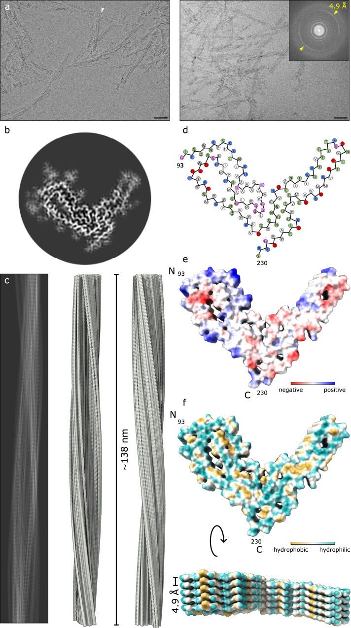
Fig. 1
Cryo-EM-based structure of aRML fibrils.
a 2D cryo-EM images of aRML fibrils. Baru2009=u200950u2009nm. Inset depicts associated Fastu00a0Fourier transform showing signals from regular 4.9u2009u00c5 spacings (yellow arrows). The white arrowhe...
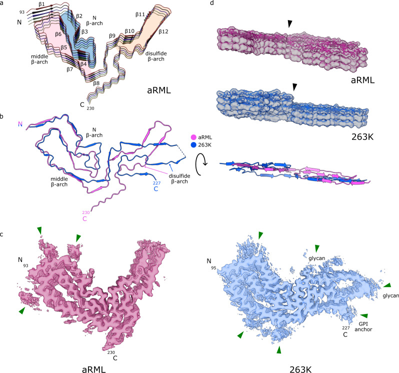
Fig. 2
Comparison of aRML and 263K prions.
a Ribbon diagram of aRML core (stack of 5), with structural motifs as colored. b Overlay of aRML and 263K cores. c Contour EM density maps of aRML and 263K. Green arrows indicate peripheral unassigned...
Figure images are served from the NIH/NLM PubMed Central Open Access Subset or Europe PMC; copyright remains with the publishers and authors.
💬 Discussion
0 commentsNo comments yet. Be the first to start a discussion!
Leave a Comment