Abstract
The lipid scramblase TMEM16F initiates blood coagulation by catalyzing the exposure of phosphatidylserine in platelets. The protein is part of a family of membrane proteins, which encompasses calcium-activated channels for ions and lipids. Here, we reveal features of murine TMEM16F (mTMEM16F) that underlie its function as a lipid scramblase and an ion channel. The cryo-EM data of mTMEM16F in absence and presence of Ca2+ define the ligand-free closed conformation of the protein and the structure of a Ca2+-bound intermediate. Both conformations resemble their counterparts of the scrambling-incompetent anion channel mTMEM16A, yet with distinct differences in the region of ion and lipid permeation. In conjunction with functional data, we demonstrate the relationship between ion conduction and lipid scrambling. Although activated by a common mechanism, both functions appear to be mediated by alternate protein conformations that are at equilibrium in the ligand-bound state.
🔬 Techniques
💻 Software
✨ Fluorophores
🧪 Sample Preparation
🔬 Cell Lines
🏭 Microscope Brands
🧪 Reagent Suppliers
💻 Software Details
💾 Data Repositories
🏛️ Research Organizations (ROR)
Affiliated research institutions:
📋 Methods
Key resources table
Reagent type (species) or resource Designation Source or reference Identifier Additional information Antibody Mouse monoclonal Anti-c-Myc Millipore Sigma Cat#M4439; Clone#9E10 Dilution – 1:5000 Antibody Peroxidase Affinipure goat anti-mouse IgG Jackson Immunoresearch Cat#115-035-146 Dilution – 1:10000 Chemical compound, drug FuGENE six transfection reagent Promega Cat# E2691 Chemical compound, drug Hygromycin B Gibco, Thermo Fisher Scientific Cat#10687010 Chemical compound, drug Tetracycline hydrochloride Millipore Sigma Cat#T7660 Chemical compound, drug n - dodecyl - β - d - maltopyranoside, Solgrade Anatrace Cat#D310S Chemical compound, drug EX-CELL 293 Serum-Free medium Millipore Sigma Cat#14571C Chemical compound, drug Fetal bovine serum Millipore Sigma Cat#F7524 Chemical compound, drug L-glutamine Millipore Sigma Cat#G7513 Chemical compound, drug Penicillin-streptomycin Millipore Sigma Cat#P0781 Chemical compound, drug Valproic acid Millipore Sigma Cat#P4543 Chemical compound, drug HyClone HyCell TransFx-H medium GE Healthcare Cat#SH30939.02 Chemical compound, drug Poloxamer 188 Millipore Sigma Cat#P5556 Chemical compound, drug Polyethylenimine MAX 40 K Polysciences Cat# 24765–1 Chemical compound, drug Dulbecco’s Modified Eagle’s Medium - high glucose Millipore Sigma D5671 Chemical compound, drug Dulbecco’s Phosphate Buffered Saline Millipore Sigma D8537 Chemical compound, drug Digitonin Reagent USP PanReac AppliChem Cat#A1905 Chemical compound, drug Calcium nitrate tetrahydrate Millipore Sigma Cat#C4955 Chemical compound, drug Sodium chloride Millipore Sigma Cat#71380 Chemical compound, drug HEPES Millipore Sigma Cat#H3375 Chemical compound, drug Ethylene glycol-bis(2- aminoethylether)-N,N,N′,N′- tetraacetic acid Millipore Sigma Cat#03777 Chemical compound, drug cOmplete, EDTA-free Protease Inhibitor Cocktail Roche Cat#5056489001 Chemical compound, drug Digitonin, High Purity - Calbiochem EMD Millipore Cat#300410 Chemical compound, drug Biotin Millipore Sigma Cat#B4501 Recombinant protein PNGase F Raimund Dutzler laboratory NA Commercial assay or kit Amicon Ultra-4–100 KDa cutoff EMD Millipore Cat#UFC8100 Commercial assay or kit 0.22 μm Ultrafree-MC Centrifugal Filter EMD Millipore Cat#UFC30GV Chemical compound, drug 1-palmitoyl-2-oleoyl -glycero-3-phosphocholine Avanti Polar Lipids, Inc Cat#850457C Chemical compound, drug 1-palmitoyl-2-oleoyl-sn- glycero-3-phospho-(1'-rac- glycerol) Avanti Polar Lipids, Inc Cat#840457C Chemical compound Diethyl ether Millipore Sigma Cat#296082 Commercial assay or kit Bio-Beads SM-2 Adsorbents Bio-Rad Cat# 1523920 Chemical compound, drug Soybean Polar Lipid Extract Avanti Polar Lipids, Inc Cat#541602C Chemical compound, drug Cholesterol Millipore Sigma Cat#C8667 Chemical compound 18:1-06:0 NBD-PE Avanti Polar Lipids, Inc Cat#810155C Chemical compound, drug 18:1-06:0 NBD-PS Avanti Polar Lipids, Inc Cat#810194C Chemical compound, drug 14:0 NBD-PE Avanti Polar Lipids, Inc Cat#810143 C Chemical compound, drug Potassium chloride Millipore Sigma Cat#746436 Chemical compound, drug Triton X-100 Millipore Sigma Cat#T9284 Chemical compound, drug Sodium dithionite Millipore Sigma Cat#157953 Chemical compound, drug N-Methyl-D-glucamine Millipore Sigma Cat#66930 Chemical compound, drug Sulphuric acid 95–97% EMD Millipore Cat# 1.00731.1000 Commercial assay or kit Borosilicate glass capilliary with filament Sutter Instrument Cat#BF150-86-10HP Commercial assay or kit Pierce Streptavidin Plus UltraLink Resin Thermo Fisher Scientific Cat#53117 Commercial assay or kit Superose 6 10/300 GL GE Healthcare Cat#17-5172-01 Commercial assay or kit Flp-In T-Rex Core Kit ThermoFisher Scientific Cat#K650001 Commercial assay or kit Whatman Nuclepore Track-Etched Membranes diam.19mm, pore size 0.4μm, polycarbonate Millipore Sigma Cat#WHA800282 Commercial assay or kit Microforge Narishige NA Commercial assay or kit Axopatch 200B amplifer Molecular Devices NA Commercial assay or kit Digidata 1440 Molecular Devices NA Commercial assay or kit 300 mesh Au 1.2/1.3 cryo-EM grids Quantifoil Cat#N1-C14nAu30-01 Cell line (Human) Flp-In T-REx 293 ThermoFisher Scientific Cat# R78007 Cell line (Human) HEK-293T ATCC Cat#CRL-1573 Cell line (Human) HEK293S GnTI - ATCC Cat#CRL-3022 Recombinant DNA Mouse mTMEM16F open reading frame Dharmacon - Horizon Discovery GenBank# BC060732 Recombinant DNA Mammalian expression vector with C-terminal 3C protease cleavage site, Myc tag and streptavidin binding peptide Raimund Dutzler laboratory NA Recombinant DNA Mammalian expression vector with C-terminal 3C protease cleavage site, Venus and Myc tags and streptavidin binding peptide Raimund Dutzler laboratory NA Recombinant DNA Synthesized mTMEM16F SCRD cDNA GenScript NA Recombinant DNA Membrane scaffold protein (MSP) 2N2 Stephen Sligar laboratory Addgene:Cat#29520 Software, algorithm WEBMAXC calculator Bers et al., 2010 http://maxchelator.stanford.edu/webmaxc/webmaxcS.htm Software, algorithm Axon Clampfit 10.7 Molecular Devices NA Software, algorithm Axon Clampex 10.6 Molecular Devices NA Software, algorithm Focus 1.1.0 Biyani et al. (2017) https://focus.c-cina.unibas.ch/about.php Software, algorithm MotionCorr2 1.1.0 Zheng et al. (2017) http://msg.ucsf.edu/em/software/motioncor2.html Software, algorithm CTFFIND 4.1 Rohou and Grigorieff (2015) http://grigoriefflab.janelia.org/ctf Software, algorithm Relion v 2.1 and 3.0 Kimanius et al., 2016 Zivanov et al., 2018 https://www2.mrc-lmb.cam.ac.uk/relion/ Software, algorithm Phenix 1.13 Adams et al. (2010) http://http://phenix-online.org/ Software, algorithm Coot 0.8.9.1 Emsley and Cowtan (2004) https://www2.mrc-lmb.cam.ac.uk/personal/pemsley/coot/ Software, algorithm Pymol 2.0 Schrodinger LLC https://pymol.org/2/ Software, algorithm Chimera 1.12 Pettersen et al. (2004) https://www.cgl.ucsf.edu/chimera/ Cell lines Flp-In T-REx 293 cell lines stably expressing mTMEM16F were adapted to suspension cultures and were grown at 37°C and 5% CO 2 in EX-CELL 293 Serum-free medium supplemented with 1% fetal bovine serum, 6 mM L-glutamine and 100 U/ml penicillin–streptomycin. GnTI - cells were grown in HyClone HyCell TransFx-H medium supplemented with 1% fetal bovine serum, 4 mM L-glutamine, 1 g/l poloxamer 188 and 100 U/ml penicillin–streptomycin. Adherent HEK293T cells were grown in DMEM medium supplemented with 10% fetal bovine serum, 2 mM L-glutamine, 1 mM Sodium pyruvate and 100 U/ml penicillin–streptomycin. Mycoplasma test performed on the used cells was negative.
Show full methods section
Key resources table
Reagent type (species) or resource Designation Source or reference Identifier Additional information Antibody Mouse monoclonal Anti-c-Myc Millipore Sigma Cat#M4439; Clone#9E10 Dilution – 1:5000 Antibody Peroxidase Affinipure goat anti-mouse IgG Jackson Immunoresearch Cat#115-035-146 Dilution – 1:10000 Chemical compound, drug FuGENE six transfection reagent Promega Cat# E2691 Chemical compound, drug Hygromycin B Gibco, Thermo Fisher Scientific Cat#10687010 Chemical compound, drug Tetracycline hydrochloride Millipore Sigma Cat#T7660 Chemical compound, drug n - dodecyl - β - d - maltopyranoside, Solgrade Anatrace Cat#D310S Chemical compound, drug EX-CELL 293 Serum-Free medium Millipore Sigma Cat#14571C Chemical compound, drug Fetal bovine serum Millipore Sigma Cat#F7524 Chemical compound, drug L-glutamine Millipore Sigma Cat#G7513 Chemical compound, drug Penicillin-streptomycin Millipore Sigma Cat#P0781 Chemical compound, drug Valproic acid Millipore Sigma Cat#P4543 Chemical compound, drug HyClone HyCell TransFx-H medium GE Healthcare Cat#SH30939.02 Chemical compound, drug Poloxamer 188 Millipore Sigma Cat#P5556 Chemical compound, drug Polyethylenimine MAX 40 K Polysciences Cat# 24765–1 Chemical compound, drug Dulbecco’s Modified Eagle’s Medium - high glucose Millipore Sigma D5671 Chemical compound, drug Dulbecco’s Phosphate Buffered Saline Millipore Sigma D8537 Chemical compound, drug Digitonin Reagent USP PanReac AppliChem Cat#A1905 Chemical compound, drug Calcium nitrate tetrahydrate Millipore Sigma Cat#C4955 Chemical compound, drug Sodium chloride Millipore Sigma Cat#71380 Chemical compound, drug HEPES Millipore Sigma Cat#H3375 Chemical compound, drug Ethylene glycol-bis(2- aminoethylether)-N,N,N′,N′- tetraacetic acid Millipore Sigma Cat#03777 Chemical compound, drug cOmplete, EDTA-free Protease Inhibitor Cocktail Roche Cat#5056489001 Chemical compound, drug Digitonin, High Purity - Calbiochem EMD Millipore Cat#300410 Chemical compound, drug Biotin Millipore Sigma Cat#B4501 Recombinant protein PNGase F Raimund Dutzler laboratory NA Commercial assay or kit Amicon Ultra-4–100 KDa cutoff EMD Millipore Cat#UFC8100 Commercial assay or kit 0.22 μm Ultrafree-MC Centrifugal Filter EMD Millipore Cat#UFC30GV Chemical compound, drug 1-palmitoyl-2-oleoyl -glycero-3-phosphocholine Avanti Polar Lipids, Inc Cat#850457C Chemical compound, drug 1-palmitoyl-2-oleoyl-sn- glycero-3-phospho-(1'-rac- glycerol) Avanti Polar Lipids, Inc Cat#840457C Chemical compound Diethyl ether Millipore Sigma Cat#296082 Commercial assay or kit Bio-Beads SM-2 Adsorbents Bio-Rad Cat# 1523920 Chemical compound, drug Soybean Polar Lipid Extract Avanti Polar Lipids, Inc Cat#541602C Chemical compound, drug Cholesterol Millipore Sigma Cat#C8667 Chemical compound 18:1-06:0 NBD-PE Avanti Polar Lipids, Inc Cat#810155C Chemical compound, drug 18:1-06:0 NBD-PS Avanti Polar Lipids, Inc Cat#810194C Chemical compound, drug 14:0 NBD-PE Avanti Polar Lipids, Inc Cat#810143 C Chemical compound, drug Potassium chloride Millipore Sigma Cat#746436 Chemical compound, drug Triton X-100 Millipore Sigma Cat#T9284 Chemical compound, drug Sodium dithionite Millipore Sigma Cat#157953 Chemical compound, drug N-Methyl-D-glucamine Millipore Sigma Cat#66930 Chemical compound, drug Sulphuric acid 95–97% EMD Millipore Cat# 1.00731.1000 Commercial assay or kit Borosilicate glass capilliary with filament Sutter Instrument Cat#BF150-86-10HP Commercial assay or kit Pierce Streptavidin Plus UltraLink Resin Thermo Fisher Scientific Cat#53117 Commercial assay or kit Superose 6 10/300 GL GE Healthcare Cat#17-5172-01 Commercial assay or kit Flp-In T-Rex Core Kit ThermoFisher Scientific Cat#K650001 Commercial assay or kit Whatman Nuclepore Track-Etched Membranes diam.19mm, pore size 0.4μm, polycarbonate Millipore Sigma Cat#WHA800282 Commercial assay or kit Microforge Narishige NA Commercial assay or kit Axopatch 200B amplifer Molecular Devices NA Commercial assay or kit Digidata 1440 Molecular Devices NA Commercial assay or kit 300 mesh Au 1.2/1.3 cryo-EM grids Quantifoil Cat#N1-C14nAu30-01 Cell line (Human) Flp-In T-REx 293 ThermoFisher Scientific Cat# R78007 Cell line (Human) HEK-293T ATCC Cat#CRL-1573 Cell line (Human) HEK293S GnTI - ATCC Cat#CRL-3022 Recombinant DNA Mouse mTMEM16F open reading frame Dharmacon - Horizon Discovery GenBank# BC060732 Recombinant DNA Mammalian expression vector with C-terminal 3C protease cleavage site, Myc tag and streptavidin binding peptide Raimund Dutzler laboratory NA Recombinant DNA Mammalian expression vector with C-terminal 3C protease cleavage site, Venus and Myc tags and streptavidin binding peptide Raimund Dutzler laboratory NA Recombinant DNA Synthesized mTMEM16F SCRD cDNA GenScript NA Recombinant DNA Membrane scaffold protein (MSP) 2N2 Stephen Sligar laboratory Addgene:Cat#29520 Software, algorithm WEBMAXC calculator Bers et al., 2010 http://maxchelator.stanford.edu/webmaxc/webmaxcS.htm Software, algorithm Axon Clampfit 10.7 Molecular Devices NA Software, algorithm Axon Clampex 10.6 Molecular Devices NA Software, algorithm Focus 1.1.0 Biyani et al. (2017) https://focus.c-cina.unibas.ch/about.php Software, algorithm MotionCorr2 1.1.0 Zheng et al. (2017) http://msg.ucsf.edu/em/software/motioncor2.html Software, algorithm CTFFIND 4.1 Rohou and Grigorieff (2015) http://grigoriefflab.janelia.org/ctf Software, algorithm Relion v 2.1 and 3.0 Kimanius et al., 2016 Zivanov et al., 2018 https://www2.mrc-lmb.cam.ac.uk/relion/ Software, algorithm Phenix 1.13 Adams et al. (2010) http://http://phenix-online.org/ Software, algorithm Coot 0.8.9.1 Emsley and Cowtan (2004) https://www2.mrc-lmb.cam.ac.uk/personal/pemsley/coot/ Software, algorithm Pymol 2.0 Schrodinger LLC https://pymol.org/2/ Software, algorithm Chimera 1.12 Pettersen et al. (2004) https://www.cgl.ucsf.edu/chimera/ Cell lines Flp-In T-REx 293 cell lines stably expressing mTMEM16F were adapted to suspension cultures and were grown at 37°C and 5% CO 2 in EX-CELL 293 Serum-free medium supplemented with 1% fetal bovine serum, 6 mM L-glutamine and 100 U/ml penicillin–streptomycin. GnTI - cells were grown in HyClone HyCell TransFx-H medium supplemented with 1% fetal bovine serum, 4 mM L-glutamine, 1 g/l poloxamer 188 and 100 U/ml penicillin–streptomycin. Adherent HEK293T cells were grown in DMEM medium supplemented with 10% fetal bovine serum, 2 mM L-glutamine, 1 mM Sodium pyruvate and 100 U/ml penicillin–streptomycin. Mycoplasma test performed on the used cells was negative.
Construct preparation
The gene encoding mTMEM16F (GenBank: BC060732 ) was obtained from Dharmacon-Horizon Discovery and stably inserted into a tetracycline-inducible HEK293T cell line using the Flp-In T-Rex System. For that purpose, the mTMEM16F sequence was cloned into two pcDNA5/FRT expression vectors, one with C-terminal Rhinovirus 3C protease recognition site, Myc and Streptavidin-binding peptide (SBP) tags and the other with an extra Venus tag before the Myc and SBP tags. Both cell lines were created in the same manner: briefly, 1 µg of mTMEM16F expression vector and 9 µg pOG44 recombinase were co-transfected after mixing with 30 µg Fugene6 transfection reagent. Hygromycin (50 µg/ml in the first week and 100 µg/ml afterwards) was used as selection marker and resistant foci appeared after 2 weeks. Per cell line, 15 foci were expanded and mTMEM16F expression was assayed after tetracycline induction (2 µg/ml) for 48 hr, extraction with 2% n - dodecyl - β - d - maltopyranoside (DDM) and Western blot analysis using a mouse monoclonal anti-Myc and a Peroxidase Affinipure goat anti-mouse IgG antibodies ( Figure 1—figure supplement 1A ). For mutagenesis, the sequence of mTMEM16F was cloned into FX-cloning compatible pcDNA3.1 vectors ( Geertsma and Dutzler, 2011 ) and point mutations were introduced with the non-overlapping primers modified QuikChange method ( Zheng et al., 2004 ). The sequence was not codon optimized except for the removal of a SapI restriction site. For experiments requiring protein purification, the sequence was cloned into an expression vector with C-terminal 3C recognition site, Myc and SBP tags. For electrophysiology experiments, an expression vector with either a Venus (in the case of WT and mutants E624Q, E667Q and Y563S) or a GFP tag (in the case of mutants Q559K, G615A, R478A and mTMEM16F SCRD ) between the 3C site and Myc-SBP was used instead. All constructs were verified by sequencing. The mTMEM16F SCRD construct, with residues 525–559 mutated to their equivalent in mTMEM16A, was synthesized by GenScript. The plasmid used for the expression of the membrane scaffold protein (MSP) 2N2 was obtained from Addgene (plasmid #29520).
Protein expression and purification
Wild-type mTMEM16F was expressed by tetracycline induction (2 µg/ml) of the stably transfected mTMEM16F-3C-Myc-SBP cell line for 48–70 hr. After induction, the medium was further supplemented with 3.5 mM valproic acid. Mutant mTMEM16F constructs were expressed by transient transfection of GnTI – cells. For cell transfection, DNA was complexed in a 1:2.5 ratio with Polyethylenimine MAX 40 K in non-supplemented DMEM medium for 20 min before addition to the cells. After transfection, the medium was further supplemented with 3.5 mM valproic acid and the cells were collected after 48–60 hr, washed with PBS and stored at −80°C until further use. Protein purification of wild-type and mutant mTMEM16F was carried out at 4°C and was completed within 14 hr. In all cases, we have purified the protein under Ca 2+ -free conditions and added 1 mM Ca 2+ when indicated during cryo-EM sample preparation. Cells were resuspended in 2% digitonin (PanReac AppliChem), 150 mM NaCl, 20 mM HEPES, pH 7.5, 5 mM EGTA and protease inhibitors and the membranes were solubilized by gentle mixing for 2 hr. Solubilized proteins were isolated by centrifugation at 85 000 g for 30 min and allowed to bind to streptavidin UltraLink resin beads for 2 hr under gentle agitation. The beads were loaded onto a gravity column and washed with 60 column volumes (CV) of SEC buffer containing 0.1% digitonin (EMD Millipore), 150 mM NaCl, 20 mM HEPES, pH 7.5 and 2 mM EGTA. Bound protein was eluted with 3 CV of SEC buffer supplemented with 4 mM biotin. Samples used for cryo-EM were digested with PNGaseF for 2 hr. Protein was concentrated using a 100 kDa cutoff filter, filtered through a 0.22 µm filter and loaded onto a Superose 6 10/300 GL column pre-equilibrated with SEC buffer. Protein-containing peak fractions were pooled, concentrated, filtered and immediately used for either cryo-EM sample preparation or reconstitution into nanodiscs or liposomes. The MSP 2N2 protein was expressed and purified as described ( Ritchie et al., 2009 ). In this case, the N-terminal poly-histidine tag was not cleaved after purification. Nanodisc reconstitution was performed as described ( Gao et al., 2016 ) with minor modifications. Purified protein was incorporated into nanodiscs composed of a1-palmitoyl-2-oleoyl-glycero-3-phosphocholine (POPC) and 1-palmitoyl-2-oleoyl-sn-glycero-3-phospho-(1'-rac-glycerol) (POPG) mixture at a molar ratio of 3:1. After mixing, the chloroform-dissolved lipids were initially dried under a nitrogen stream, washed with diethyl ether and again dried under nitrogen stream and vacuum desiccation overnight. The lipid mix was solubilized in 30 mM DDM, at a final lipid concentration of 10 mM. A molar ratio of 2:10:2200 of mTMEM16F:MSP 2N2:lipids was used for reconstitution. Purified protein was incubated with the lipid mix on ice for 40 min. Subsequently, 2N2 was added to the mixture and further incubated for 40 min on ice. Detergent was removed overnight after addition of 50 mg of SM-II biobeads per mg of DDM. Biobeads were removed from the clear sample by column filtration and the nanodisc suspension was incubated with streptavidin UltraLink resin for 2 hr. Further purification proceeded as described for the digitonin-solubilized protein, except that no detergent was present in any of the buffers. Liposome reconstitution and scrambling experiments For the characterization of lipid scrambling, purified mTMEM16F was reconstituted into liposomes containing trace amounts (0.5% weight/weight) of fluorescently (nitrobenzoxadiazole, NBD) labeled lipids. The lipid mix used for the majority of scrambling experiments was soybean polar lipids extract with 20% cholesterol (mol/mol) and 0.5% 18:1-06:0 NBD-PE. To investigate whether scrambling proceeds independently of the chemical nature of the headgroup of the labeled lipid or the location of the fluorophore, the tail labeled 18:1-06:0 NBD-PE was substituted by 18:1-06:0 NBD-PS or head-labeled 14:0 NBD-PE while maintaining the other liposome components unchanged. To characterize the protein activity in the lipid mix used for the preparation of nanodisc samples, we reconstituted mTMEM16F into liposomes composed of 3 POPC:1 POPG: 0.5% 18:1-06:0 NBD-PE. Lipids were treated similarly as the ones used for nanodisc reconstitution until solubilization. Liposomes were prepared as described ( Geertsma et al., 2008 ). For liposomes, the lipids were solubilized in 20 mM HEPES pH 7.5, 300 mM KCl and 2 mM EGTA (buffer A) in a final concentration of 20 mg/ml, sonicated, subjected to three freeze-thaw cycles in liquid N 2 and stored at −80°C. The liposomes were protected from direct light as much as possible. Prior to reconstitution, the lipids were extruded 21 times through a 400 nm pore polycarbonate membrane and diluted to 4 mg/ml in buffer A. Liposomes were destabilized by addition of Triton X-100 aliquots in 0.02% steps until the onset of lipid solubilization monitored by the decrease of the scattering at 540 nm in a spectrophotometer. After addition of another aliquot of 0.1% Triton X-100, the protein was added at a lipid-to-protein ratio of 100:1 (weight/weight) unless stated otherwise to the destabilized liposomes and the mixture was incubated at room temperature (RT) for 15 min under gentle agitation. Subsequently, 20 mg of SM-II biobeads per mg of lipids were added four times to completely remove digitonin and Triton X-100. After 45 min, the sample was moved to 4°C. Proteoliposomes were harvested after 24 hr by filtration to remove the biobeads followed by centrifugation at 150,000 g for 30 min. The pelleted proteoliposomes were resuspended in buffer A with solutions containing the appropriate amount of Ca(NO 3 ) 2 to obtain the indicated concentrations of free Ca 2+ calculated with the online WEBMAXC calculator ( Bers et al., 2010 ). The liposomes were resuspended at either 10 or 20 mg/ml and subjected to three freeze-thaw cycles in liquid N 2 . All remaining steps were carried out at RT. After extrusion through a 400 nm membrane pre-equilibrated in buffer A containing the desired amount of free Ca 2+ , proteoliposomes were diluted to 0.2 mg/ml in 80 mM HEPES pH 7.5, 300 mM KCl, 2 mM EGTA plus Ca 2+ (buffer B). Scrambling was performed essentially as described for other scramblases of the TMEM16 family ( Brunner et al., 2014 ; Malvezzi et al., 2013 ). Scrambling was monitored by spectrofluorimeter measurements of the NBD fluorophore with an excitation wavelength of 470 nm and emission wavelength of 530 nm. After 60 s of recording, 30 mM of the membrane-impermeable reducing agent sodium dithionite was added to irreversibly bleach exposed NBD groups. The total recording time was of 400 s unless stated otherwise and the sample was stirred during the entire measurement. The NBD-fluorescence decay was plotted as F/F max . To investigate the ligand-dependence of the scrambling rate, we measured the NBD-fluorescence value 120 s after the addition of dithionite at different Ca 2+ concentrations and quantified scrambling activity as 1-F Ca2+ /F 0Ca2+ since we were unable to detect any pronounced scrambling activity for mTMEM16F in the absence of Ca 2+ . The effect of Ca 2+ on the scrambling rate was studied using symmetric buffer conditions, with the same Ca 2+ concentration inside and outside the liposomes. To investigate the delay time between calcium addition and scrambling activation we reconstituted mTMEM16F in calcium-free liposomes and recorded the NBD-fluorescence decay upon addition of 100 µM Ca(NO 3 ) 2 to the outside buffer, 120 s after dithionite addition, and monitored fluorescence for extra 420 s. The same batch of liposomes was incubated in buffer B containing 100 µM Ca(NO 3 ) 2 for 10 min prior to measurement as control. In empty liposomes, addition of dithionite causes a decrease of the fluorescence to about half of its initial value, due to the bleaching of the fraction of fluorescent lipids located in the outer leaflet of the bilayer ( Figure 1—figure supplement 1C ). In contrast, proteoliposomes containing an active scramblase show a stronger time-dependent decrease of the fluorescence to a plateau, which corresponds to the fraction of fluorescent lipids that are not accessible to dithionite since they either reside in liposomes not containing a reconstituted scramblase or they are located on the inside of a multi-lamellar vesicle ( Malvezzi et al., 2018 ; Ploier and Menon, 2016 ) ( Figure 1A ; Figure 1—figure supplement 1C–F ). For different reconstitutions, we find similar plateau values for empty liposomes and proteoliposomes in Ca 2+ -free conditions. In contrast, the plateau of fully activated samples is consistent for different constructs using the same batch of destabilized lipids but it varies between different reconstitutions, with fluorescence levels ranging from 18% to 30%. We attribute this difference to the efficiency of reconstitution that is dependent on the batch of destabilized liposomes. Independent of the plateau, all reconstitutions showed a very similar Ca 2+ -dependence. Due to the variable reconstitution efficiency, all the mutants were purified in parallel with wild type (WT) mTMEM16F and reconstituted using the same batch of lipids in equivalent reconstitution conditions. Protein incorporation into liposomes for WT and mutants was verified by Western blot against the Myc tag after solubilization of proteoliposomes with DDM. With the exception of mTMEM16F SCRD , all mutants reconstituted with an efficiency similar to WT ( Figure 6—figure supplement 2G ). mTMEM16F SCRD behaved well during purification but reconstituted with somewhat lower efficiency. For that purpose, it was compared to WT reconstituted at lower lipid to protein ratio of 150:1. For this case proteoliposomes contained similar protein levels ( Figure 6—figure supplement 2G ).
Electrophysiology
For electrophysiology, HEK293T cells were transfected with plasmids at a concentration of 8 µg DNA per 100 mm culture dish with 25 µl FuGENE 6 transfection reagent. Transfected cells were identified by Venus or GFP fluorescence and used for patch clamp experiments within 24–72 hr of transfection. WT currents were also recorded from cells stably expressing mTMEM16F without fluorescent fusion protein used for purification. All recordings were performed in the inside-out configuration ( Hamill et al., 1981 ) at RT (20–22°C). Inside-out patches were excised from HEK293T cells expressing the mTMEM16F construct of interest after the formation of a gigaohm seal. Seal resistance was typically 4–8 GΩ or higher. Patch pipettes were pulled from borosilicate glass capillaries (OD 1.5 mm, ID 0.86 mm) and were fire-polished using a microforge. Pipette resistance was typically 3–8 MΩ when filled with pipette solutions. Voltage-clamp recordings were performed using the Axopatch 200B amplifier controlled by the Clampex 10.6 software through Digidata 1440. The data were sampled at 10 kHz and filtered at 1 kHz. Solution exchange was achieved using a double-barreled theta glass pipette mounted on an ultra-high speed piezo-driven stepper (Siskiyou). Liquid junction potential was not corrected. Step-like solution exchange was elicited by analogue voltage signals delivered through Digidata 1440. All recordings were measured in symmetrical NaCl solutions except for selectivity experiments. The background current was recorded in Ca 2+ -free solution and subtracted prior to analysis. Ca 2+ -free intracellular solution contained 150 mM NaCl, 5 mM EGTA, and 10 mM HEPES, pH 7.40. High intracellular Ca 2+ solution, with a free concentration of 1 mM, contained 150 mM NaCl, 5.99 mM Ca(OH) 2 , 5 mM EGTA, and 10 mM HEPES, pH 7.40. The pH was adjusted using 1 M NMDG-OH solution. Intermediate Ca 2+ solutions were obtained by mixing high Ca 2+ and Ca 2+ -free intracellular solutions at the ratio calculated according to the WEBMAXC calculator ( Bers et al., 2010 ), free Ca 2+ concentrations above 1 mM (up to 10 mM) were adjusted by adding CaCl 2 from a 1 M stock solution. The Ca 2+ -free intracellular solution was also used as the pipette solution. For permeability experiments using (NMDG) 2 SO 4 to compensate for the ionic strength, the appropriate NaCl concentration was adjusted by mixing NaCl stock solutions and (NMDG) 2 SO 4 stock solutions at the required ratios. Stock NaCl solutions were the same as above. Stock (NMDG) 2 SO 4 solutions contained 100 mM (NMDG) 2 SO 4 , 5.99 mM Ca(OH) 2 , 5 mM EGTA, 10 mM HEPES, pH 7.40, and 100 mM (NMDG) 2 SO 4 , 5 mM EGTA, and 10 mM HEPES, pH 7.40. Current of mTMEM16F runs down in the excised patch configuration. In order to obtain a more accurate EC 50 of Ca 2+ -activation, rundown correction was performed using a previously described method ( Lim et al., 2016 ). In short, a reference Ca 2+ pulse was applied at a regular time interval before and after the test pulse. The magnitude of the test pulse was normalized to the average magnitude of the pre- and post-reference pulses. The normalized concentration–response data were fitted to the Hill equation. To correct for rundown during rectification experiments, the instantaneous and steady-state current at each voltage step was divided by the ratio of the remaining current at the 80 mV pre-pulse and expressed as normalized current (I/I 80mV ). The rectification indices of instantaneous currents were calculated as the ratio between the current measured at 80 mV, 100 ms before the voltage jump, and 6 ms after changing to −80 mV at which the capacitive transients have already decayed. For calculation of the rectification index of steady-state currents, the value at −80 mV was taken once the current response has reached a plateau (typically 150–200 ms after the voltage change). The significance of differences between the constructs was determined with a one-way ANOVA and a Tukey-Kramer post-hoc test. Values were considered significantly different if p
📊 Figures
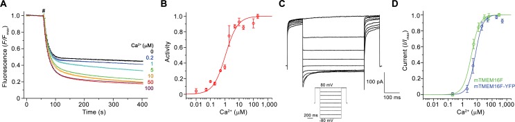
Figure 1.
Functional characterization of mTMEM16F.
( A ) Ca 2+ -dependence of scrambling activity in mTMEM16F-containing proteoliposomes. Traces depict fluorescence decrease of tail-labeled NBD-PE lipids after addition of dithionite (#) at different C...
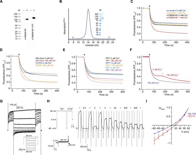
Figure 1u2014figure supplement 1.
Biochemistry and functional characterization.
( A ) Western blot of stable cell-lines expressing mTMEM16F before (u2013) and after (+) induction with tetracycline.u00a0mTMEM16F and a C-terminal YFP fusion (mTMEM16F-YFP) were detected with a mouse...
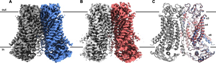
Figure 2.
mTMEM16F structures.
Cryo-EM map of mTMEM16F in digitonin in presence ( A ) and absence ( B ) of calcium at 3.2 u00c5 and 3.6 u00c5, respectively. ( C ) Cartoon representation of a superposition of the Ca 2+ -bound (blue ...
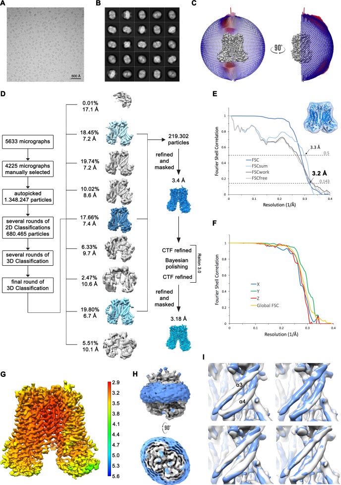
Figure 2u2014figure supplement 1.
Structure determination of mTMEM16F in complex with Ca 2+ in digitonin.
( A ) Representative cryo-EM image and ( B ) 2D-class averages of vitrified mTMEM16F in a Ca 2+ -bound state in digitonin. ( C ) Angular distribution plot of particles included in the final C2-symmetr...
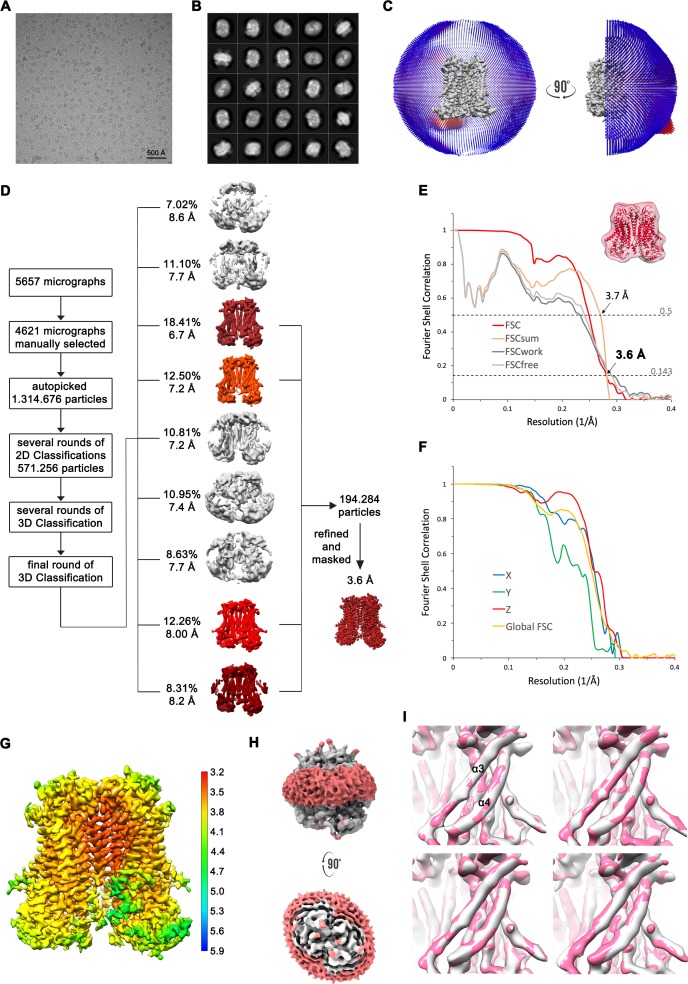
Figure 2u2014figure supplement 2.
Structure determination of mTMEM16F in absence of Ca 2+ in digitonin.
( A ) Representative cryo-EM image and ( B ) 2D-class averages of vitrified mTMEM16F in a Ca 2+ -free state in digitonin. ( C ) Angular distribution plot of particles included in the final C2-symmetri...
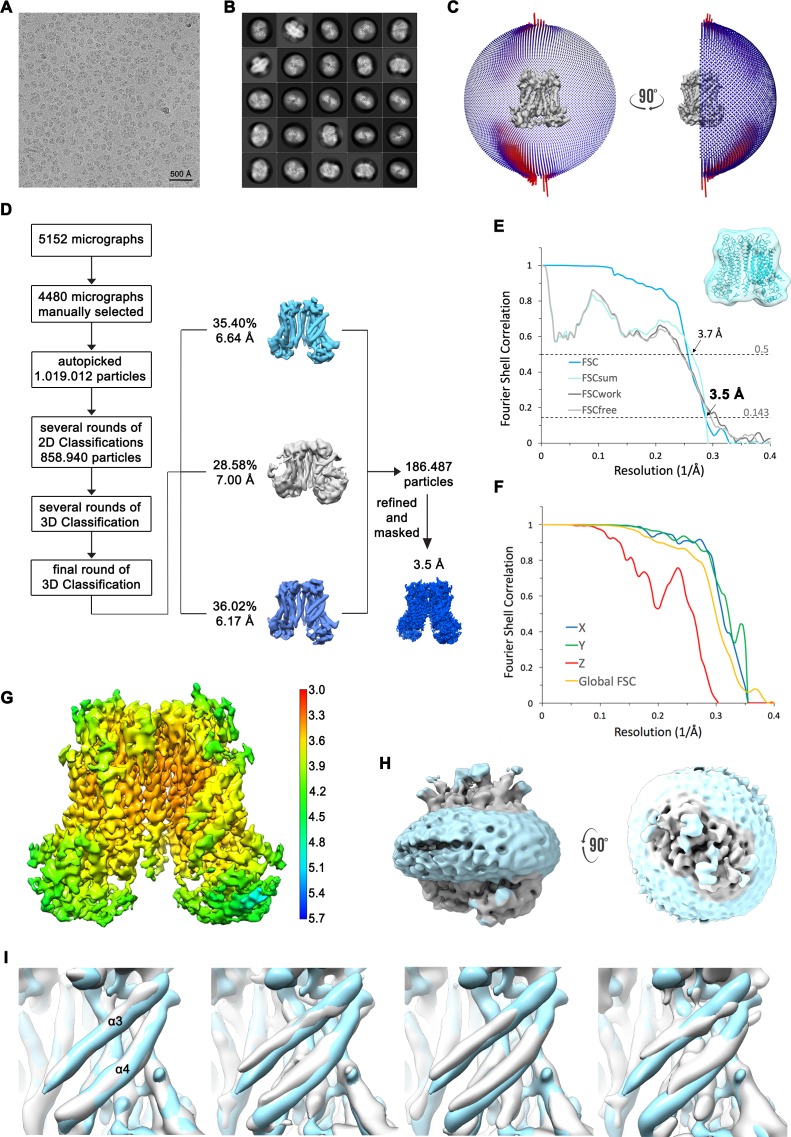
Figure 2u2014figure supplement 3.
Structure determination of mTMEM16F in complex with Ca 2+ in nanodisc.
( A ) Representative cryo-EM image and ( B ) 2D-class averages of vitrified mTMEM16F in a Ca 2+ -bound state in nanodiscs. ( C ) Angular distribution plot of particles included in the final C2-symmetr...
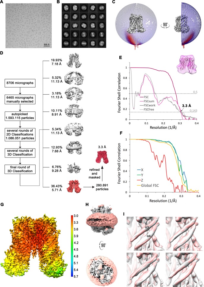
Figure 2u2014figure supplement 4.
Structure determination of mTMEM16F in absence of Ca 2+ in nanodiscs.
( A ) Representative cryo-EM image and ( B ) 2D-class averages of vitrified mTMEM16F in a Ca 2+ -free state in nanodiscs. ( C ) Angular distribution plot of particles included in the final C2-symmetri...
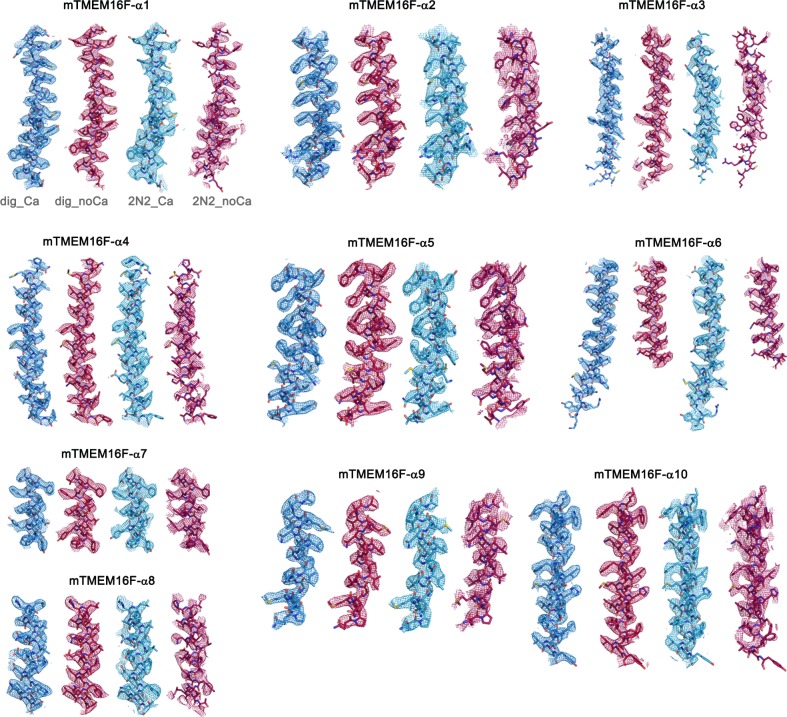
Figure 2u2014figure supplement 5.
Cryo-EM Density Maps.
Sections of the cryo-EM density of all four mTMEM16F maps superimposed on the respective refined models. Models are shown as sticks and structural elements are labelled. mTMEM16F Ca 2+ -bound in digit...
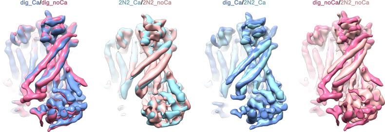
Figure 2u2014figure supplement 6.
Superpositions of the four final cryo-EM maps.
mTMEM16F in presence of calcium is shown in dark blue in digitonin and in light blue when reconstituted in nanodiscs. mTMEM16F in absence of calcium is shown in magenta in digitonin and in coral when ...
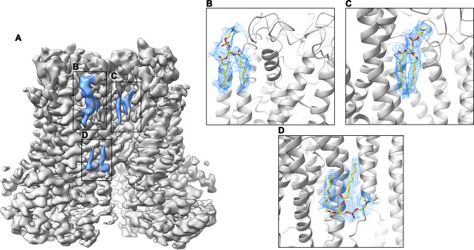
Figure 2u2014figure supplement 7.
Cryo-EM density of lipids at the dimer interface.
( A ) Cryo-EM map of mTMEM16F in digitonin in the presence of Ca 2+ (grey). Residual density at the dimer interface is highlighted in blue. ( Bu2013D ) Close-up of putative lipid densities (blue mesh)...
Video 1.
Structure of mTMEM16F in complex with Ca 2+ .
Shown is the cryo-EM density map of mTMEM16F obtained in complex with Ca 2+ in digitonin superimposed on the refined structure. For clarity, only a single subunit is displayed. The cryo-EM map is depi...
Video 2.
Structure of mTMEM16F in absence of Ca 2+ .
Shown is the cryo-EM density map of the mTMEM16F obtained in a Ca 2+ -free state in digitonin superimposed on the refined structure. For clarity, only a single subunit is displayed. The cryo-EM map is...
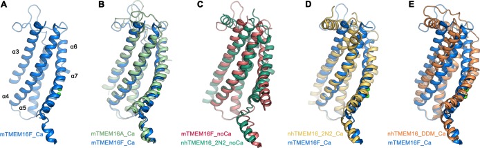
Figure 3.
Comparison of the u2018subunit cavityu2019 of TMEM16 homologues.
( A ) u2018Subunit cavityu2019 in the Ca 2+ -bound mTMEM16F structure composed of u03b1-helices 3u20137 (blue). ( B ) Superposition of the u2018subunit cavityu2019 between the Ca 2+ -bound structures ...
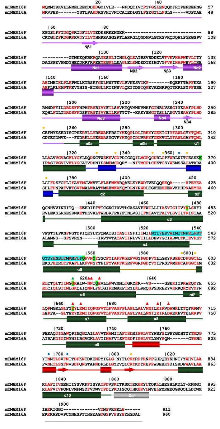
Figure 3u2014figure supplement 1.
Sequence alignment.
Protein sequences of murine TMEM16F (UniProt: Q6P9J9 .1) and murine TMEM16A (UniProt: Q8BHY3 .2) were aligned with Clustal Omega ( Sievers et al., 2011 ). Numbering corresponds to mTMEM16F. Secondary ...
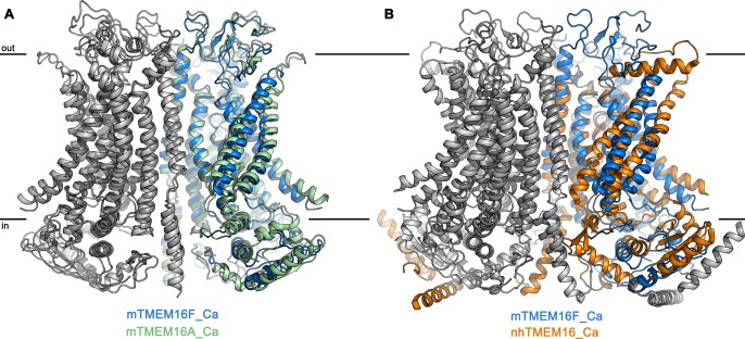
Figure 3u2014figure supplement 2.
Superposition of TMEM16 dimers.
( A ) Superposition of the Ca 2+ -bound structures of mTMEM16F in digitonin (blue and grey) and the anion channel mTMEM16A (PDBID 5OYB ( Paulino et al., 2017a ), green and grey). ( B ) Superposition o...
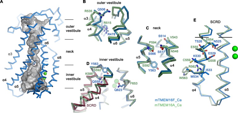
Figure 4.
Pore region of mTMEM16F.
( A ) u2018Subunit cavityu2019 in the Ca 2+ -bound state of mTMEM16F showing the putative ion conduction pore. Grey mesh shows pore surface (sampled with probe radius of 1 u00c5). ( B to D ) Superposi...
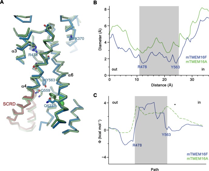
Figure 4u2014figure supplement 1.
Structural features of the pore region and electrostatics.
( A ) View of the catalytic regions in a superposition of mTMEM16F (blue) and mTMEM16A (green, remodeled version of PDBID 5OYB, see Materials and methods). The SCRD is colored in brown. Selected resid...
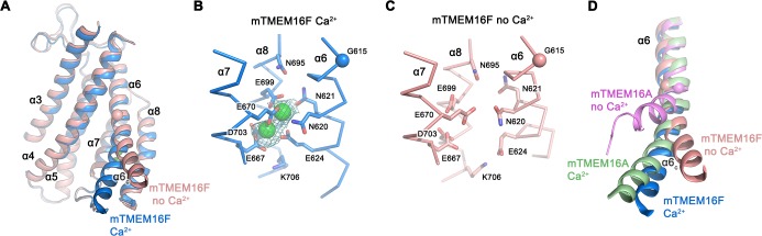
Figure 5.
Ca 2+ -binding site and conformational changes.
( A ) Superposition of u03b1-helices 3u20138 of mTMEM16F in the presence (blue) and absence of Ca 2+ (coral). ( B and C ) Close-up of the Ca 2+ -binding site in complex ( B ) or in absence of Ca 2+ ( ...
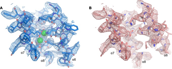
Figure 5u2014figure supplement 1.
Cryo-EM density of the Ca 2+ -binding site.
Close-up of Ca 2+ -binding site of the Ca 2+ -bound ( A ) and Ca 2+ -free ( B ) state of mTMEM16F in digitonin as depicted in Figure 5B . A, B, The respective cryo-EM density is shown as mesh, all res...
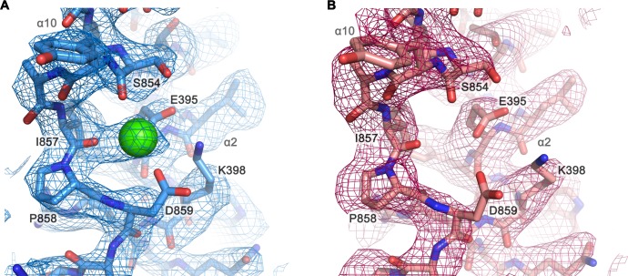
Figure 5u2014figure supplement 2.
Cryo-EM density of a putative Ca 2+ -binding site located at the end of u03b110.
Close-up of a putative Ca 2+ -binding site in the Ca 2+ -bound state of mTMEM16F in digitonin ( A ) and in the Ca 2+ -free state of mTMEM16F in digitonin ( B ). The respective cryo-EM densities are sh...
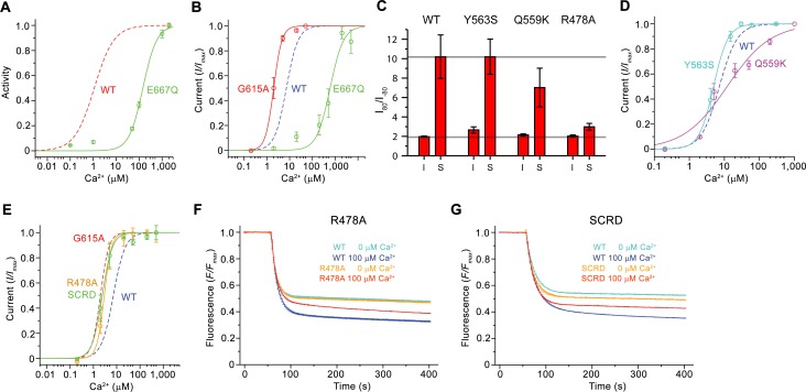
Figure 6.
Functional properties of mutants.
( A ) Ca 2+ concentration-response relationship of lipid scrambling in the binding site mutant E667Q. Data show average of either six (0, 1, 10, 100, 1000 u00b5M) or three (other concentrations) indep...
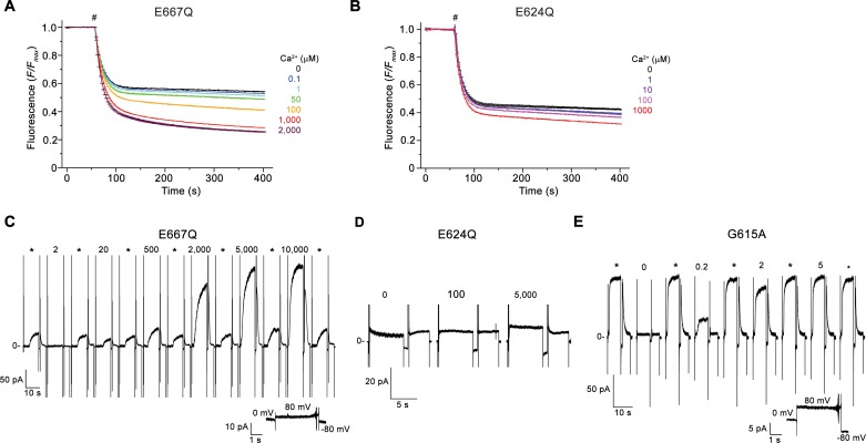
Figure 6u2014figure supplement 1.
Electrophysiology and lipid scrambling data of mutants of the Ca 2+ -binding site and the gating hinge.
( A and B ) Ca 2+ -dependence of scrambling activity in proteoliposomes containing the Ca 2+ -binding site mutants E667Q ( A ) and E624Q ( B ) at indicated ligand concentrations. ( C, D and E ) Repres...
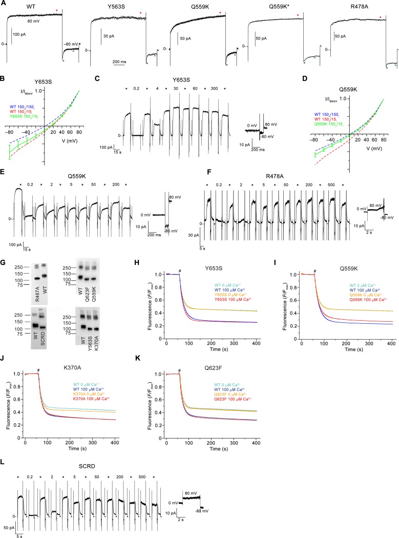
Figure 6u2014figure supplement 2.
Electrophysiology and lipid scrambling data of mutants of the pore region.
( A ) Representative current traces of WT and mutants used for calculation of the rectification index (I 80mV /I u201380mV ) of instantaneous currents (6 ms after the voltage change) and at steady-sta...
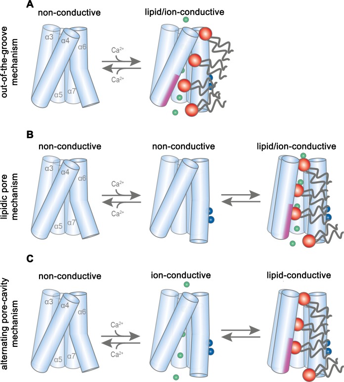
Figure 7.
Potential transport mechanisms in mTMEM16F.
( A ) u2018Out-of-the-groove mechanismu2019: upon calcium-activation the subunit cavity remains closed, ions and lipids permeate separate parts of the protein. ( B ) u2018Lipidic pore mechanismu2019: ...
Figure images are served from the NIH/NLM PubMed Central Open Access Subset or Europe PMC; copyright remains with the publishers and authors.
💬 Discussion
0 commentsNo comments yet. Be the first to start a discussion!
Leave a Comment