Abstract
AbstractSuper‐resolution microscopy (SRM) greatly benefits from the ability to install small photostable fluorescent labels into proteins. Genetic code expansion (GCE) technology addresses this demand, allowing the introduction of small labeling sites, in the form of uniquely reactive noncanonical amino acids (ncAAs), at any residue in a target protein. However, low incorporation efficiency of ncAAs and high background fluorescence limit its current SRM applications. Redirecting the subcellular localization of the pyrrolysine‐based GCE system for click chemistry, combined with DNA‐PAINT microscopy, enables the visualization of even low‐abundance proteins inside mammalian cells. This approach links a versatile, biocompatible, and potentially unbleachable labeling method with residue‐specific precision. Moreover, our reengineered GCE system eliminates untargeted background fluorescence and substantially boosts the expression yield, which is of general interest for enhanced protein engineering in eukaryotes using GCE.
🔬 Techniques
🧬 Organisms
✨ Fluorophores
🧪 Sample Preparation
🔬 Cell Lines
🏭 Microscope Brands
🏛️ Research Organizations (ROR)
Affiliated research institutions:
📊 Figures
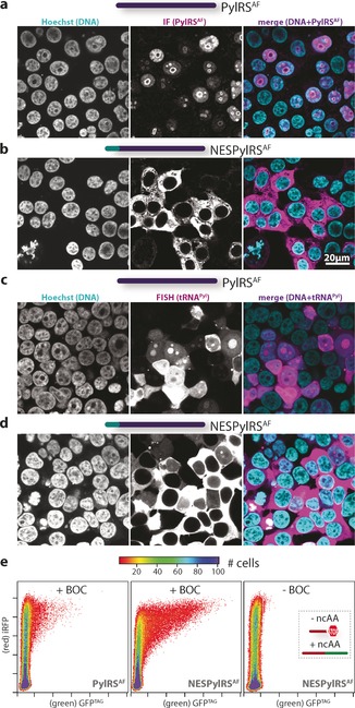
Figure 1
a,b)u2005Immunofluorescence staining of HEK cells expressing either tRNA Pyl /PylRS AF (a) or tRNA Pyl /NESPylRS AF (b). Left panel: Hoechst 33342, central panel: Ab PylRS staining, right panel: merge...
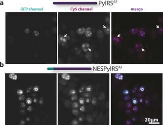
Figure 2
Confocal images of Cy5u2010tet labeled junu2010B 348TAGu2192TCO*a u2010GFP expressed in HEK cells containing either tRNA Pyl /PylRS AF (a) or tRNA Pyl /NESPylRS AF (b). Left panel: GFP, central panel:...
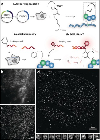
Figure 3
a)u2005Schematic representation of the Clicku2010PAINT method. The POI TAG is first expressed in mammalian cells in the presence of TCO *a when cou2010transfected with the tRNA Pyl /NESPylRS AF . Then...
Figure images are served from the NIH/NLM PubMed Central Open Access Subset or Europe PMC; copyright remains with the publishers and authors.
💬 Discussion
0 commentsNo comments yet. Be the first to start a discussion!
Leave a Comment