Abstract
This article reviews deep learning applications in biomedical optics with a particular emphasis on image formation. The review is organized by imaging domains within biomedical optics and includes microscopy, fluorescence lifetime imaging, in vivo microscopy, widefield endoscopy, optical coherence tomography, photoacoustic imaging, diffuse tomography, and functional optical brain imaging. For each of these domains, we summarize how deep learning has been applied and highlight methods by which deep learning can enable new capabilities for optics in medicine. Challenges and opportunities to improve translation and adoption of deep learning in biomedical optics are also summarized. Lasers Surg. Med. © 2021 Wiley Periodicals LLC.
🔬 Techniques
💻 Software
🧪 Sample Preparation
💻 Software Details
🏛️ Research Organizations (ROR)
Affiliated research institutions:
📊 Figures
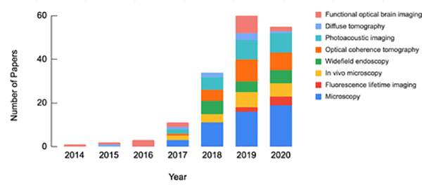
Fig 1:
Number of reviewed research papers which utilize DL in biomedical optics stratified by year and imaging domain.
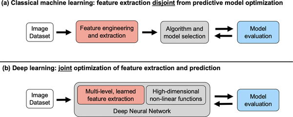
Fig 2:
(a) Classical machine learning uses engineered features and a model. (b) Deep learning uses learned features and predictors in an u201cend-to-endu201d deep neural network.
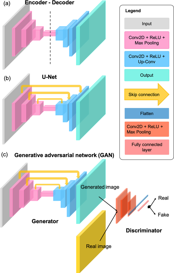
Fig 3:
Three of the most commonly-used DNN architectures in biomedical optics: (a) Encoder-decoder, (b) U-Net, and (c) GAN.
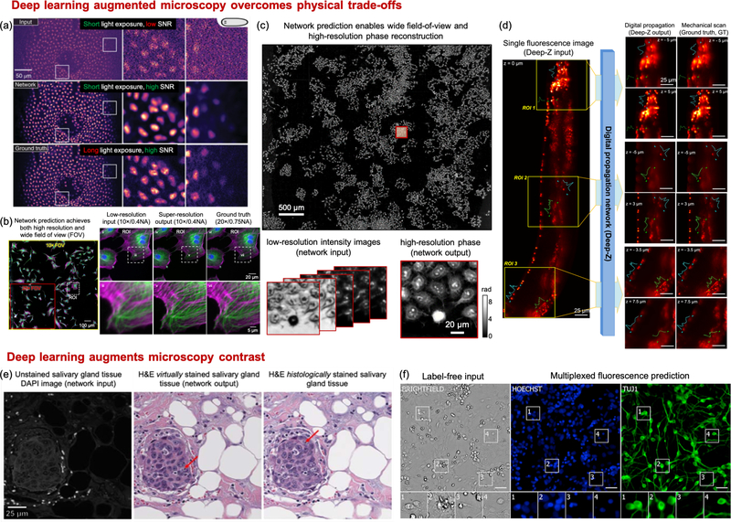
Fig 4:
DL overcomes physical tradeoffs and augments microscopy contrast. (a) CARE network achieves higher SNR with reduced light exposure (with permission from the authors [ 18 ]). (b) Cross-modality super-r...
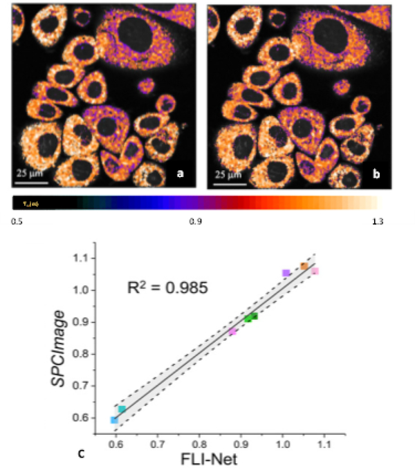
Fig 5:
Example of quantitative FLI metabolic imaging as reported by NADH tm for a breast cancer cell line (AU565) as obtained (a) with SPCImage and (b) FLI-Net. (c) Linear regression with corresponding 95% c...
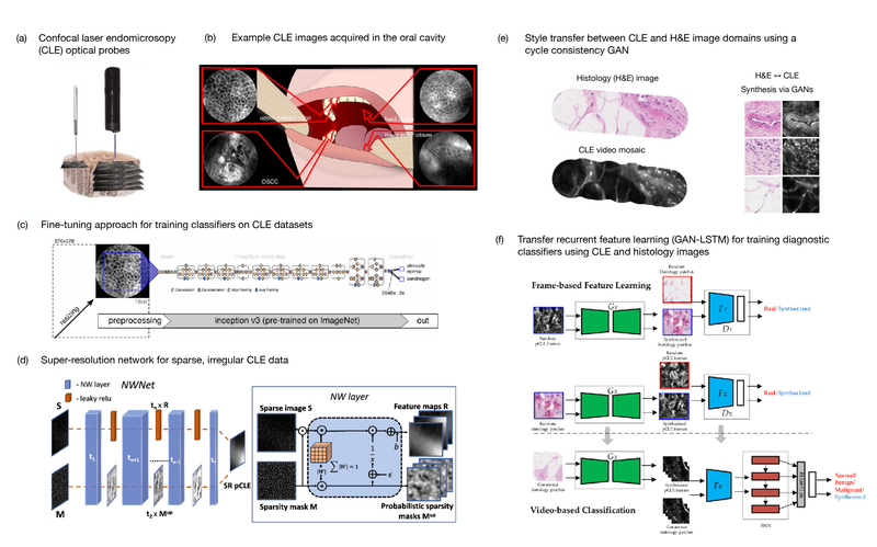
Fig 6:
DL approaches to support real-time, automated diagnostic assessment of tissues with confocal laser endomicroscopy. (a) Graphical rendering of two confocal laser endomicroscopy probes (left: Cellvizio,...
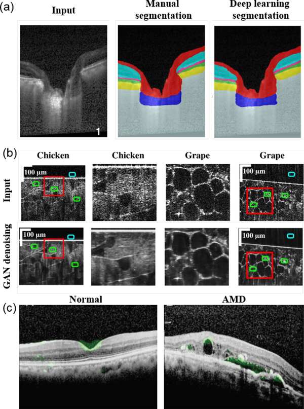
Fig 7:
(a) Example automatic retinal layer segmentation using DL compared to manual segmentation (reprinted from [ 175 ]). (b) GAN for denoising OCT images (adapted from [ 181 ]). (c) Attention map overlaid ...
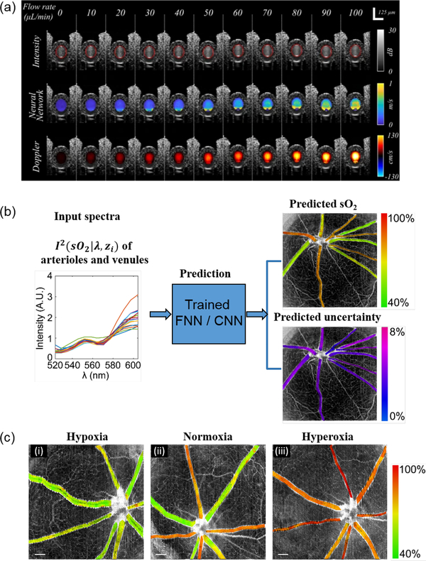
Fig 8:
(a) Examples of using DL to predict blood flow based on structural OCT image features (reprinted from [ 191 ]). (b) Example of deep spectral learning for label-free oximetry in visible light OCT (repr...
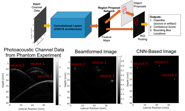
Fig 9:
Example of point source detection as a precursor to photoacoustic image formation after identifying true sources and removing reflection artifacts, modified from [ 201 ]. (u00a92018 IEEE. Adapted, wit...
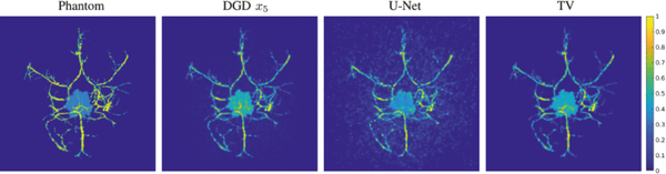
Fig 10:
Example of blood vessel and tumor phantom results with multiple DL approaches. (Reprinted from [ 205 ].)
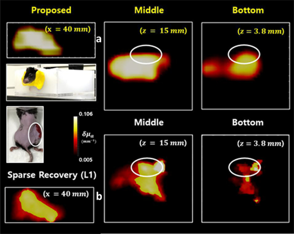
Fig 11:
Reconstruction for a mouse with tumor (right thigh) where higher absorption values are resolved (slices at z=15 and 3.8 mm) for the tumor area with the DNN in (a) compared to the L1-based inversion in...
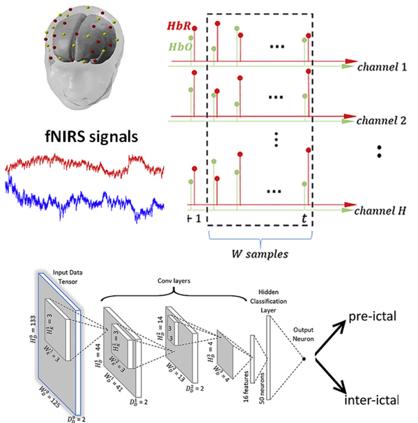
Fig 12:
Hemodynamic time series for prediction of epileptic seizure using a CNN (with permission from the authors [ 257 ]) (Computers in Biology and Medicine, 11, 2019, 103355, Rosas-Romero et al. , Predictio...
Figure images are served from the NIH/NLM PubMed Central Open Access Subset or Europe PMC; copyright remains with the publishers and authors.
💬 Discussion
0 commentsNo comments yet. Be the first to start a discussion!
Leave a Comment