Abstract
AbstractWe present a two-photon microendoscope capable of in vivo label-free deep-tissue high-resolution fast imaging through a very long optical fiber. First, an advanced light-pulse spectro-temporal shaping device optimally precompensates for linear and nonlinear distortions occurring during propagation within the endoscopic fiber. This enables the delivery of sub-40-fs duration infrared excitation pulses at the output of 5 meters of fiber. Second, the endoscopic fiber is a custom-made double-clad polarization-maintaining photonic crystal fiber specifically designed to optimize the imaging resolution and the intrinsic luminescence backward collection. Third, a miniaturized fiber-scanner of 2.2 mm outer diameter allows simultaneous second harmonic generation (SHG) and two-photon excited autofluorescence (TPEF) imaging at 8 frames per second. This microendoscope’s transverse and axial resolutions amount respectively to 0.8 μm and 12 μm, with a field-of-view as large as 450 μm. This microendoscope’s unprecedented capabilities are validated during label-free imaging, ex vivo on various fixed human tissue samples and in vivo on an anesthetized mouse kidney demonstrating an imaging penetration depth greater than 300 μm below the surface of the organ. The results reported in this manuscript confirm that nonlinear microendoscopy can become a valuable clinical tool for real-time in situ assessment of pathological states.
🔬 Techniques
💻 Software
🧪 Sample Preparation
🔬 Cell Lines
🏭 Microscope Brands
💻 Software Details
🏛️ Research Organizations (ROR)
Affiliated research institutions:
📋 Methods
TPME system
The system is fed by a standard MIRA 900 Ti:Sapphire oscillator followed by a Faraday isolator avoiding reinjection back to the oscillator. More than 400 mW of the laser power are injected in the first 0.5 m-long PM-SMF in which pulses are temporally and spectrally broadened. Three different half-wave plates allow controlling the polarization where it is necessary in the setup (see in SI Fig. S2a).
Human Tissue and Animal Preparation
The reported investigation was in accordance with the relevant guidelines for both humans and animals. The rat tail tendon was fresh while all other reported ex vivo samples were fixed in 4% paraformaldehyde and stored in phosphate buffered saline. The healthy human pulmonary tissue samples, prepared at Rouen University Hospital, were obtained from a lobectomy. They were sampled from a healthy area of a pulmonary lobe. According to the French regulation, the use of the human tissue sample for the experiment was approved by scientific committee of the Rouen University Hospital Tumor Tissue Bank, and a written informed consent obtained from the patient. Induction of UUO in mice and the preparation of animals for in vivo imaging sessions were carried out by the staff of the UMR CNRS 7276 in accordance with European regulations, applied in France by Decret No. 2013-118 of 1st February 2013 on the protection of animals used by scientists. The protocol was approved by the ethics committee for animals used by scientists registered under the code C2A2-33 by the French “Ministère de l’Education Nationale, de l’Enseignement Supérieur et de la Recherche”. During in vivo imaging sessions, the preparation and observation of the mice were made with a protocol similar to the one described by Brown et al. 12 , the main difference being the anesthesia. In our case, mice received a non-barbiturate anesthetic and a muscle relaxant sedative analgesic (ketamine at 18 mg/mL and xylazine at 0.9 mg/mL) through intraperitoneal injection. The kidney, elevated from the body, was clamped between two tongue depressors and placed on the flank of the mice, beneath the probe (see Fig. 5a ). The TPME probe was manipulated by means of a 3-axis precision motorized micromanipulator (Thorlabs, Inc., MAX 343). The micromanipulator was connected to an articulated arm (see Fig. 5a ) holding the TPME distal tip. The depth resolved imaging results reported in Fig. 4e–h and in Fig. 5d were obtained with this micromanipulator that allows precisely controlling the z coordinate.
Show full methods section
TPME system
The system is fed by a standard MIRA 900 Ti:Sapphire oscillator followed by a Faraday isolator avoiding reinjection back to the oscillator. More than 400 mW of the laser power are injected in the first 0.5 m-long PM-SMF in which pulses are temporally and spectrally broadened. Three different half-wave plates allow controlling the polarization where it is necessary in the setup (see in SI Fig. S2a).
Human Tissue and Animal Preparation
The reported investigation was in accordance with the relevant guidelines for both humans and animals. The rat tail tendon was fresh while all other reported ex vivo samples were fixed in 4% paraformaldehyde and stored in phosphate buffered saline. The healthy human pulmonary tissue samples, prepared at Rouen University Hospital, were obtained from a lobectomy. They were sampled from a healthy area of a pulmonary lobe. According to the French regulation, the use of the human tissue sample for the experiment was approved by scientific committee of the Rouen University Hospital Tumor Tissue Bank, and a written informed consent obtained from the patient. Induction of UUO in mice and the preparation of animals for in vivo imaging sessions were carried out by the staff of the UMR CNRS 7276 in accordance with European regulations, applied in France by Decret No. 2013-118 of 1st February 2013 on the protection of animals used by scientists. The protocol was approved by the ethics committee for animals used by scientists registered under the code C2A2-33 by the French “Ministère de l’Education Nationale, de l’Enseignement Supérieur et de la Recherche”. During in vivo imaging sessions, the preparation and observation of the mice were made with a protocol similar to the one described by Brown et al. 12 , the main difference being the anesthesia. In our case, mice received a non-barbiturate anesthetic and a muscle relaxant sedative analgesic (ketamine at 18 mg/mL and xylazine at 0.9 mg/mL) through intraperitoneal injection. The kidney, elevated from the body, was clamped between two tongue depressors and placed on the flank of the mice, beneath the probe (see Fig. 5a ). The TPME probe was manipulated by means of a 3-axis precision motorized micromanipulator (Thorlabs, Inc., MAX 343). The micromanipulator was connected to an articulated arm (see Fig. 5a ) holding the TPME distal tip. The depth resolved imaging results reported in Fig. 4e–h and in Fig. 5d were obtained with this micromanipulator that allows precisely controlling the z coordinate.
Supplementary Material Supplementary Information Supplementary Video S1 Supplementary Video S2 Supplementary Video S3
📊 Figures
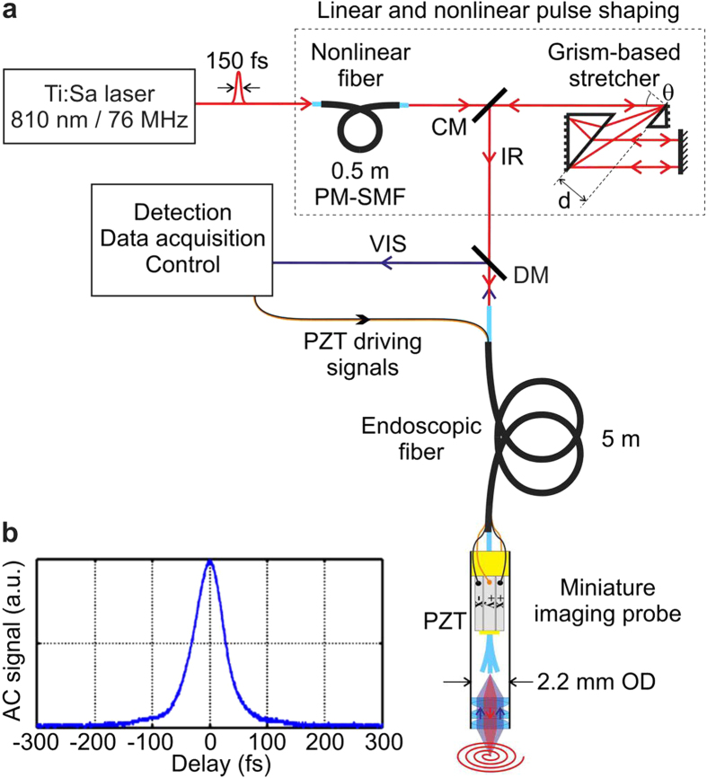
Figure 1
Scheme of the TPME system with linear and nonlinear pulse shaping.
( a ) Scheme of the experimetal setup; CM: cut mirror; DM: dichroic mirror; PZT: piezzoelectric tube. The miniature fiber-scanning imaging probe is embeded inside a 2.2u2009mm outer diameter (OD) stai...
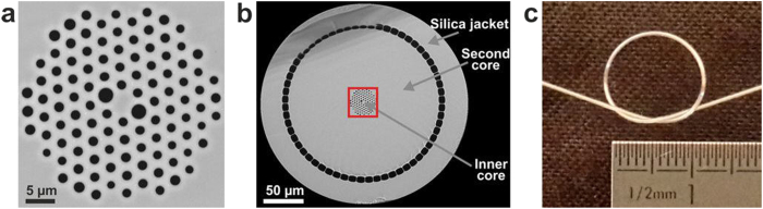
Figure 2
Custom-design air-silica DC-PCF used as the endoscopic fiber within the TPME.
( a ) Close view of the inner core of the fiber through scanning electron microscopy (SEM). Pure silica is in grey and air in black. ( b ) SEM image of the fiber cross-section without its outer polyme...
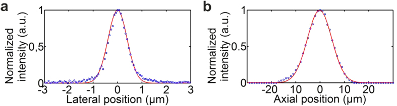
Figure 3
TPME optical resolutions. Intensity spatial distributions obtained during imaging a 0.1u2009u03bcm-diameter fluorescent bead.
Blue circle: measurements; red line: Gaussian fit. Transverse and axial resolutions were deduced from the FWHM of the Gaussian fits of the intensity distributions. ( a ) Transverse resolution: u0394xu...
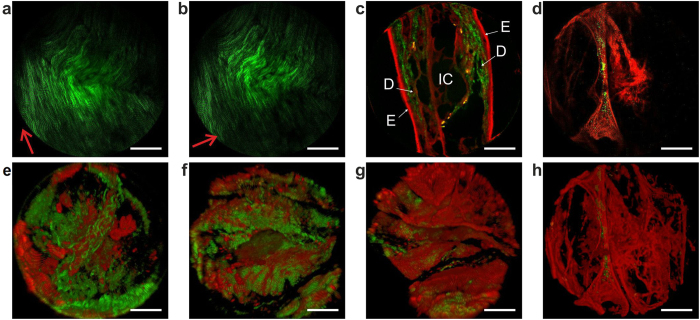
Figure 4
Label-free microendoscopy images of fixed tissue samples ex vivo .
Intrinsic TPEF in red and SHG in green. ( a u2013 d ) raw optical sections. ( e u2013 h ) perspective view from ImageJ 3D software from a set of sixty optical sections each one corresponding to a give...
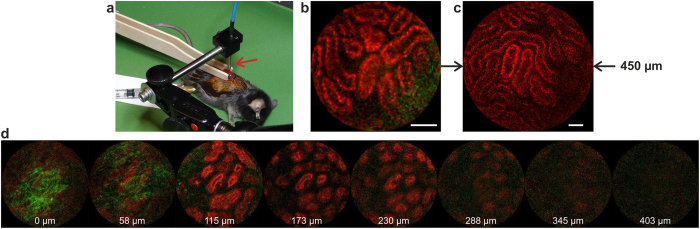
Figure 5
Label-free in vivo experiment.
( a ) Anesthetized mouse with one kidney being elevated from the body and clamped between two tongue depressors, beneath the 2.2u2009mm TPME probe (red arrow). A constant power of 30u2009mW was launch...
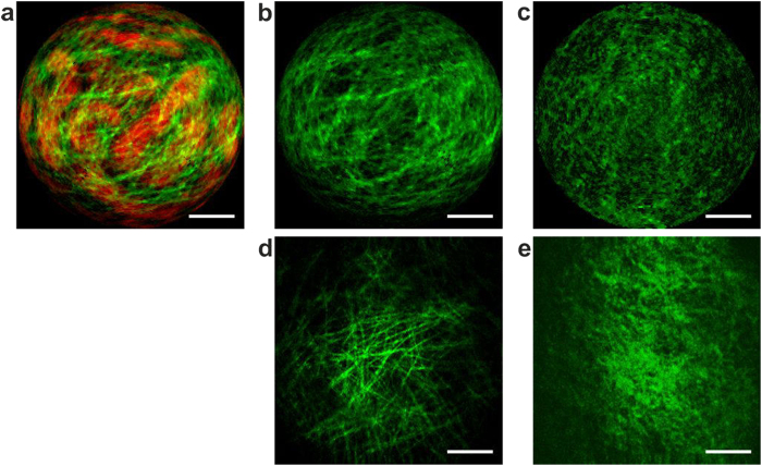
Figure 6
Comparison between healthy and fibrotic kidney capsules.
Top line: in vivo label-free raw images delivered by the TPME; SHG in green and TPEF in red. ( a , b ) correspond to the same image, without TPEF in ( b ). Bottom line: reference ex vivo SHG images of...
Figure images are served from the NIH/NLM PubMed Central Open Access Subset or Europe PMC; copyright remains with the publishers and authors.
💬 Discussion
0 commentsNo comments yet. Be the first to start a discussion!
Leave a Comment