Abstract
The human penis and clitoris develop from the ambisexual genital tubercle. To compare and contrast the development of human penis and clitoris, we used macroscopic photography, optical projection tomography, light sheet microscopy, scanning electron microscopy, histology and immunohistochemistry. The human genital tubercle differentiates into a penis under the influence of androgens forming a tubular urethra that develops by canalization of the urethral plate to form a wide diamond-shaped urethral groove (opening zipper) whose edges (urethral folds) fuse in the midline (closing zipper). In contrast, in females, without the influence of androgens, the vestibular plate (homologue of the urethral plate) undergoes canalization to form a wide vestibular groove whose edges (vestibular folds) remain unfused, ultimately forming the labia minora defining the vaginal vestibule. The neurovascular anatomy is similar in both the developing human penis and clitoris and is the key to successful surgical reconstructions.
🔬 Techniques
🔭 Microscopes
🧪 Sample Preparation
🔬 Cell Lines
🏭 Microscope Brands
🧪 Reagent Suppliers
🏛️ Research Organizations (ROR)
Affiliated research institutions:
📋 Methods
First and second-trimester human fetal specimens were collected free of patient identifiers after elective termination of pregnancy (Committee on Human Research approval at the University of California at San Francisco, IRB# 12–08813). Gender was determined by inspection with a dissecting microscope for Wolffian (mesonephric) and Müllerian (paramesonephric) ductal morphology. For the youngest specimens at the indifferent stage (8 to 10 weeks), PCR of the SRY- chromosomal sequences was used to determine sex ( Cui et al., 1994 ) ( Li et al., 2015 ). Heel-toe length was used as a determinant of gestational age ( Drey et al., 2005 ; Mercer et al., 1987 ; Mhaskar et al., 1989 ). Human fetal specimens were then examined by optical projection tomography ( Li et al., 2015 ), lightsheet fluorescence microscopy ( Isaacson et al., 2018a ), and scanning electron microscopy ( Shen et al., 2016 ) as previously described. Transverse and sagittal serial paraffin sections of human fetal specimens were prepared and stained with hematoxylin and eosin or immunostained using antibodies for cytokeratins (KRT) 6, 7, 10 and 14, androgen receptor, Foxa1, Mafb, Runx1/2/3, Notch, the proliferation marker Ki67, and the apoptotic marker caspase 3 ( Li et al., 2015 ; Overland et al., 2016 ). Immunostaining was detected using horseradish-peroxidase-based Vectastain kits (Vector Laboratories, Burlingame, CA) or by immunofluorescence. Negative controls deleted the primary antibody ( Robboy et al., 2017b ).
📊 Figures
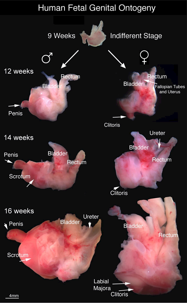
Figure 1.
Gross Human Fetal Pelvic Ontogeny: Gross ontogeny of the human fetal pelvis at 9 weeks of gestation (end of indifferent stage), 12 weeks, 14 weeks and 16 weeks of gestation. Note the divergent develop...
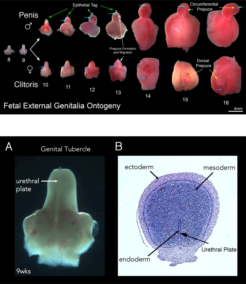
Figure 2.
Fetal External Genitalia Ontogeny: Representative ventral views of external genitalia of the human males (top row) and females (bottom row) (8u201316 weeks of gestation). Note the morphologic differen...
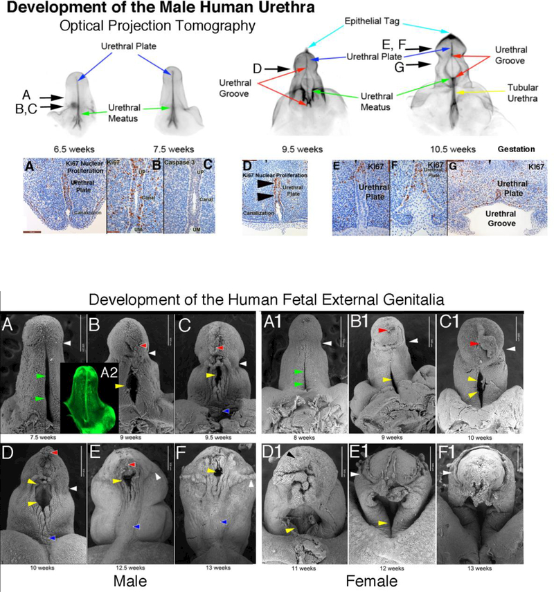
Figure 3:
Human male genital tubercle/ future human penis at 9 weeks of gestation. Note the urethral plate in the gross specimen (A). In the corresponding histologic cross section (B), the three embryonic layer...
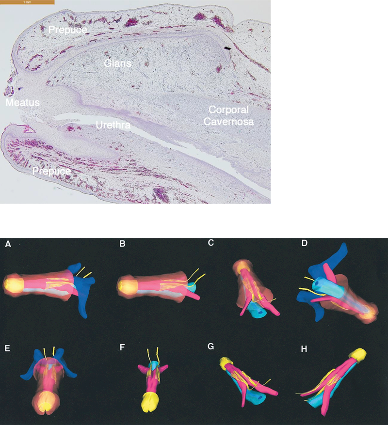
Figure 4:
Optical projection tomography of male urethral development from 6.5 to 10.5 weeks fetal age. Note the urethral plate (blue arrow) that ends within the glans. The wide-open urethral groove (red arrows)...
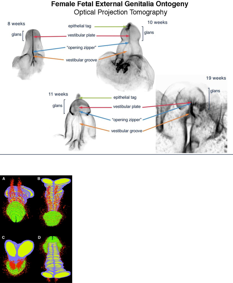
Figure 5:
Scanning electron microscopy ontogeny of the developing human fetal penis (A-F) from 7.5 u221213 weeks and developing human clitoris (A1-F1) 8u201313 weeks of gestation. White arrowheads indicate the ...
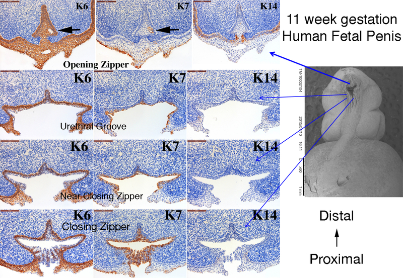
Figure 6:
Sagittal section of an 18-weeks gestation human fetal glans penis. Note that the penile epidermis meets the distal aspect of the urethral epithelium at the meatus.
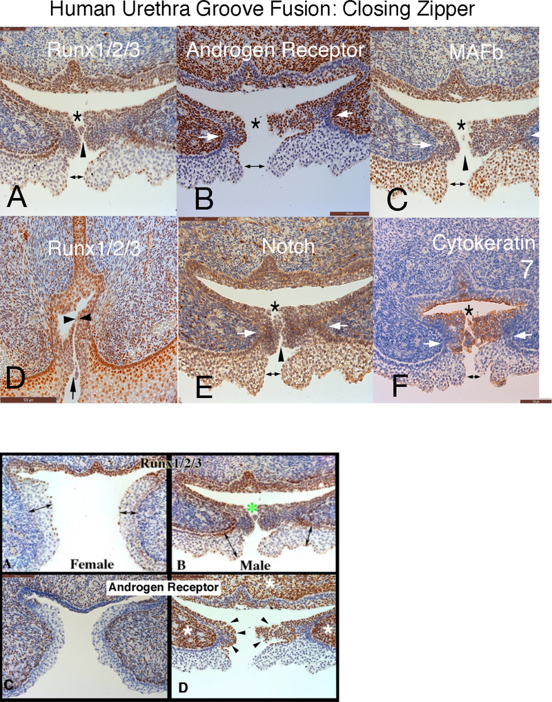
Figure 7.
3-D Neuroanatomy of the fully formed fetal penis at 17.5 weeks of gestation. A to H, multiple views of 3-D reconstruction of human fetal penis dorsal nerve neuroanatomy with respective layers removed ...
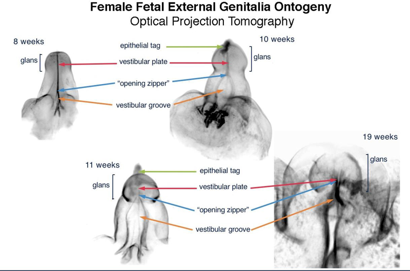
Figure 8.
Optical Projection Tomography: Fetal ontogeny of human clitoris from 8 to 19 weeks of fetal age. Note the opening zipper, which facilitates vestibular plate opening to form vestibular groove, and lack...
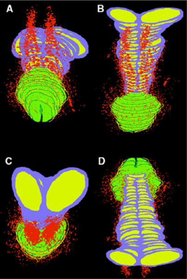
Figure 9.
Computer generated 3-D reconstruction of normal human fetal clitoris at 24 weeks of gestation. Red areas represent nerve pathway with paucity of nerves at bottom and top midline of clitori., Purple= t...
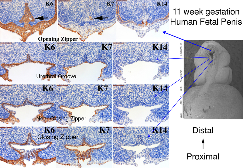
Figure 10.
Cytokeratin (K) 6, 7 and 14 expression in an 11-week human fetal penis. Representative sections from the opening zipper, urethral groove, near the closing zipper and the closing zipper. Approximate se...
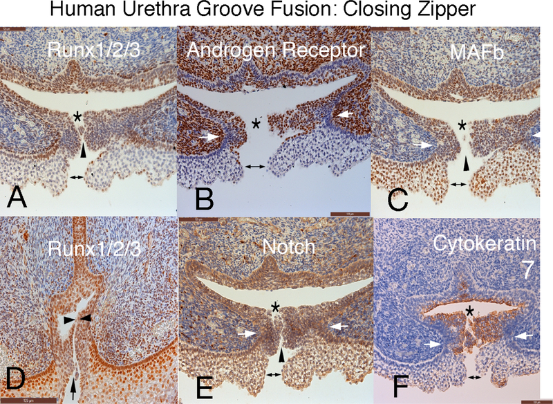
Figure 11.
Human fetal penis at 11 weeks of gestation: Closing zipper - fusion of the urethral folds. Immunohistochemical localization of: A) Runx 1/2/3. B) Androgen Receptor. C) MAFb. D) Runx 1/2/3. E) Notch. a...
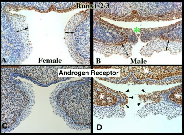
Figure 12.
Runx1/2/3 and androgen receptor Immunohistochemistry of 11-week female and male human fetal clitoris and penis at the level of the closing zipper. RUNX1/2/3-positive epithelial cells are seen in males...
Figure images are served from the NIH/NLM PubMed Central Open Access Subset or Europe PMC; copyright remains with the publishers and authors.
💬 Discussion
0 commentsNo comments yet. Be the first to start a discussion!
Leave a Comment