Abstract
Background Previous experimental studies have shown that downstream microvascular thromboinflammation is involved in brain damage from acute ischemic stroke. Using intravital microscopy, we investigated and characterized the sequence of downstream microvascular thromboinflammation in an ischemia/reperfusion acute ischemic stroke model. Methods and Results Rats underwent transient monofilament middle cerebral artery (MCA) occlusion. Cerebral microcirculation in the MCA territory was exposed through a craniotomy and analyzed using real‐time intravital imaging coupled with laser Doppler interferometry. Leukocytes, platelets, fibrinogen, and blood–brain barrier permeability were analyzed by intravenous injection of fluorescent antibodies and bovine serum albumin. MCA occlusion induced a sudden and profound drop in downstream microvascular blood flow associated with leukocyte margination in the venous compartment. Leukocyte margination fostered fibrinogen deposition and thrombosis in postcapillary venules. Either in venules or arterioles, blood flow was not fully restored after MCA recanalization. Furthermore, venular thrombi persisted despite MCA recanalization, and leukocyte extravasation continued to develop in venules in association with blood–brain barrier disruption. Finally, microhemorrhages were occasionally observed, colocalizing with thrombosed venules characterized by marked leukocyte margination. Conclusions We showed that microvascular thrombosis in transient monofilament MCA occlusion and blood–brain barrier disruption are initiated immediately after occlusion and are propagated through the venous compartment in close association with marginating leukocytes. MCA occlusion–induced downstream microvascular thromboinflammation response was responsible for incomplete reperfusion after MCA recanalization and delayed microhemorrhages.
🔬 Techniques
💻 Software
✨ Fluorophores
🏭 Microscope Brands
📷 Detectors
💻 Software Details
🏛️ Research Organizations (ROR)
Affiliated research institutions:
📋 Methods
Methods and Results Rats underwent transient monofilament middle cerebral artery (MCA) occlusion. Cerebral microcirculation in the MCA territory was exposed through a craniotomy and analyzed using real‐time intravital imaging coupled with laser Doppler interferometry. Leukocytes, platelets, fibrinogen, and blood–brain barrier permeability were analyzed by intravenous injection of fluorescent antibodies and bovine serum albumin. MCA occlusion induced a sudden and profound drop in downstream microvascular blood flow associated with leukocyte margination in the venous compartment. Leukocyte margination fostered fibrinogen deposition and thrombosis in postcapillary venules. Either in venules or arterioles, blood flow was not fully restored after MCA recanalization. Furthermore, venular thrombi persisted despite MCA recanalization, and leukocyte extravasation continued to develop in venules in association with blood–brain barrier disruption. Finally, microhemorrhages were occasionally observed, colocalizing with thrombosed venules characterized by marked leukocyte margination.
Materials and Methods
The data and analytic methods and the study materials will be made available to other researchers on request for purposes of reproducing the results or replicating the procedure. Middle Cerebral Artery Occlusion and Reperfusion Male Sprague‐Dawley rats (Janvier, France) underwent 120 minutes of transient monofilament middle cerebral artery occlusion (MCAO), as described previously. 7 All experimental procedures were declared to the French ministry of research and authorized after ethics review (no. 20160125164052; APAFIS 3792). Real‐Time Intravital Imaging Coupled With Laser Doppler Interferometry Distal branches of the middle cerebral artery (MCA) of rats were exposed through a 4×4‐mm craniotomy performed in the right temporoparietal cortex with a hand‐held drill, as described previously. 7 Cerebral microcirculation was directly visualized using a fluorescence macroscope (MacroFluo; Leica Microsystems) equipped with a heating plate with a thermostat and a ×5 objective and connected to a scientific CMOS camera (ORCA‐Flash4.0; Hamamatsu Photonics). Data acquisition and analysis were done using Metamorph software (Molecular Devices). All fluorescent markers were administered intravenously into the tail vein. Rhodamine 6G was used to label leukocytes and platelets, FITC (fluorescein isothiocyanate)–conjugated polyclonal rabbit anti–human fibrinogen was used to stain fibrin(ogen), Alexa 555–conjugated hamster anti–rat CD42d was used to stain platelets, and Alexa Fluor 647–conjugated BSA was used to assess vascular permeability. Fluorescent BSA was injected intravenously 5 minutes before recanalization. Cortical vessel diameter before MCA occlusion and 1 hour after recanalization was measured in intravital video microscopy images using ZEN software (Zeiss). Blood cell velocity in microvessels was measured using a single‐point laser Doppler vibrometer with an integrated CCD video camera (CLV‐2534; Polytec) and mounted on the macroscope to monitor laser spot positioning. To allow specific measurement of blood cell velocity, breathing‐related and flow‐induced vessel wall vibrations were identified by their bidirectional associated signal and eliminated by applying a low‐pass filter set at 2 kHz. Frequencies due to the unidirectional out‐of‐plane vibrations caused by circulating blood cells were converted to speed according to the formula v=(Δf×λ)/(2×cosα), where v is the blood cell velocity, Δf is the Doppler‐frequency shift, λ is the wavelength of the emitted wave (633 nm), and α is the angle between the blood cell direction and the incident laser beam, which was estimated at 80° (Figure 1 ). Data were recorded and treated using the Polytec Vibrometer Software. Figure 1 Schematic representation of the experimental design used for intravital imaging and laser Doppler vibrometry of pial microvessels downstream of the middle cerebral artery (MCA). Blood flow and cell interactions in cortical pial microvessels downstream of the MCA were analyzed by intravital microscopy and laser Doppler interferometry through a cranial window. To enable measurement of red blood cell velocity, a low‐pass filter set at 2 kHz was applied to eliminate environmental noise, including breathing‐related movement. Using these settings, cell interactions and blood flow were monitored before MCA occlusion, during occlusion, and after MCA recanalization.
Show full methods section
Methods and Results Rats underwent transient monofilament middle cerebral artery (MCA) occlusion. Cerebral microcirculation in the MCA territory was exposed through a craniotomy and analyzed using real‐time intravital imaging coupled with laser Doppler interferometry. Leukocytes, platelets, fibrinogen, and blood–brain barrier permeability were analyzed by intravenous injection of fluorescent antibodies and bovine serum albumin. MCA occlusion induced a sudden and profound drop in downstream microvascular blood flow associated with leukocyte margination in the venous compartment. Leukocyte margination fostered fibrinogen deposition and thrombosis in postcapillary venules. Either in venules or arterioles, blood flow was not fully restored after MCA recanalization. Furthermore, venular thrombi persisted despite MCA recanalization, and leukocyte extravasation continued to develop in venules in association with blood–brain barrier disruption. Finally, microhemorrhages were occasionally observed, colocalizing with thrombosed venules characterized by marked leukocyte margination.
Materials and Methods
The data and analytic methods and the study materials will be made available to other researchers on request for purposes of reproducing the results or replicating the procedure. Middle Cerebral Artery Occlusion and Reperfusion Male Sprague‐Dawley rats (Janvier, France) underwent 120 minutes of transient monofilament middle cerebral artery occlusion (MCAO), as described previously. 7 All experimental procedures were declared to the French ministry of research and authorized after ethics review (no. 20160125164052; APAFIS 3792). Real‐Time Intravital Imaging Coupled With Laser Doppler Interferometry Distal branches of the middle cerebral artery (MCA) of rats were exposed through a 4×4‐mm craniotomy performed in the right temporoparietal cortex with a hand‐held drill, as described previously. 7 Cerebral microcirculation was directly visualized using a fluorescence macroscope (MacroFluo; Leica Microsystems) equipped with a heating plate with a thermostat and a ×5 objective and connected to a scientific CMOS camera (ORCA‐Flash4.0; Hamamatsu Photonics). Data acquisition and analysis were done using Metamorph software (Molecular Devices). All fluorescent markers were administered intravenously into the tail vein. Rhodamine 6G was used to label leukocytes and platelets, FITC (fluorescein isothiocyanate)–conjugated polyclonal rabbit anti–human fibrinogen was used to stain fibrin(ogen), Alexa 555–conjugated hamster anti–rat CD42d was used to stain platelets, and Alexa Fluor 647–conjugated BSA was used to assess vascular permeability. Fluorescent BSA was injected intravenously 5 minutes before recanalization. Cortical vessel diameter before MCA occlusion and 1 hour after recanalization was measured in intravital video microscopy images using ZEN software (Zeiss). Blood cell velocity in microvessels was measured using a single‐point laser Doppler vibrometer with an integrated CCD video camera (CLV‐2534; Polytec) and mounted on the macroscope to monitor laser spot positioning. To allow specific measurement of blood cell velocity, breathing‐related and flow‐induced vessel wall vibrations were identified by their bidirectional associated signal and eliminated by applying a low‐pass filter set at 2 kHz. Frequencies due to the unidirectional out‐of‐plane vibrations caused by circulating blood cells were converted to speed according to the formula v=(Δf×λ)/(2×cosα), where v is the blood cell velocity, Δf is the Doppler‐frequency shift, λ is the wavelength of the emitted wave (633 nm), and α is the angle between the blood cell direction and the incident laser beam, which was estimated at 80° (Figure 1 ). Data were recorded and treated using the Polytec Vibrometer Software. Figure 1 Schematic representation of the experimental design used for intravital imaging and laser Doppler vibrometry of pial microvessels downstream of the middle cerebral artery (MCA). Blood flow and cell interactions in cortical pial microvessels downstream of the MCA were analyzed by intravital microscopy and laser Doppler interferometry through a cranial window. To enable measurement of red blood cell velocity, a low‐pass filter set at 2 kHz was applied to eliminate environmental noise, including breathing‐related movement. Using these settings, cell interactions and blood flow were monitored before MCA occlusion, during occlusion, and after MCA recanalization.
Statistical Analysis
Red blood cell velocities were compared using the nonparametric Wilcoxon signed rank test for paired samples. Values of P< 0.05 were considered statistically significant.
Supporting information Video S1. Microvascular consequences of middle cerebral artery (MCA) occlusion and recanalization. Intravital microscopy observation of pial microvessels downstream of the MCA during MCA occlusion and after MCA recanalization. Circulating leukocytes and platelets were labeled by intravenous injection of rhodamine 6G. Note that MCA occlusion causes a drop in venous blood flow and inversions of blood flow direction in downstream arterioles. Both phenomena are associated with leukocyte margination. Although recanalization partially corrected blood flow anomalies, leukocyte margination and extravasation continued to develop in venules. A indicates arterioles; V, venules. Click here for additional data file.
📊 Figures
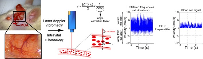
Figure 1
Schematic representation of the experimental design used for intravital imaging and laser Doppler vibrometry of pial microvessels downstream of the middle cerebral artery (MCA). Blood flow and cell in...
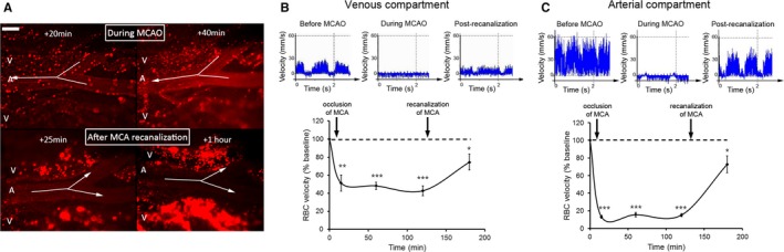
Figure 2
Early microvascular consequences of transient middle cerebral artery occlusion (MCAO). A, Fluorescent intravital microscopy images illustrating the marked adhesion of rhodamine 6Gu2013labeled leukocyt...
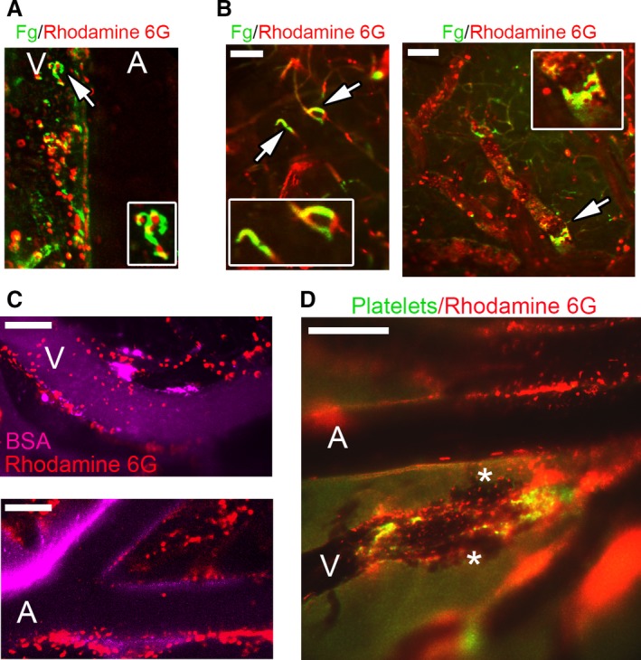
Figure 3
Leukocyte margination in the ischemic brain is associated with fibrinogen accumulation, thrombosis, increased permeability, and microhemorrhage. A and B, Fluorescent intravital microscopy images showi...
Figure images are served from the NIH/NLM PubMed Central Open Access Subset or Europe PMC; copyright remains with the publishers and authors.
💬 Discussion
0 commentsNo comments yet. Be the first to start a discussion!
Leave a Comment