Abstract
Abstract Background Advances in optical imaging modalities and the continued evolution of genetically-encoded fluorescent proteins are coming together to facilitate the study of cell behavior at high resolution in living organisms. As a result, imaging using autofluorescent protein reporters is gaining popularity in mouse transgenic and targeted mutagenesis applications. Results We have used embryonic stem cell-mediated transgenesis to label cells at sub-cellular resolution in vivo, and to evaluate fusion of a human histone protein to green fluorescent protein for ubiquitous fluorescent labeling of nucleosomes in mice. To this end we have generated embryonic stem cells and a corresponding strain of mice that is viable and fertile and exhibits widespread chromatin-localized reporter expression. High levels of transgene expression are maintained in a constitutive manner. Viability and fertility of homozygous transgenic animals demonstrates that this reporter is developmentally neutral and does not interfere with mitosis or meiosis. Conclusions Using various optical imaging modalities including wide-field, spinning disc confocal, and laser scanning confocal and multiphoton excitation microscopy, we can identify cells in various stages of the cell cycle. We can identify cells in interphase, cells undergoing mitosis or cell death. We demonstrate that this histone fusion reporter allows the direct visualization of active chromatin in situ. Since this reporter segments three-dimensional space, it permits the visualization of individual cells within a population, and so facilitates tracking cell position over time. It is therefore attractive for use in multidimensional studies of in vivo cell behavior and cell fate.
🔬 Techniques
🔭 Microscopes
✨ Fluorophores
🧪 Sample Preparation
🔬 Cell Lines
🏭 Microscope Brands
🧪 Reagent Suppliers
🔎 Objectives
💻 Software Details
🏛️ Research Organizations (ROR)
Affiliated research institutions:
📋 Methods
The coding sequence for the human histone H2B gene ( X57127 ) was amplified from genomic DNA by PCR using Pfx Polymerase (Invitrogen). The resulting product was cloned into pCR4 TOPO (Invitrogen) to generate pH2B. The H2B fragment was then cloned into plasmids pEGFP-N1, pDsRed2-N1 pDsRedExpress-N1 (BD Biosciences, Inc) in order to generate plasmids pH2B-EGFP, pH2B-DsRed2 and pH2B-DsRedExpress (oligonucleotide sequences are available upon request). The resulting fusions were then re-amplified by PCR and cloned into the XhoI site of pCAGGS [ 19 ] to generate pCX-H2B-EGFP, pCX-H2B-DsRed2 and pCX-H2B-DsRedExpress. All vectors were tested by transient transfection of Cos-7 cells and R1 ES cells using Fugene 6 Transfection Reagent as per manufacturer's recommendations (Roche) and electroporation respectively. The H2B-DsRed2 and H2B-DsRedExpress fusions failed to produce sustained homogenous levels of fluorescent signal, however the H2B-EGFP fusion gave strong nuclear-localized fluorescence throughout the extended culture period without perturbing cell morphology, the rate of proliferation, or the mitotic index (Fig. 2 and data not shown). Transgenic ES cell lines constitutively expressing H2B-EGFP were generated by co-electroporation of the linearized reporter construct and a circular PGK-Puro-pA plasmid [ 34 ] conferring transient puromycin resistance. Puromycin selection was carried out as described previously [ 20 , 22 ]. Briefly, drug selection was initiated 24–36 hours after electroporation, maintained for 5 days, after which time it was replaced with non-selection media for a further 24–48 hours. Fluorescent colonies were identified and picked under an epifluorescence microscope (Nikon SMZ1500). Clones were passaged in 96-well plates, and scored for maintenance and extent of fluorescence. Those exhibiting homogeneous and robust transgene expression in vitro under both stem cell conditions and conditions employed to promote their differentiation were maintained further. For stem cell conditions ES cells were grown on gelatin in the presence of LIF. For differentiation, ES cells were grown on bacteriological Petri dishes in the absence of LIF for 2–5 days to promote embryoid body formation. Thereafter embryoid bodies were re-plated onto tissue culture dishes in the presence of factors promoting directed differentiation. To assess whether an H2B fusion can continue to be widely expressed and transmitted through the germline of mice we used H2B-EGFP expressing ES cells for chimera generation by injection into C57BL/6 blastocysts using standard procedures [ 21 ]. Chimeras were bred to outbred ICR and inbred 129/Tac mice (Taconic, Germantown, NY) for germline transmission and subsequent maintenance of the lines. Two independent clones were taken germline giving indistinguishable results. We therefore focused on one of the transgenic lines. After germline transmission, this transgene was maintained at homozygosity, suggesting that the site of integration is not perturbing essential gene function. All animals retained widespread homogenous fluorescence for at least five subsequent generations. Homozygotes were distinguished from heterozygotes either by increased fluorescence in newborn (unpigmented) animal tails, by breeding, or by intensity of an EGFP hybridizing fragment on a Southern blot. Embryos and organs were dissected in HEPES buffered DMEM media containing 10% fetal calf serum, then cultured either in a standard tissue culture incubator or on a microscope stage under standard conditions promoting the culture of mouse embryos [ 35 , 36 ] in 50% rat serum: 50% DMEM buffered with bicarbonate and maintained under physiological conditions in a closed temperature-controlled, humidified and oxygenated (95% air, 5% CO 2 ) chamber (Bioptechs Inc. or Solent Sci Ltd. or home-made). For cytoplasmic staining, samples were incubated in Cell Tracker Orange (Molecular Probes; 1:500 dilution in dissecting or culture media) for 10–20 minutes. Embryos were kept in a standard tissue culture incubator at 37°C during staining. Samples were then washed twice with warm dissecting or culture media prior to imaging. All images shown (except Fig. 7 ) are of living hemizygous (Tg/+) embryos or freshly dissected (unfixed) tissues obtained from Tg/+ adults and maintained under physiological conditions. Increased fluorescence and a higher signal-to-noise ratio was observed in homozygous (Tg/Tg) specimens. The embryo presented in Figure 7 was fixed in 4% paraformaldehyde at 4°C for 72 hours, then washed, stored and imaged in PBS. Similar results were obtained in embryos fixed for up to two weeks. Wide-field images were acquired on a Nikon SMZ1500 stereo-dissecting microscope or Nikon Eclipse 5000 inverted microscope equipped with epifluorescent illumination. Spinning disc confocal data was acquired on an UltraView RS3 (Perkin-Elmer Systems) fitted on a Zeiss Axiovert 200M microscope with illumination from a 488 nm Argon laser (Melles Griot). Laser scanning confocal and multiphoton excitation data were taken on a Zeiss LSM510 NLO on a Zeiss Axioscop 2 FS MOT microscope. Objective lenses used on the Axiovert 200M and Axioscop 2 were plan-apochromat 63x/NA1.4, C-apochromat 40x/NA1.2, a plan-apochromat 20x/NA0.75 and a fluar 5x/NA0.25. For laser scanning microscopy GFP was excited using either a 488 nm Argon laser (Lassos, Inc) at 488 nm (for single-photon excitation) or a Titanium:Sapphire laser (Coherent Mira 900F with Verdi 5W pump laser) tuned between 860 and 890 nm (for two-photon excitation). Cell Tracker Orange was excited using a 543 nm HeNe laser (for single-photon use). Images were acquired as z -stacks comprising sequential x - y sections taken at 0.1–2 μm z -intervals. Raw data was processed using a variety of packages including Zeiss AIM software (Carl Zeiss Microsystems at ), Image J (NIH at ) and Volocity (Improvision at ). Each image series was re-animated using software to make the time-lapse movies that are available as additional files. Both rendered volume and time-lapse movies were assembled in QuickTime Player (Apple Computer, Inc at ).
Show full methods section
The coding sequence for the human histone H2B gene ( X57127 ) was amplified from genomic DNA by PCR using Pfx Polymerase (Invitrogen). The resulting product was cloned into pCR4 TOPO (Invitrogen) to generate pH2B. The H2B fragment was then cloned into plasmids pEGFP-N1, pDsRed2-N1 pDsRedExpress-N1 (BD Biosciences, Inc) in order to generate plasmids pH2B-EGFP, pH2B-DsRed2 and pH2B-DsRedExpress (oligonucleotide sequences are available upon request). The resulting fusions were then re-amplified by PCR and cloned into the XhoI site of pCAGGS [ 19 ] to generate pCX-H2B-EGFP, pCX-H2B-DsRed2 and pCX-H2B-DsRedExpress. All vectors were tested by transient transfection of Cos-7 cells and R1 ES cells using Fugene 6 Transfection Reagent as per manufacturer's recommendations (Roche) and electroporation respectively. The H2B-DsRed2 and H2B-DsRedExpress fusions failed to produce sustained homogenous levels of fluorescent signal, however the H2B-EGFP fusion gave strong nuclear-localized fluorescence throughout the extended culture period without perturbing cell morphology, the rate of proliferation, or the mitotic index (Fig. 2 and data not shown). Transgenic ES cell lines constitutively expressing H2B-EGFP were generated by co-electroporation of the linearized reporter construct and a circular PGK-Puro-pA plasmid [ 34 ] conferring transient puromycin resistance. Puromycin selection was carried out as described previously [ 20 , 22 ]. Briefly, drug selection was initiated 24–36 hours after electroporation, maintained for 5 days, after which time it was replaced with non-selection media for a further 24–48 hours. Fluorescent colonies were identified and picked under an epifluorescence microscope (Nikon SMZ1500). Clones were passaged in 96-well plates, and scored for maintenance and extent of fluorescence. Those exhibiting homogeneous and robust transgene expression in vitro under both stem cell conditions and conditions employed to promote their differentiation were maintained further. For stem cell conditions ES cells were grown on gelatin in the presence of LIF. For differentiation, ES cells were grown on bacteriological Petri dishes in the absence of LIF for 2–5 days to promote embryoid body formation. Thereafter embryoid bodies were re-plated onto tissue culture dishes in the presence of factors promoting directed differentiation. To assess whether an H2B fusion can continue to be widely expressed and transmitted through the germline of mice we used H2B-EGFP expressing ES cells for chimera generation by injection into C57BL/6 blastocysts using standard procedures [ 21 ]. Chimeras were bred to outbred ICR and inbred 129/Tac mice (Taconic, Germantown, NY) for germline transmission and subsequent maintenance of the lines. Two independent clones were taken germline giving indistinguishable results. We therefore focused on one of the transgenic lines. After germline transmission, this transgene was maintained at homozygosity, suggesting that the site of integration is not perturbing essential gene function. All animals retained widespread homogenous fluorescence for at least five subsequent generations. Homozygotes were distinguished from heterozygotes either by increased fluorescence in newborn (unpigmented) animal tails, by breeding, or by intensity of an EGFP hybridizing fragment on a Southern blot. Embryos and organs were dissected in HEPES buffered DMEM media containing 10% fetal calf serum, then cultured either in a standard tissue culture incubator or on a microscope stage under standard conditions promoting the culture of mouse embryos [ 35 , 36 ] in 50% rat serum: 50% DMEM buffered with bicarbonate and maintained under physiological conditions in a closed temperature-controlled, humidified and oxygenated (95% air, 5% CO 2 ) chamber (Bioptechs Inc. or Solent Sci Ltd. or home-made). For cytoplasmic staining, samples were incubated in Cell Tracker Orange (Molecular Probes; 1:500 dilution in dissecting or culture media) for 10–20 minutes. Embryos were kept in a standard tissue culture incubator at 37°C during staining. Samples were then washed twice with warm dissecting or culture media prior to imaging. All images shown (except Fig. 7 ) are of living hemizygous (Tg/+) embryos or freshly dissected (unfixed) tissues obtained from Tg/+ adults and maintained under physiological conditions. Increased fluorescence and a higher signal-to-noise ratio was observed in homozygous (Tg/Tg) specimens. The embryo presented in Figure 7 was fixed in 4% paraformaldehyde at 4°C for 72 hours, then washed, stored and imaged in PBS. Similar results were obtained in embryos fixed for up to two weeks. Wide-field images were acquired on a Nikon SMZ1500 stereo-dissecting microscope or Nikon Eclipse 5000 inverted microscope equipped with epifluorescent illumination. Spinning disc confocal data was acquired on an UltraView RS3 (Perkin-Elmer Systems) fitted on a Zeiss Axiovert 200M microscope with illumination from a 488 nm Argon laser (Melles Griot). Laser scanning confocal and multiphoton excitation data were taken on a Zeiss LSM510 NLO on a Zeiss Axioscop 2 FS MOT microscope. Objective lenses used on the Axiovert 200M and Axioscop 2 were plan-apochromat 63x/NA1.4, C-apochromat 40x/NA1.2, a plan-apochromat 20x/NA0.75 and a fluar 5x/NA0.25. For laser scanning microscopy GFP was excited using either a 488 nm Argon laser (Lassos, Inc) at 488 nm (for single-photon excitation) or a Titanium:Sapphire laser (Coherent Mira 900F with Verdi 5W pump laser) tuned between 860 and 890 nm (for two-photon excitation). Cell Tracker Orange was excited using a 543 nm HeNe laser (for single-photon use). Images were acquired as z -stacks comprising sequential x - y sections taken at 0.1–2 μm z -intervals. Raw data was processed using a variety of packages including Zeiss AIM software (Carl Zeiss Microsystems at ), Image J (NIH at ) and Volocity (Improvision at ). Each image series was re-animated using software to make the time-lapse movies that are available as additional files. Both rendered volume and time-lapse movies were assembled in QuickTime Player (Apple Computer, Inc at ).
Supplementary Material Additional File 1 Rotating 3D projection of a whole live CAG::H2B-EGFP Tg/+ blastocyst. Supplementary to stills presented in Fig. 3 . This file was assembled at 6 frames/second. Click here for file Additional File 2 Rotating 3D projection of a (electronic) half blastocyst. Generated from the same raw data set used to compile Supplementary File 1. Supplementary to stills presented in Fig. 3 . This file was assembled at 6 frames/second. Click here for file Additional File 3 Rotating 3D projection the node region of a live CAG::H2B-EGFP Tg/+ E7.5 embryo. Supplementary to stills presented in Fig. 4 . This file was assembled at 6 frames/second. Click here for file Additional File 4 Rotating 3D projection of the notochord of a live CAG::H2B-EGFP Tg/+ E10.5 embryo. Supplementary to Fig. 4 . This file was assembled at 6 frames/second. Click here for file Additional File 5 Rotating 3D projection of the branchial region of a live CAG::H2B-EGFP Tg/+ E10.5 embryo. Supplementary to stills presented in Fig. 4 . This file was assembled at 6 frames/second. Click here for file Additional File 6 Time-lapse sequence of CAG::H2B-EGFP Tg/+ ES cells. Supplementary to stills presented in Fig. 6 . This file was assembled at 12 frames/second. Click here for file Additional File 7 Time-lapse sequence of CAG::H2B-EGFP Tg/+ preimplantation embryos. Supplementary to stills presented in Fig. 6 . This file was assembled at 6 frames/second. Click here for file Additional File 8 Time-lapse sequence of a CAG::H2B-EGFP Tg/+ postimplantation embryo. Supplementary to stills presented in Fig. 6 . This file was assembled at 6 frames/second. Click here for file Additional File 9 Rotating 3D projection of a fixed CAG::H2B-EGFP Tg/+ E8.5 embryo. Supplementary to stills presented in Fig. 7 . This file was assembled at 6 frames/second. Click here for file Additional File 10 High-magnification rotating 3D projection of the headfold region of the same fixed CAG::H2B-EGFP Tg/+ E8.5 embryo as shown in Movie 7. Supplementary to stills presented in Fig. 7 . This file was assembled at 6 frames/second. Click here for file Additional File 11 High-magnification rotating 3D projection of the allantois and posterior tail region of the same fixed CAG::H2B-EGFP Tg/+ E8.5 embryo as shown in Movie 7. Supplementary to stills presented in Fig. 7 . This file was assembled at 6 frames/second. Click here for file
📊 Figures
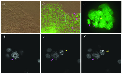
Figure 1
Imaging chromatin in living transgenic ES cells constitutively expressing a H2B-EGFP fusion protein. ( a ) Bright-field and ( b ) dark-field micrographs of a CAG::H2B-EGFP ES cell colony. The inset sh...
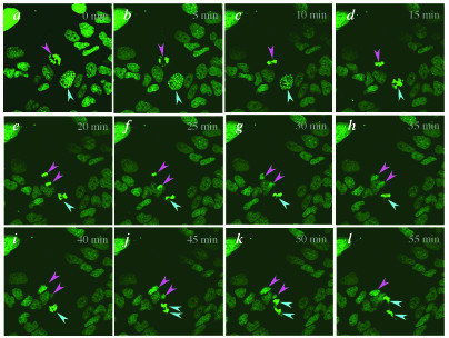
Figure 2
Live imaging the progression through mitosis. Laser scanning confocal x - y images taken at a single z -plane at five minute intervals for one hour. Note that not all green fluorescence (corresponding...
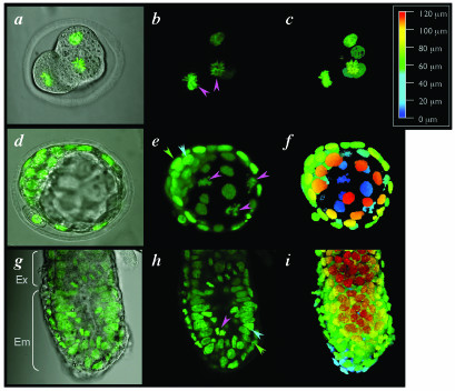
Figure 3
Live embryo imaging of preimplantation and early postimplantation mouse embryos hemizygous for a constitutively expressed H2B-EGFP fluorescent fusion. (a) Single confocal optical section fluorescence ...
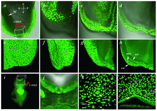
Figure 4
Live imaging H2B-EGFP in postimplantation mouse embryos. (a) Lateral view of the embryonic region of an E7.5 embryo (anterior to the left) with box depicting the region imaged in b and double-headed a...
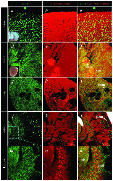
Figure 5
High resolution live imaging of the organs of CAG::H2B-EGFP adult mice. Confocal images of freshly isolated organs from a 6 week old adult male hemizygous CAG::H2B-EGFP Tg/+ animal illustrate the wide...
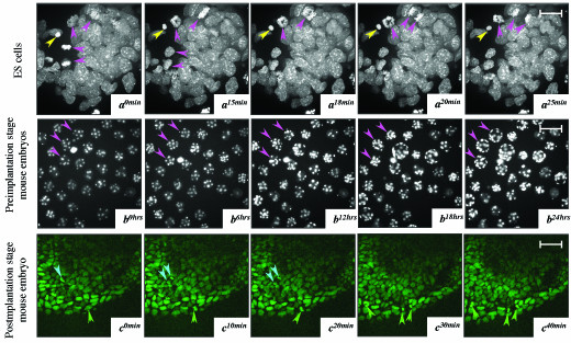
Figure 6
Dynamic time-lapse imaging of mouse CAG::H2B-EGFP transgenic ES cells, preimplantation and postimplantation embryos using different imaging modalities. ( a ) Rendered confocal stacks of transgenic ES ...
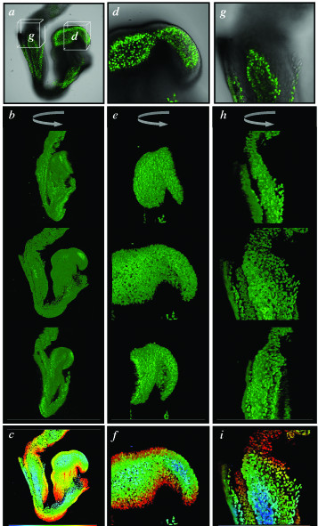
Figure 7
High-resolution 3-dimensional imaging of fixed CAG::H2B-EGFP transgenic embryos. Confocal images of an E8.5 CAG::H2B-EGFP transgenic embryo fixed in 4% paraformaldehyde for 72 hours, then washed, stor...
Figure images are served from the NIH/NLM PubMed Central Open Access Subset or Europe PMC; copyright remains with the publishers and authors.
💬 Discussion
0 commentsNo comments yet. Be the first to start a discussion!
Leave a Comment