Abstract
The signal transducer and activator of transcription-3 (STAT3) induces transcription of genes that control differentiation, inflammation, proliferation, and tumor cell invasion. Cytokines such as interleukin-6 and interferon stimulate the specific tyrosine phosphorylation of STAT3, which confers its ability to bind consensus DNA targets. In addition, unphosphorylated STAT3 has been demonstrated to induce specific gene expression. STAT3 must gain entrance to the nucleus to impact transcription, however access to the nucleus is a tightly regulated process. Because nuclear trafficking is critical to the function of STAT3, we investigated the molecular mechanisms by which STAT3 is imported to the nucleus. Live cell imaging techniques were used with STAT3 tagged with green fluorescence protein (GFP) or photoactivatable GFP to follow the cellular dynamics of both unphosphorylated and tyrosine phosphorylated forms. Cytokine activation did not alter the rate of STAT3 nuclear import or nuclear export. In addition, Förster resonance energy transfer experiments revealed homomeric interaction of unphosphorylated STAT3 dependent on its amino terminus, but this dimerization is not necessary for its nuclear import. Previous work demonstrated the adapter importin-α3 binds to STAT3 and is required for nuclear import. To determine whether STAT3 nuclear import is mediated by the importin-α/importin-β1 heterodimer, the effects of siRNA to importin-β1 were evaluated. Results indicate STAT3 nuclear import is dependent on the function of importin-β1. Since the Ran GTPase is necessary to bind importin-β1 in the nucleus for release of importin-α-cargo, the effect of a GTPase deficient mutant of Ran was tested. Expression of the Ran interfering mutant inhibited STAT3 nuclear import. This study defines importin-α/importin-β1/Ran as the molecular mechanism by which STAT3 traffics to the nucleus.
🔬 Techniques
🔭 Microscopes
💻 Software
✨ Fluorophores
🧪 Sample Preparation
🔬 Cell Lines
🏭 Microscope Brands
🧪 Reagent Suppliers
🔴 Lasers
💻 Software Details
🏛️ Research Organizations (ROR)
Affiliated research institutions:
📋 Methods
Cell Culture, Transfection, and Cytokine Treatment HeLa, Hep3B, and 293FT cells (ATCC) were cultured in DMEM with 10% fetal bovine serum, 1 mM L-glutamine, and 1% v/v penicillin/streptomycin. For microscopy, cells were seeded in glass bottom tissue culture dishes (Mattek Corp.) or on glass coverslips. Transfections were performed with TransIT-LT1 reagent (Mirus Bio LLC). Cells were cultured in serum-free media for 12 hours prior to experiments. Cells were treated with 20 ng/ml IL-6 (BioSource International), IFNα 1000 units/ml (gift from Roche, Nutley, NJ), or TNF (Invitrogen PHC3015).
Plasmids
STAT3 (gift of James Darnell, Jr., The Rockefeller University) was cloned into the vectors pEGFP-N1 (STAT3-GFP), pEYFP-N1 (STAT3-YFP), and pECFP-N1 (STAT3-CPF) (Clontech). STAT3 DNA-binding mutant (VVV) was cloned into pEGFP-N1 [63] . The N-terminus deletion mutant construct (STAT3-135-770-GFP) was generated previously [22] . The vector encoding photoactivatable GFP (PA-GFP) was a gift of Jennifer Lippincott-Schwartz (NIH) and used to generate STAT3-PA-GFP [38] . pDsRed-N1 was obtained from Clontech.
Human Ran and mutant Ran
Q69L were gifts from Colin Dingwall (Kings College, London) and were sub-cloned in pECFP-C1 (CFP-Ran and CFP-Ran Q69L) [64] . T7-tagged-Rac1-N17 and Rac1 were gifts of Linda Van Aelst (Cold Spring Harbor Laboratory). Harvey Ras-V12 was a gift of Dafna Bar-Sagi (New York University) [65] and was subcloned into pEYFP-C1 (Clontech). The luciferase reporter gene regulated by the STAT-responsive gamma IFN activated site has been described previously [58] . The NF-κB-responsive luciferase reporter gene was obtained from Stratagene, and the Renilla luciferase gene was obtained from Promega. RNA interference Cells were treated with small interfering RNA (siRNA) corresponding to importin-β1 or vimentin control (Qiagen) as described [66] . Briefly, siRNAs were transfected into cells using X-tremeGENE siRNA transfection reagent (Roche) and 24 hours later the cells were transfected with STAT3-GFP plasmid. STAT3-GFP fluorescence was evaluated by confocal microscopy 48 hours after siRNA transfection. RT-PCR was used to quantify endogenous importin-β1 mRNA and GAPDH mRNA as described previously [66] .
Show full methods section
Cell Culture, Transfection, and Cytokine Treatment HeLa, Hep3B, and 293FT cells (ATCC) were cultured in DMEM with 10% fetal bovine serum, 1 mM L-glutamine, and 1% v/v penicillin/streptomycin. For microscopy, cells were seeded in glass bottom tissue culture dishes (Mattek Corp.) or on glass coverslips. Transfections were performed with TransIT-LT1 reagent (Mirus Bio LLC). Cells were cultured in serum-free media for 12 hours prior to experiments. Cells were treated with 20 ng/ml IL-6 (BioSource International), IFNα 1000 units/ml (gift from Roche, Nutley, NJ), or TNF (Invitrogen PHC3015).
Plasmids
STAT3 (gift of James Darnell, Jr., The Rockefeller University) was cloned into the vectors pEGFP-N1 (STAT3-GFP), pEYFP-N1 (STAT3-YFP), and pECFP-N1 (STAT3-CPF) (Clontech). STAT3 DNA-binding mutant (VVV) was cloned into pEGFP-N1 [63] . The N-terminus deletion mutant construct (STAT3-135-770-GFP) was generated previously [22] . The vector encoding photoactivatable GFP (PA-GFP) was a gift of Jennifer Lippincott-Schwartz (NIH) and used to generate STAT3-PA-GFP [38] . pDsRed-N1 was obtained from Clontech.
Human Ran and mutant Ran
Q69L were gifts from Colin Dingwall (Kings College, London) and were sub-cloned in pECFP-C1 (CFP-Ran and CFP-Ran Q69L) [64] . T7-tagged-Rac1-N17 and Rac1 were gifts of Linda Van Aelst (Cold Spring Harbor Laboratory). Harvey Ras-V12 was a gift of Dafna Bar-Sagi (New York University) [65] and was subcloned into pEYFP-C1 (Clontech). The luciferase reporter gene regulated by the STAT-responsive gamma IFN activated site has been described previously [58] . The NF-κB-responsive luciferase reporter gene was obtained from Stratagene, and the Renilla luciferase gene was obtained from Promega. RNA interference Cells were treated with small interfering RNA (siRNA) corresponding to importin-β1 or vimentin control (Qiagen) as described [66] . Briefly, siRNAs were transfected into cells using X-tremeGENE siRNA transfection reagent (Roche) and 24 hours later the cells were transfected with STAT3-GFP plasmid. STAT3-GFP fluorescence was evaluated by confocal microscopy 48 hours after siRNA transfection. RT-PCR was used to quantify endogenous importin-β1 mRNA and GAPDH mRNA as described previously [66] .
Antibodies
Western blots were performed as described previously using the following antibodies: anti-phospho-tyrosine (p705) STAT3 (SantaCruz, Sc-8059/clone B7), anti-STAT3 (Santa Cruz sc-482), anti-importin-β1 (Santa Cruz H-7 sc-137016), anti-tubulin (Sigma B5-1-2), anti-rabbit (Alexa-labeled Invitrogen A21109), and anti-mouse HRP (Amersham Biosciences, NA931V) [22] . Antibodies for immunofluorescence were anti-T7 (Novagen) and rhodamine-conjugated anti-mouse (Jackson Laboratory).
Confocal Microscopy
Live cell imaging was performed using a Zeiss LSM 510 META NLO Two-Photon Laser Scanning Confocal Microscope System and cell chamber system with 37°C temperature control (Temperature Control 37-2, and Heating Insert P from Zeiss) and CO 2 control (CTI Controller 3700, and Incubator S from Zeiss) [66] , [67] . Images were captured and analyzed using the imaging software Zeiss LSM 510 Meta version 3.2 and Image J. Images are presented using Adobe Photoshop graphic software. GraphPad Prism software was used for curve-fitting analyses.
Fluorescence recovery after photobleaching
(FRAP) with STAT3-GFP or STAT3-YFP was performed by bleaching a region of interest (ROI) in the nucleus at 100% power of an argon laser (488 nm for GFP, 514 nm for YFP) for a duration of time ranging from 60 to 120 seconds.
Fluorescence loss in photobleaching
(FLIP) was performed by bleaching a ROI in the cytoplasm, repeatedly every 12 seconds at maximum laser intensity. Photoactivation experiments were performed using the two photon laser system Chameleon XR Laser System, with the following settings: 800 nm laser, laser power of 10–20%, and 50–100 laser continual iterations. Förster resonance energy transfer (FRET) experiments were performed using the method of “FRET after acceptor photobleaching” using CFP and YFP fluorophore pairs [47] , [68] . HeLa cells were grown on glass coverslips and transfected with DNA ratio 1∶3 between the CFP constructs and YFP constructs. 2 days post transfection the cells were washed twice in cold PBS and fixed with 4% paraformaldehyde. The coverslips were washed in PBS and mounted onto microscope glass slides using mounting media Vectashield (Vector Laboratories) and sealed. STAT3-YFP was excited and bleached and the resultant energy transfer to STAT3-CFP was quantified. The microscope settings were as follows: objective lens 63× C-Apochromat, laser 458 nm for CFP excitation, laser 514 nm for YFP excitation and bleaching, filter lambda mode for CFP emission 458–510 nm, and filter lambda mode for YFP emission 530–630 nm. The FRET efficiencies were calculated by curve fitting analysis of CFP fluorescence versus YFP fluorescence using the mathematical approach of Amiri et al. [47] . For STAT3-GFP mitochondrial localization, live cells were treated with 50 nM of the cell permeable MitoTracker Orange fluorescence dye (Molecular Probes, Invitrogen) for 1 hour and subsequently washed with media. Live cell imaging was performed using the following microscope settings: objective lens 63× C-Apochromat, laser 488 nm for GFP excitation, laser 543 nm for mitotracker orange, and laser 514 nm for YFP, filter BP 500–550 IR for GFP emission, filter BP 565–615 IR for MitoTracker Orange emission and filter BP 535–590 for YFP emission. Different fluorescence channel images were acquired sequentially. In order to achieve a satisfactory vertical resolution (
📊 Figures
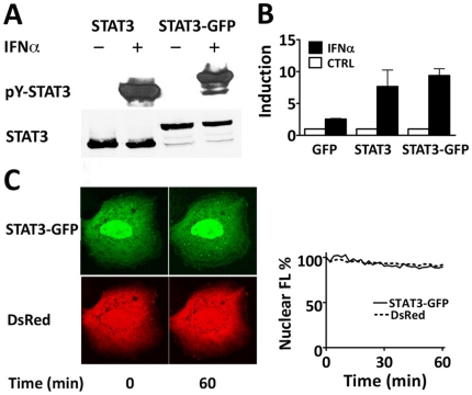
Figure 1
Prominent nuclear localization of STAT3-GFP independent of tyrosine phosphorylation.
A ) Western blot of cell lysates demonstrate tyrosine phosphorylation of STAT3 and STAT3-GFP in response to cytokine. 293 cells were transfected with untagged STAT3 or STAT3-GFP expression plasmids, s...
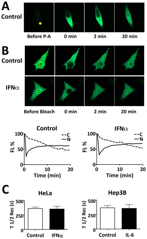
Figure 2
Time-lapse imaging with photoactivation or photobleaching reveals STAT3-GFP continuous nuclear import.
A ) Nuclear import of photoactivatable STAT3 (STAT3-PA-GFP). HeLa cells expressing STAT3-PA-GFP were serum-starved. A region in the cytoplasm (solid dot) was subjected to continuous high-intensity las...
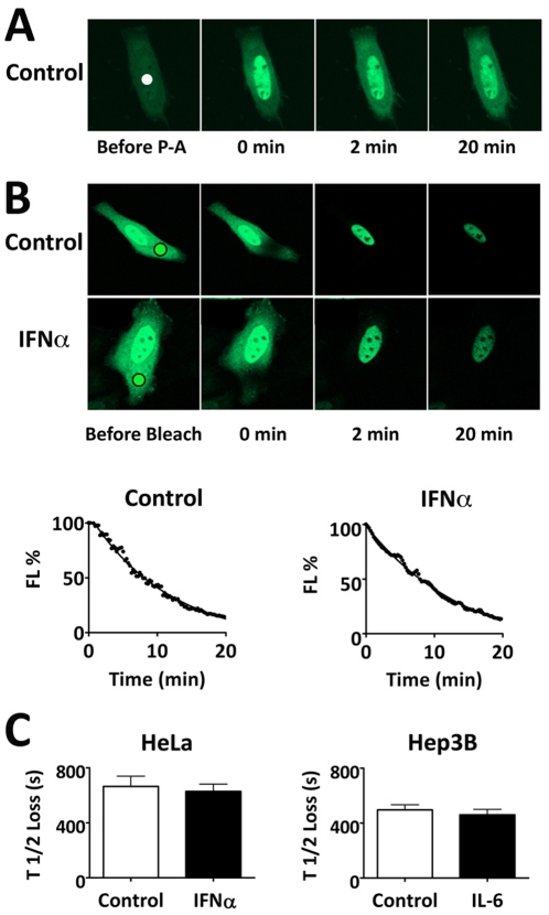
Figure 3
STAT3-GFP nuclear export independent of cytokine stimulation.
A ) Photoactivation of nuclear STAT3-PA-GFP in HeLa cells. A ROI in the nucleus (solid dot) of serum-starved cells was subjected to high intensity laser activation with 2-photon laser microscopy. B ) ...
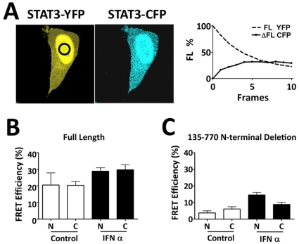
Figure 4
STAT3-STAT3 protein interaction in unphosphorylated and tyrosine phosphorylated states analyzed by FRET.
A ) HeLa cells co-expressing STAT3-YFP and STAT3-CFP were serum starved, fixed, and analyzed for STAT3-STAT3 interactions by FRET. A ROI in the nucleus (black circle) was subjected to laser for gradua...
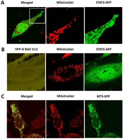
Figure 5
STAT3-GFP is excluded from mitochondria.
A ) HeLa cells expressing STAT3-GFP were stained with MitoTracker Orange and the localization of STAT3-GFP and mitochondria was captured with live cell imaging. B ) HeLa cells co-expressing STAT3-GFP ...
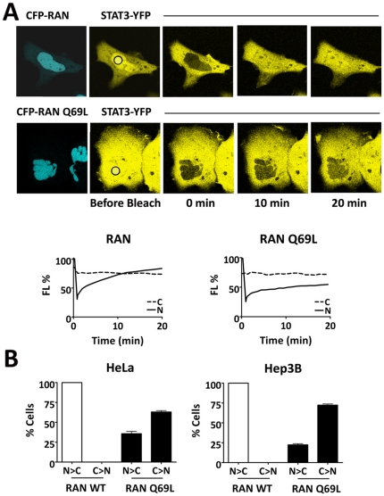
Figure 6
STAT3 nuclear import is dependent on Ran.
A ) Nuclear FRAP experiments were performed to photobleach STAT3-YFP in the nucleus of Hep3B cells co-expressing either CFP-Ran wild type (CFP image top left panel) or CFP-Ran Q69L (CFP image bottom l...
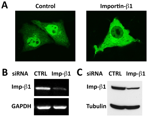
Figure 7
Knockdown of importin-u03b21 inhibits STAT3-GFP nuclear import.
HeLa cells were transfected with vimentin siRNA (control) or importin-u03b21 siRNA and STAT3-GFP. A ) Images of STAT3-GFP localization. Four independent experiments indicated 100% of cells expressing ...
Figure images are served from the NIH/NLM PubMed Central Open Access Subset or Europe PMC; copyright remains with the publishers and authors.
💬 Discussion
0 commentsNo comments yet. Be the first to start a discussion!
Leave a Comment