Abstract
Unlike most cells, protozoa in the phylum Apicomplexa divide by a distinctive process in which multiple daughters are assembled within the mother (schizogony or endodyogeny), using scaffolding known as the inner membrane complex (IMC). The IMC underlies the plasma membrane during interphase, but new daughters develop in the cytoplasm, as cytoskeletal filaments associate with flattened membrane cisternae (alveolae), which elongate rapidly to encapsulate subcellular organelles. Newly assembled daughters acquire their plasma membrane as they emerge from the mother, leaving behind vestiges of the maternal cell. Although the maternal plasma membrane remains intact throughout this process, the maternal IMC disappears - is it degraded, or recycled to form the daughter IMC? Exploiting fluorescently tagged IMC markers, we have used live-cell imaging, fluorescence recovery after photobleaching (FRAP) and mEos2 photoactivation to monitor the dynamics of IMC biogenesis and turnover during the replication of Toxoplasma gondii tachyzoites. These studies reveal that the formation of the T. gondii IMC involves two distinct steps - de novo assembly during daughter IMC elongation within the mother cell, followed by recycling of maternal IMC membranes after the emergence of daughters from the mother cell.
🔬 Techniques
💻 Software
✨ Fluorophores
🧪 Sample Preparation
🏭 Microscope Brands
🧪 Reagent Suppliers
📷 Detectors
🔎 Objectives
🎨 Filters
💻 Software Details
🏛️ Research Organizations (ROR)
Affiliated research institutions:
📋 Methods
Cells and parasites Human foreskin fibroblasts (HFFs) were cultivated at 37°C under a humidified atmosphere containing 5% CO 2 , as previously described ( Roos et al., 1995 ), in a 5∶1 mixture of high glucose Dulbecco's Modified Eagle's Medium (DMEM, Life Technologies, Grand Island, NY) to Medium 199 (Life Technologies), supplemented with 10% newborn calf serum (NBS, Thermo Scientific, Waltham, MA), 50 U/ml penicillin, 50 µg/ml streptomycin and 25 µg/ml gentamicin (Life Technologies). Immediately prior to inoculation of the cells with T. gondii tachyzoites, this growth medium was replaced with Minimal Essential Medium (MEM, Life Technologies) supplemented with 2 mM Glutamax (Life Technologies), 1% heat-inactivated fetal bovine serum (FBS, Thermo Scientific) and antibiotics (as above).
Plasmids
The allelic replacement plasmid pLic GAP40YFP- dhfr HXGPRT was engineered by PCR amplification of 1358-bp-spanning Tg GAP40 ( Tg ME49_249850; ToxoDB), using the primers shown in supplementary material Table S2 , and integration into the Lic sequences in p YFP.Lic.HXG [kindly provided by Vern Carruthers, University of Michigan ( Huynh and Carruthers, 2009 )].
Plasmid pLic IMC1mCherry- dhfr
DHFR was engineered similarly, using a 1950-bp fragment from the 3′ end of Tg IMC1 ( Tg ME49_231640) integrated into p mCherry.Lic.DHFR. All plasmids were confirmed by restriction digestion and sequencing. After linearization with Kas I (for GAP40) or BsiW I (for IMC1), 15×10 6 freshly harvested RHΔKu80ΔHXGPRT strain T. gondii tachyzoites ( Huynh and Carruthers, 2009 ) were electroporated with 50 µg of plasmid and were selected in 25 µg/ml mycophenolic acid with 50 µg/ml xanthine (GAP40) or 1 µM pyrimethamine (IMC1) ( Roos et al., 1995 ). Clonal plaques were isolated by limiting dilution and screened by fluorescence microscopy for transgene expression.
Show full methods section
Cells and parasites Human foreskin fibroblasts (HFFs) were cultivated at 37°C under a humidified atmosphere containing 5% CO 2 , as previously described ( Roos et al., 1995 ), in a 5∶1 mixture of high glucose Dulbecco's Modified Eagle's Medium (DMEM, Life Technologies, Grand Island, NY) to Medium 199 (Life Technologies), supplemented with 10% newborn calf serum (NBS, Thermo Scientific, Waltham, MA), 50 U/ml penicillin, 50 µg/ml streptomycin and 25 µg/ml gentamicin (Life Technologies). Immediately prior to inoculation of the cells with T. gondii tachyzoites, this growth medium was replaced with Minimal Essential Medium (MEM, Life Technologies) supplemented with 2 mM Glutamax (Life Technologies), 1% heat-inactivated fetal bovine serum (FBS, Thermo Scientific) and antibiotics (as above).
Plasmids
The allelic replacement plasmid pLic GAP40YFP- dhfr HXGPRT was engineered by PCR amplification of 1358-bp-spanning Tg GAP40 ( Tg ME49_249850; ToxoDB), using the primers shown in supplementary material Table S2 , and integration into the Lic sequences in p YFP.Lic.HXG [kindly provided by Vern Carruthers, University of Michigan ( Huynh and Carruthers, 2009 )].
Plasmid pLic IMC1mCherry- dhfr
DHFR was engineered similarly, using a 1950-bp fragment from the 3′ end of Tg IMC1 ( Tg ME49_231640) integrated into p mCherry.Lic.DHFR. All plasmids were confirmed by restriction digestion and sequencing. After linearization with Kas I (for GAP40) or BsiW I (for IMC1), 15×10 6 freshly harvested RHΔKu80ΔHXGPRT strain T. gondii tachyzoites ( Huynh and Carruthers, 2009 ) were electroporated with 50 µg of plasmid and were selected in 25 µg/ml mycophenolic acid with 50 µg/ml xanthine (GAP40) or 1 µM pyrimethamine (IMC1) ( Roos et al., 1995 ). Clonal plaques were isolated by limiting dilution and screened by fluorescence microscopy for transgene expression.
Plasmid ptub GAP40YFPHA- sag
CAT was engineered by replacing the ACP sequences in ptub ACP-YFP-HA/ sag CAT sag ( Nishi et al., 2008 ) with GAP40 ( Bgl II– Avr II).
Plasmid ptub GAP40mEos2- sag
CAT was engineered by replacing the YFP-HA in ptub GAP40YFPHA- sag CAT with mEos2 amplified as an Avr II– Afl II fragment from the construct mEos-vinculin [kindly provided by Michael Davidson, Florida State University ( Kanchanawong et al., 2010 )]. Parasites were transfected with 50 µg of plasmid as above and transient transfectants were examined at ∼18 h post-transfection. All transgenes (YFP, mCherry, mEos2) utilized standard (non-optimized) coding sequences.
Immunofluorescence microscopy
HFF cells were grown to confluence on 22-mm glass coverslips, infected with T. gondii tachyzoites and incubated at 37°C for a further 18–24 h. Coverslips were then fixed for 15–20 min (4% formaldehyde and 0.05% glutaraldehyde in PBS), permeabilized for 15 min (0.25% Triton X-100 in PBS) and blocked for 1 h at room temperature in 3% bovine serum albumin (BSA) fraction V plus 0.25% Triton X-100. After incubation for 1 h with murine monoclonal anti-SAG1 [1∶400 in blocking solution; kindly provided by Lloyd Kasper, Dartmouth College ( Mineo et al., 1993 )] or anti-IMC1 [1∶2000, kindly provided by Gary Ward, University of Vermont ( Mann and Beckers, 2001 ; Wichroski et al., 2002 )] coverslips were washed three times with 0.25% Triton X-100 in PBS and stained for 1 h in Alexa-Fluor-594-conjugated goat anti-mouse-IgG antibody (1∶5000; Life Technologies). For DNA labeling, samples were then incubated for 10 min with 4′,6-diamidino-2-phenylindole dihydrochloride (DAPI, EMD Millipore, Billerica, MA) at a final concentration of 0.5 µg/µl in PBS, washed twice with 0.25% Triton X-100 and once with PBS and mounted on glass slides in Fluoromount-G (Southern Biotech, Birmingham, AL). Imaging was performed on an Olympus IX70 inverted microscope equipped with a UPlanSApo 100× oil-immersion objective (NA1.4), 300 W xenon arc lamp and a CoolSNAP HQ monochrome cooled-CCD camera. The excitation and emission filters used for DAPI were 360/40 nm and 455/50 nm, respectively; for GFP were 470/40 nm and 520/40 nm, and for mCherry or RFP were 572/35 nm and 632/60 nm. Image stacks were captured using DeltaVision SoftWorx software (Applied Precision, Issaquah, WA) and deconvolved ( Figs 1 , 6 only) using the constrained iterative algorithm, to minimize the effects of out-of-focus fluorescence. Step size was 0.1 or 0.2 µm and acquisition depth was ∼2–3 µm, satisfying Nyquist sampling. Images were further analyzed using open-source Fiji software ( Schindelin et al., 2012 ) and were imported into PowerPoint for figure preparation.
Time-lapse microscopy Confluent
HFF cell monolayers were cultivated in 35-mm glass-bottomed dishes (Ibidi, Verona, WI), infected with T. gondii tachyzoites at a multiplicity of infection (MOI) of ∼2∶1 [in DMEM lacking Phenol Red (Life Technologies), supplemented with 1% FBS, 1 mM sodium pyruvate, 2 mM glutamine, 100 U/ml penicillin, 100 µg/ml streptomycin and 50 µg/ml gentamicin], and incubated for 12–16 h at 37°C. Prior to imaging, cultures were rinsed with warm PBS lacking divalent cations (to remove extracellular parasites) and incubated in fresh Phenol-Red-free DMEM (as above) supplemented with 10% FBS (to minimize laser phototoxicity) and 25 mM HEPES pH 7. Samples were then transferred to a Chamlide TC stage-top environmental chamber (Live Cell Instruments, Guelph, ON, Canada) that was equipped with a digital temperature, CO 2 and humidity control unit and equilibrated ∼2 h before data acquisition. Time-lapse imaging was performed on an Olympus IX-71 spinning-disk confocal microscope equipped with a UPlanSApo 100× oil-immersion objective (NA1.4), CSU-10 scanner (Yokogawa, Newnan, GA), and C9100-13 EMCCD camera (Hamamatsu, Bridgewater, NJ). 5 µm image stacks (26 planes×0.2 µm steps) were acquired by excitation at 488 nm and 561 nm (1% laser power) every 15 or 30 min, using the emission filters ET525/50 for GFP and ET630/75 for mCherry or RFP (Spectral Applied Research). Data was collected using MetaMorph 7.7.4 (Molecular Devices, Downingtown, PA), and processed using MetaMorph and Fiji software. Some images were contrast enhanced for figure presentation. For quantification of IMC development ( Fig. 2 ), 9–12 time-lapse image stacks (26 planes, as above) were collected over a continuous 9 h session for each of 15 vacuoles in eight fields. At each time-point (for each parasitophorous vacuole) the central image plane was selected from the stack and Fiji software was used to collect perimeter length and total fluorescence (after background subtraction) for all maternal, daughter and/or grand-daughter IMCs, based on manually drawn lines representing all distinctly resolvable IMC structures ( supplementary material Table S1a for images and S1b for quantification). Individual time-lapse series were aligned based on the estimated time of daughter parasite initiation (‘Offset’ column in supplementary material Table S1 and Fig. S2A ). Offset values can be reliably estimated to
📊 Figures
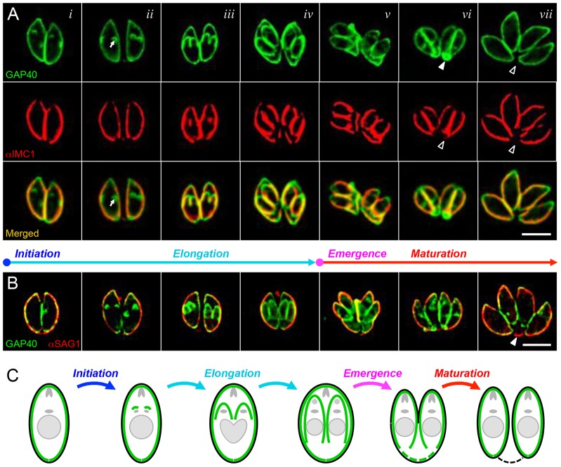
Fig. 1.
Stages of the IMC cycle in T. gondii . Colocalization of the integral membrane protein GAP40 (green, A and B) with the cytoskeletal alveolin IMC1 (red, A) and the plasma membrane protein SAG1 (red, B)...
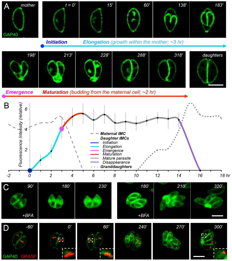
Fig. 2.
Time-lapse imaging and quantitative dynamics of GAP40. (A) Time-lapse imaging of GAP40u2013YFP-expressing transgenic parasites (C-terminally tagged at the endogenous genomic locus); see supplementary ...
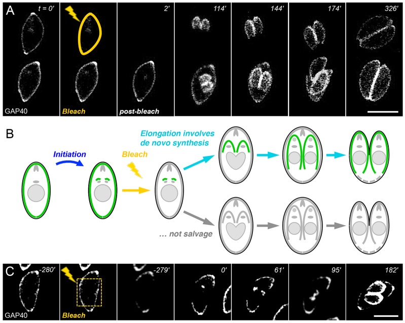
Fig. 3.
The daughter IMC is assembled de novo . (A) Laser photobleaching of the maternal IMC (yellow line) has no impact on daughter IMC initiation or elongation, indicating that the daughter IMC is synthesiz...
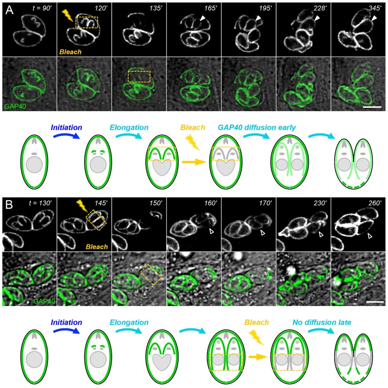
Fig. 4.
GAP40 redistribution declines during daughter elongation. GAP40 fluorescence in daughter parasites recovers rapidly at early stages of elongation (closed arrowheads), owing to a combination of de novo...
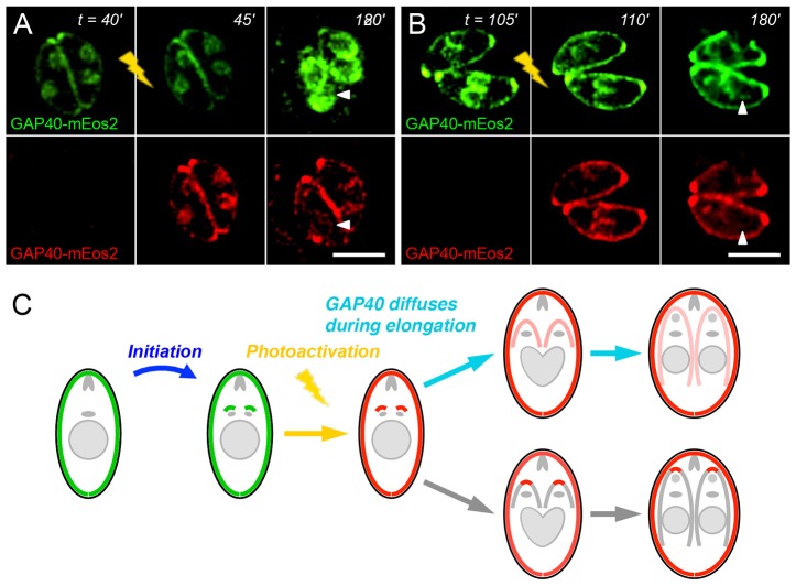
Fig. 5.
Photoactivation of GAP40u2013mEos2 indicates protein movement within the elongating IMC. Exposure of transiently expressed GAP40u2013mEos2 to violet light at various times after initiation (A,B) conve...
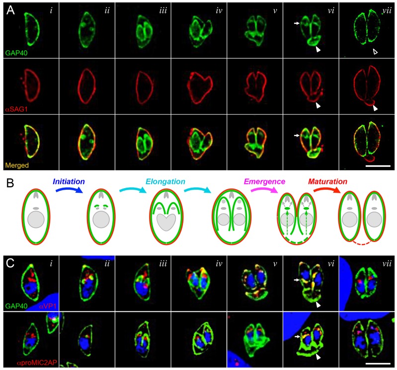
Fig. 6.
GAP40 is lost from the residual body and appears in the ELC during daughter parasite emergence and maturation. (A) Colocalization of the IMC protein GAP40 (green) and plasma membrane protein SAG1 (red...
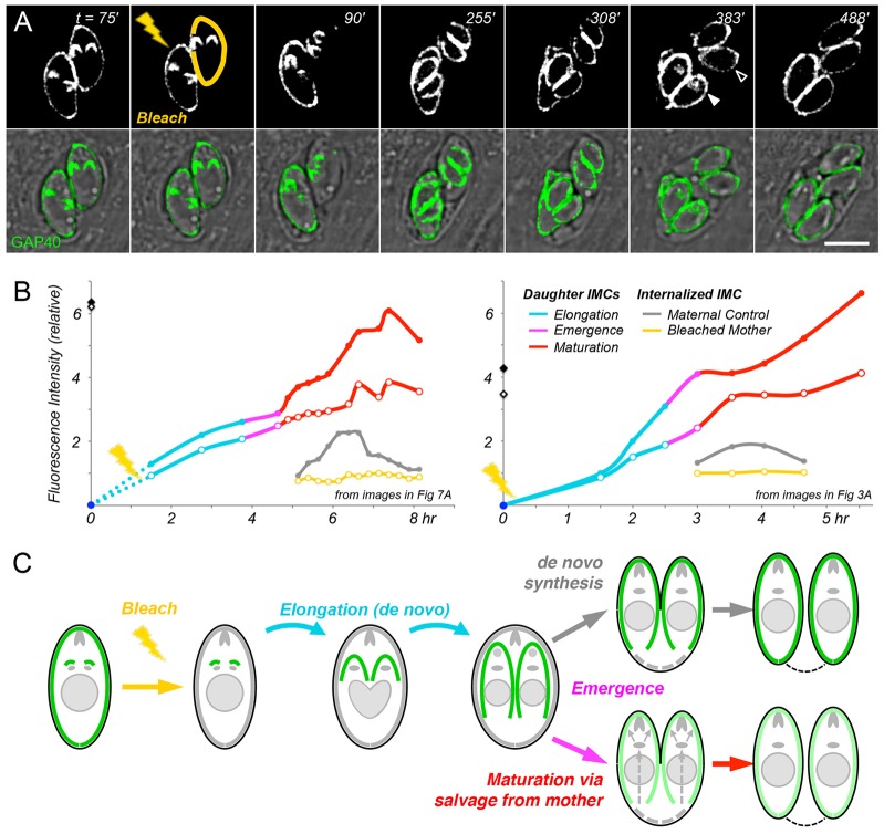
Fig. 7.
Maternal IMC is recycled into developing daughters after their emergence. (A) The entire IMC of one parasite was photobleached (yellow), whereas that of its sister was not (providing an internal contr...
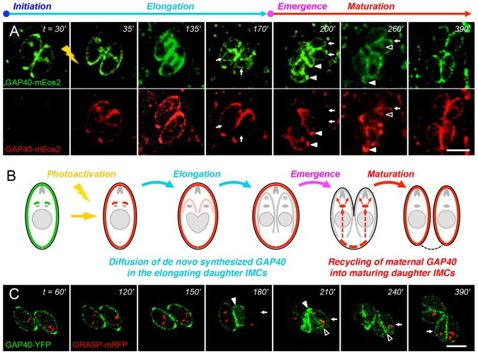
Fig. 8.
Photoactivation confirms recycling of maternal GAP40 into daughters during maturation. (A) Activating GAP40u2013mEos2 at the onset of daughter formation (lightning) converts a portion of GAP40 from gr...
Figure images are served from the NIH/NLM PubMed Central Open Access Subset or Europe PMC; copyright remains with the publishers and authors.
💬 Discussion
0 commentsNo comments yet. Be the first to start a discussion!
Leave a Comment