Abstract
Local edema, a cardinal sign of inflammation associates closely with neutrophil emigration. Neutrophil emigration has been described to occur primarily through endothelial junctions (paracellular) and more rarely directly through endothelial cells (transcellular). Recently, we reported that unlike in wild-type (wt) mice, Mac-1-/- (CD11b) neutrophils predominantly emigrated transcellularly and was significantly delayed taking 20-30 min longer than the paracellular emigration (wt). In the present study we noted significant anatomical disruption of the endothelium and hypothesized that transcellular emigration would greatly increase vascular permeability. Surprisingly, despite profound disruption of the endothelial barrier as the neutrophils moved through the cells, the changes in vascular permeability during transcellular emigration (Mac-1-/-) were not increased more than in wt mice. Instead increased vascular permeability completely tracked the number of emigrated cells and as such, permeability changes were delayed in Mac-1-/- mice. However, by 60 min neutrophils from both sets of mice were emigrating in large numbers. Electron-microscopy and spinning disk multichannel fluorescence confocal microscopy revealed endothelial docking structures that progressed to dome-like structures completely covering wt and Mac-1-/- neutrophils. These domes completely enveloped the emigrating neutrophils in both wt and Mac-1-/- mice making the mode of emigration underneath these structures extraneous to barrier function. In conclusion, predominantly paracellular versus predominantly transcellular emigration does not affect vascular barrier integrity as endothelial dome-like structures retain barrier function.
🔬 Techniques
💻 Software
✨ Fluorophores
🧪 Sample Preparation
🔬 Cell Lines
🏭 Microscope Brands
🧪 Reagent Suppliers
📷 Detectors
💻 Software Details
🏛️ Research Organizations (ROR)
Affiliated research institutions:
📋 Methods
Animals Male C57BL/6 mice were obtained from The Jackson Laboratories and Mac-1 deficient male mice were originally made by Dr Ballantyne (Baylor College of Medicine). All mice weighed between 18 and 33 grams. All experimental procedures were approved by the University of Calgary Animal Care Committee and conformed to the guidelines established by the Canadian Council for Animal Care. Intravital video-microscopy The mice were anesthetized with an i.p. injection of a mixture of 10 mg kg −1 xylazine (Animal Health; Bayer Inc.) and 200 mg kg −1 ketamine hydrochloride (Biomeda-MTC, Animal Health Inc., ON). For all protocols, the right jugular vein was cannulated to administer additional anesthetic. The mouse cremaster muscle preparation was used to study the behavior of leukocytes in the microcirculation and adjacent connective tissue as described previously [27] , [28] . In brief, an incision was made in the scrotal skin to expose the left cremaster muscle, which was then carefully dissected free of the associated fascia and continuously kept moist with 37°C bicarbonate-buffered saline. The cremaster muscle was cut longitudinally with a cautery. The testicle and epididymis were separated from the underlying muscle and were gently pushed into the abdominal cavity. The muscle was held flat on an optically clear viewing pedestal and was secured along the edges with five 4.0 sutures. A coverslip with silicon grease on two of the edges was gently placed over the muscle without touching it, to avoid drying of the tissue. An intravital microscope (Optiphot-2; Nikon Inc., Mississauga, ON, Canada) with a ×10 eyepiece and a ×25 objective (Weltzlar L25/0.35; E. Leitz Inc.) was used to examine the microcirculation in the muscle. A video camera (5100 HS; Panasonic) was used to project the images onto a monitor, and the images were recorded for playback analysis using a videocassette recorder. Rolling, adhesion and emigration Single unbranched venules (25–40 µm in diameter) were selected, and to minimize variability, the same section of vessel was observed throughout the experiments. The number of rolling, adherent, and emigrated leukocytes was determined offline during video playback analysis. Rolling leukocytes were defined as those cells moving at a velocity less than that of erythrocytes within the vessel. The flux of rolling cells was measured as the number of rolling leukocytes passing by a given point in the venule per minute. Leukocyte rolling velocity was calculated from the average time required for 10 leukocytes to roll along 100 µm of the vessel. A leukocyte was considered to be adherent if it remained stationary for more than 30 sec, and total leukocyte adhesion was quantified as the number of adherent cells within a 100 µm length of venule over 5 min. Leukocyte emigration was defined as the number of cells in the extra vascular space within a 200×300 µm area (0.06 mm 2 ). Only cells adjacent to and clearly outside the vessel under study were counted as emigrated. To induce chemotaxis, the superfusion buffer was changed to MIP-2 superfusion in a concentration of 0.5 or 5 nmol/L for 60 min after the control period.
Show full methods section
Animals Male C57BL/6 mice were obtained from The Jackson Laboratories and Mac-1 deficient male mice were originally made by Dr Ballantyne (Baylor College of Medicine). All mice weighed between 18 and 33 grams. All experimental procedures were approved by the University of Calgary Animal Care Committee and conformed to the guidelines established by the Canadian Council for Animal Care. Intravital video-microscopy The mice were anesthetized with an i.p. injection of a mixture of 10 mg kg −1 xylazine (Animal Health; Bayer Inc.) and 200 mg kg −1 ketamine hydrochloride (Biomeda-MTC, Animal Health Inc., ON). For all protocols, the right jugular vein was cannulated to administer additional anesthetic. The mouse cremaster muscle preparation was used to study the behavior of leukocytes in the microcirculation and adjacent connective tissue as described previously [27] , [28] . In brief, an incision was made in the scrotal skin to expose the left cremaster muscle, which was then carefully dissected free of the associated fascia and continuously kept moist with 37°C bicarbonate-buffered saline. The cremaster muscle was cut longitudinally with a cautery. The testicle and epididymis were separated from the underlying muscle and were gently pushed into the abdominal cavity. The muscle was held flat on an optically clear viewing pedestal and was secured along the edges with five 4.0 sutures. A coverslip with silicon grease on two of the edges was gently placed over the muscle without touching it, to avoid drying of the tissue. An intravital microscope (Optiphot-2; Nikon Inc., Mississauga, ON, Canada) with a ×10 eyepiece and a ×25 objective (Weltzlar L25/0.35; E. Leitz Inc.) was used to examine the microcirculation in the muscle. A video camera (5100 HS; Panasonic) was used to project the images onto a monitor, and the images were recorded for playback analysis using a videocassette recorder. Rolling, adhesion and emigration Single unbranched venules (25–40 µm in diameter) were selected, and to minimize variability, the same section of vessel was observed throughout the experiments. The number of rolling, adherent, and emigrated leukocytes was determined offline during video playback analysis. Rolling leukocytes were defined as those cells moving at a velocity less than that of erythrocytes within the vessel. The flux of rolling cells was measured as the number of rolling leukocytes passing by a given point in the venule per minute. Leukocyte rolling velocity was calculated from the average time required for 10 leukocytes to roll along 100 µm of the vessel. A leukocyte was considered to be adherent if it remained stationary for more than 30 sec, and total leukocyte adhesion was quantified as the number of adherent cells within a 100 µm length of venule over 5 min. Leukocyte emigration was defined as the number of cells in the extra vascular space within a 200×300 µm area (0.06 mm 2 ). Only cells adjacent to and clearly outside the vessel under study were counted as emigrated. To induce chemotaxis, the superfusion buffer was changed to MIP-2 superfusion in a concentration of 0.5 or 5 nmol/L for 60 min after the control period.
Electron microscopy
The cremaster muscle in wt or Mac-1-/- mice was superfused for 60 min with 0.5 nmol/L MIP-2, cut out and processed for transmission electron microscopy. Briefly, samples were fixed with 2.5% glutaraldehyde in 0.1 mol/L cacodylate buffer (pH 7.4) for 2 hours, washed three times with the same buffer, and postfixed with 1% osmium tetroxide in 0.1 mol/L cacodylate buffer (pH 7.4) for 1 hour at room temperature. Then the samples were dehydrated in a graded series of ethanol and embedded in Spurr's resin. Ultrathin sections were obtained with a diamond knife on a ultramicrotome (Ultracut E,, Reichert-Jung, Vienna, Austria), stained with uranyl acetate and lead citrate and then viewed with a Hitachi H-7000 transmission electron microscope (TEMHitachi H-7000, Tokyo, Japan) at 75kV.
Microvascular permeability measurement
The degree of vascular albumin leakage from cremasteric venules of Mac-1-/- and wt mice was quantified as described previously by both our laboratory [18] and others [19] , [20] . In brief, 25 mg kg −1 FITC-labeled BSA (Sigma-Aldrich) was administered to the mice i.v. at the start of the experiment, and FITC-derived fluorescence (excitation wavelength, 450–490 nm; emission wavelength, 520 nm) was detected using a silicon-intensified fluorescence camera (model C-2400-80; Hamamatsu Photonics).
Image analysis software
(ImageJ, the National Institutes of Health, http://rsb.info.nih.gov/ij/ ), was used to determine the intensity of FITC-albumin-derived fluorescence within the lumen of the venule and in the adjacent perivascular tissue. Background was defined as the fluorescence intensity before FITC-albumin administration. The exposed cremaster muscle was superfused with 0.5 or 5.0 nmol/L MIP-2 in 37°C warmed bicarbonate-buffered saline. The index of vascular albumin leakage (permeability index) at different time points before and after MIP-2 superfusion was determined according to the following ratio expressed as a percentage: (mean interstitial intensity–background)/(venular intensity–background).
Spinning Disk Confocal Microscopy
The microcirculation of the cremaster muscle was prepared for microscopy as described above and was visualized using a spinning disk confocal microscope. Images were acquired with an Olympus BX51 (Olympus, Center Valley, PA, USA) upright microscope using a 40×/0.75 Plan Fl water immersion objective. The microscope was equipped with a confocal light path (WaveFx, Quorum, Guelph, On. Canada) based on a modified Yokagawa CSU-10 head (Yokagawa Electric Corporation, Tokyo, Japan). Neutrophils were visualized with anti-mouse Ly-6G (Gr-1; BD Biosciences; 2ug/mouse) conjugated with Alexa Fluor 555 (Molecular Probes) injected iv into Tie-2 GFP mice (Background C57Bl/6) to image the neutrophils and endothelium simultaneously. Both 488 and 561 nm laser excitation (Cobalt, Stockholm, Sweden) was used in rapid succession and visualized with the appropriate long pass filters (Semrock, Rochester, New York, USA). Typical exposure times for both excitation wavelengths were 400 ms. A 512×512 pixel back thinned EMCCD camera (C9100, Hamamatsu, Bridgewater, NJ, USA) was used for fluorescence detection. Volocity Acquisition software (Improvision, Lexington, MA, USA) was used to drive the confocal microscope and to remove noise from the captured images. Images captured using the spinning disk were processed and analyzed in Volocity 4.20. The image brightness was adjusted equally in all micrographs to improve visibility.
Statistics
All data are presented as mean±standard error of the mean (SEM). ANOVA, single-factor, non-repeated measures followed by Fisher protected least significant difference test was performed for multiple comparisons.
📊 Figures
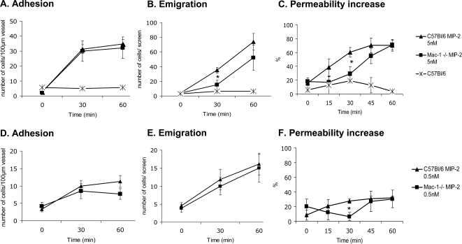
Figure 1
Leukocyte recruitment and vascular permeability, 5 nmol/L MIP-2 (Au2013C) and 0.5 nmol/L MIP-2 (Du2013F) superfusion.
Leukocyte adhesion (A and D), emigration (B and E) and microvascular permeability increase (C and F) before (time 0) and after addition of MIP-2 (5 nmol/L in Au2013C and 0.5 nmol/L in Du2013F) in the ...
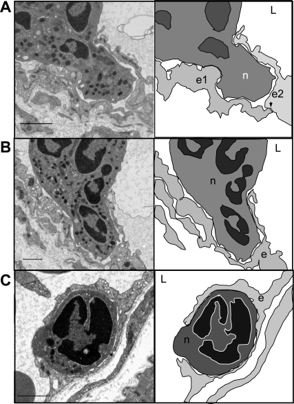
Figure 2
Endothelial transmigratory cup formation.
Electron micrographs and cartoons of transmigrating wild-type neutrophils where the endothelium forms a transmigratory cup (A, B and C). The arrowheads mark the junctions and L marks the lumen of the ...
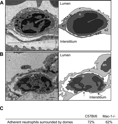
Figure 3
Endothelial enscapsulation of transmigrating neutrophils.
Electron micrographs and cartoons of transmigrating wild-type (A) and Mac-1 deficient neutrophils (B). The arrowheads mark the junctions seen in A, the thin endothelial sheet that covers the transmigr...
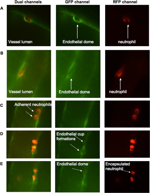
Figure 4
Endothelial dome formation using spinning disk confocal microscopy.
Spinning disk confocal images of MIP-2 superfused cremaster muscle of mice with GFP positive endothelium (green) and GR-1 Alexa Fluor 555 stained neutrophils (red). Panels A and B show adherent neutro...
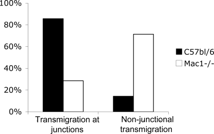
Figure 5
Transmigration in respect to endothelial junctions.
The distribution of transmigrating wild-type (nu200a=u200a40) and Mac-1-/- neutrophils (nu200a=u200a24) in respect to junctions on electron micrographs.
Figure images are served from the NIH/NLM PubMed Central Open Access Subset or Europe PMC; copyright remains with the publishers and authors.
💬 Discussion
0 commentsNo comments yet. Be the first to start a discussion!
Leave a Comment