Abstract
Matrix metalloproteinase enzymes, overexpressed in HT-1080 human fibrocarcinoma tumors, were used to guide the accumulation and retention of an enzyme-responsive nanoparticle in a xenograft mouse model. The nanoparticles were prepared as micelles from amphiphilic block copolymers bearing a simple hydrophobic block and a hydrophilic peptide brush. The polymers were end-labeled with Alexa Fluor 647 dyes leading to the formation of labeled micelles upon dialysis of the polymers from DMSO/DMF to aqueous buffer. This dye-labeling strategy allowed the presence of the retained material to be visualized via whole animal imaging in vivo and in ex vivo organ analysis following intratumoral injection into HT-1080 xenograft tumors. We propose that the material is retained by virtue of an enzyme-induced accumulation process whereby particles change morphology from 20 nm spherical micelles to micrometer-scale aggregates, kinetically trapping them within the tumor. This hypothesis is tested here via an unprecedented super-resolution fluorescence analysis of ex vivo tissue slices confirming a particle size increase occurs concomitantly with extended retention of responsive particles compared to unresponsive controls.
🔬 Techniques
✨ Fluorophores
🏛️ Research Organizations (ROR)
Affiliated research institutions:
📊 Figures
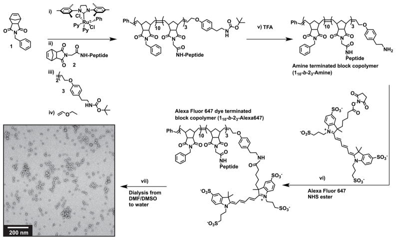
Figure 1
Preparation of enzyme-responsive Alexa Fluor 647 labeled micellar nanoparticles. L -amino acid based norbornyl-peptide substrates were polymerized to generate PPA-L ( L -amino acid PPA) for assembly t...
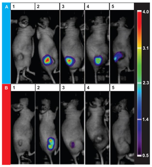
Figure 2
Intratumoral injection to determine relative levels of retention of enzyme-responsive nanoparticles vs control particles with HT-1080 tumors. A) M injected. B) M D injected. 1) Background prior to inj...
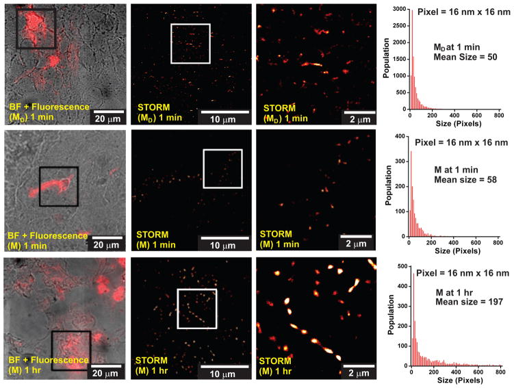
Figure 3
Confocal and super resolution fluorescence microscopy images of tissue slices from M - and M D -intratumorally injected mice. TOP row: M D -injected mice sacrificed at t = 1 min post-injection. MIDDLE...
Figure images are served from the NIH/NLM PubMed Central Open Access Subset or Europe PMC; copyright remains with the publishers and authors.
💬 Discussion
0 commentsNo comments yet. Be the first to start a discussion!
Leave a Comment