Abstract
STUDY QUESTION: Does intrauterine biosynthesis of estrogen play an important role in early pregnancy by altering the function of uterine natural killer (uNK) cells? SUMMARY ANSWER: Estrogens directly regulate the function of human uNK cells by increasing uNK cell migration and secretion of uNK cell-derived chemokine (C-C motif) ligand 2 (CCL2) that critically facilitates uNK-mediated angiogenesis. WHAT IS KNOWN ALREADY: uNK cells are a phenotypically distinct population of tissue-resident immune cells that regulate vascular remodelling within the endometrium and decidua. Recently we discovered that decidualisation of human endometrial stromal cells results in the generation of an estrogen-rich microenvironment in areas of decidualised endometrium. We hypothesize that intrauterine biosynthesis of estrogens plays an important role in early pregnancy by altering the function of uNK cells. STUDY DESIGN, SIZE, DURATION: This laboratory-based study used primary human uNK cells which were isolated from first trimester human decidua (n = 32). PARTICIPANTS/MATERIALS, SETTING, METHODS: Primary uNK cells were isolated from first trimester human decidua using magnetic cell sorting. The impact of estrogens on uNK cell function was assessed. Isolated uNK cells were treated with estrone (E1, 10(-8) M) or estradiol (E2, 10(-8) M) alone or in combination with the anti-estrogen ICI 182 780 (ICI, 10(-6) M). uNK cell motility was assessed by transwell migration assay and time-lapse microscopy. Expression of chemokine receptors was assessed by quantitative PCR (qPCR) and immunohistochemistry, and angiogenic factors were assessed by qPCR and cytokine array. Concentrations of CCL2 in supernatants were measured by enzyme-linked immunosorbent assay. Angiogenesis was assessed in a human endometrial endothelial cell network formation assay. MAIN RESULTS AND THE ROLE OF CHANCE: Treatment with either E1 or E2 increased uNK cell migration (P = 0.0092 and P = 0.0063, respectively) compared with control. Co-administration of the anti-estrogen ICI blocked the effects of E1 and E2 on cell migration. Concentrations of C-X-C chemokine receptor type 4 (CXCR4) mRNA in uNK cells were increased by E2 treatment. The network formation assay revealed that conditioned media from uNK cells treated with E2 significantly increased human endometrial endothelial cell (HEEC) angiogenesis (P = 0.0029 versus control). Analysis of media from uNK cells treated with E2 using an antibody array identified CCL2 as the most abundant cytokine. Validation assays confirmed concentrations of CCL2 mRNA and protein were increased by E2 in uNK cells (P < 0.05 versus controls). Compared with the control, recombinant human CCL2 was found to increase HEEC network formation (P < 0.05) and neutralization of CCL2 in uNK conditioned media significantly decreased E2-dependent uNK-mediated network formation (P = 0.0006). LIMITATIONS, REASONS FOR CAUTION: Our results are based on in vitro responses of primary human cells and we cannot be certain that similar mechanisms occur in vivo in humans. Primary human uNK cells were isolated from first trimester decidua at a range of gestations (8-12 weeks), which may be a source of variation. Primary human uNK cells from non-pregnant endometrium were not assessed and therefore the responses of uNK cells to E2 treatment described in this study may be distinct to uNK cells from first trimester decidua. WIDER IMPLICATIONS OF THE FINDINGS: E2 is an essential regulator of reproductive competence. This study demonstrates a critical role for E2 in regulating cellular cross-talk within the endometrium during early pregnancy. We provide the first evidence that E2 directly regulates the function of human uNK cells by altering uNK cell migration and the secretion of uNK-derived angiogenic factors. We describe a novel mechanism of estrogen-dependent secretion of CCL2 which critically mediates uNK-dependent endometrial angiogenesis. Dysregulation of uNK cell function has been implicated in the aetiology of early implantation disorders and disorders of pregnancy. These novel findings provide unique insight into the regulation of uNK cell activity during the establishment of pregnancy in women and highlight key processes which may be targeted in future therapeutic strategies. STUDY FUNDING/COMPETING INTERESTS: Studies undertaken in the authors' laboratory were supported by MRC Programme Grant G1100356/1 to P.T.K.S. The authors have no conflicts of interest to disclose.
🔬 Techniques
🔭 Microscopes
✨ Fluorophores
🧪 Sample Preparation
🔬 Cell Lines
🏭 Microscope Brands
🧪 Reagent Suppliers
💻 Software Details
🏛️ Research Organizations (ROR)
Affiliated research institutions:
📋 Methods
PARTICIPANTS/MATERIALS, SETTING, METHODS Primary uNK cells were isolated from first trimester human decidua using magnetic cell sorting. The impact of estrogens on uNK cell function was assessed. Isolated uNK cells were treated with estrone (E1, 10 −8 M) or estradiol (E2, 10 −8 M) alone or in combination with the anti-estrogen ICI 182 780 (ICI, 10 −6 M). uNK cell motility was assessed by transwell migration assay and time-lapse microscopy. Expression of chemokine receptors was assessed by quantitative PCR (qPCR) and immunohistochemistry, and angiogenic factors were assessed by qPCR and cytokine array. Concentrations of CCL2 in supernatants were measured by enzyme-linked immunosorbent assay. Angiogenesis was assessed in a human endometrial endothelial cell network formation assay.
Materials and Methods Isolation of uNK cells
Decidual samples ( n = 32) were obtained from women requesting surgical termination of pregnancy procedure with a mean gestation of 10 weeks (range 8–12 weeks, Supplementary Table SI ) dated according to the woman's reported last menstrual period. All women had an ultrasound scan to confirm viability of pregnancy and gestational age; none were over 12 weeks gestation. Local ethical committee approval was granted and written informed patient consent was obtained prior to tissue collection by a dedicated research nurse. (Ethical approval held by HODC; LREC/05/51104/12 and LREC/10/51402/59). Primary human uNK cells were isolated from human first trimester decidua as described previously ( Kane et al. , 2009 ) using magnetic separation and the MACS ® system (Miltenyi Biotech, Germany). uNK cells were isolated following CD3 depletion and CD56 positive selection using the appropriate antibody-coated magnetic microbeads. Isolated uNK cells were immuno-phenotyped using flow cytometry ( Supplementary Fig. S1 and Supplementary Table SII ). uNK cell migration assay Migration of uNK cells was assessed using a modified transwell migration assay. Isolated uNK cells were treated with vehicle control (dimethyl sulfoxide, DMSO) or either 10 −8 M E1 or 10 −8 M E2, alone or in combination with 10 −6 M ICI 182 780 (ICI) for 1 h prior to assay ( n = 6 per group). Following treatment, cells were pelleted and resuspended in culture media (Phenol red-free RPMI 1640 plus 10% charcoal stripped fetal calf serum, Invitrogen) prior to migration assay. Cell culture plates were coated with a 1.6 mg/ml collagen gel (GIBCO/Invitrogen, A10644-01); serum-free, Phenol red-free RPMI 1640 media was added to the well on top of the collagen matrix. Cell culture plates were collagen coated prior to assay to provide an adherent surface for attachment of migrated uNK cells. Cell culture inserts with a 5 µm membrane pore (Costar/Corning, 3421, NY, USA) were then placed into the well into which the uNK suspension was added (2 × 10 5 cells per insert). Cells were treated in duplicate and left in the incubator to migrate for 1 h. Migrated cells were counted by imaging 8 random fields using Axiovert 200 Inverted Fluorescent Microscope (Zeiss) and the mean number of migrated cells was calculated for each treatment. The fold change compared with the vehicle control was plotted.
Show full methods section
PARTICIPANTS/MATERIALS, SETTING, METHODS Primary uNK cells were isolated from first trimester human decidua using magnetic cell sorting. The impact of estrogens on uNK cell function was assessed. Isolated uNK cells were treated with estrone (E1, 10 −8 M) or estradiol (E2, 10 −8 M) alone or in combination with the anti-estrogen ICI 182 780 (ICI, 10 −6 M). uNK cell motility was assessed by transwell migration assay and time-lapse microscopy. Expression of chemokine receptors was assessed by quantitative PCR (qPCR) and immunohistochemistry, and angiogenic factors were assessed by qPCR and cytokine array. Concentrations of CCL2 in supernatants were measured by enzyme-linked immunosorbent assay. Angiogenesis was assessed in a human endometrial endothelial cell network formation assay.
Materials and Methods Isolation of uNK cells
Decidual samples ( n = 32) were obtained from women requesting surgical termination of pregnancy procedure with a mean gestation of 10 weeks (range 8–12 weeks, Supplementary Table SI ) dated according to the woman's reported last menstrual period. All women had an ultrasound scan to confirm viability of pregnancy and gestational age; none were over 12 weeks gestation. Local ethical committee approval was granted and written informed patient consent was obtained prior to tissue collection by a dedicated research nurse. (Ethical approval held by HODC; LREC/05/51104/12 and LREC/10/51402/59). Primary human uNK cells were isolated from human first trimester decidua as described previously ( Kane et al. , 2009 ) using magnetic separation and the MACS ® system (Miltenyi Biotech, Germany). uNK cells were isolated following CD3 depletion and CD56 positive selection using the appropriate antibody-coated magnetic microbeads. Isolated uNK cells were immuno-phenotyped using flow cytometry ( Supplementary Fig. S1 and Supplementary Table SII ). uNK cell migration assay Migration of uNK cells was assessed using a modified transwell migration assay. Isolated uNK cells were treated with vehicle control (dimethyl sulfoxide, DMSO) or either 10 −8 M E1 or 10 −8 M E2, alone or in combination with 10 −6 M ICI 182 780 (ICI) for 1 h prior to assay ( n = 6 per group). Following treatment, cells were pelleted and resuspended in culture media (Phenol red-free RPMI 1640 plus 10% charcoal stripped fetal calf serum, Invitrogen) prior to migration assay. Cell culture plates were coated with a 1.6 mg/ml collagen gel (GIBCO/Invitrogen, A10644-01); serum-free, Phenol red-free RPMI 1640 media was added to the well on top of the collagen matrix. Cell culture plates were collagen coated prior to assay to provide an adherent surface for attachment of migrated uNK cells. Cell culture inserts with a 5 µm membrane pore (Costar/Corning, 3421, NY, USA) were then placed into the well into which the uNK suspension was added (2 × 10 5 cells per insert). Cells were treated in duplicate and left in the incubator to migrate for 1 h. Migrated cells were counted by imaging 8 random fields using Axiovert 200 Inverted Fluorescent Microscope (Zeiss) and the mean number of migrated cells was calculated for each treatment. The fold change compared with the vehicle control was plotted.
Immunofluorescence
Expression of CD56, CXCR4 and C-X-C motif chemokine 12 (CXCL12) in sections of human decidua (chosen because they have a very high abundance of CD56+ cells) was determined by immunohistochemistry using standard protocols and tyramide signal amplification detection system (Perkin Elmer). Primary antibodies; CXCL12 (mouse monoclonal antibody; R&D systems MAB350), CXCR4 (mouse monoclonal antibody; R&D systems MAB172) and CD56 (mouse monoclonal antibody; Zymed 18-0152) were incubated overnight at 4°C. Fluorescent images were examined using Zeiss LSM 510 Meta-Confocal microscope. uNK cell chemokinesis The chemokinesis of uNK cells was assessed using Ibidi µ-Slide Chemotaxis 3D chamber slides (Ibidi, 80 326, supplied by Thistle Scientific Ltd, Uddingston, UK). Isolated uNK cells were suspended in a collagen matrix (as above) and the response to E2 treatment was measured using time-lapse microscopy. Chamber slides were set up containing serum-free phenol red-free RPMI 1640 media in both reservoirs and uNK cells were treated ±E2. Cells were imaged every 5 min for 4 h ( n = 30 per treatment) using an Axiovert 200 Inverted Fluorescent Microscope (Zeiss). Data were analysed using ImageJ (manual cell tracking plug-in) and chemotaxis and migration tool software (Ibidi).
Network formation assay
The formation of networks by telomerase immortalized human endometrial endothelial cells (HEEC, gifted from Yale University, New Haven, CT, USA; ( Schatz et al. , 2000 )) was assessed as described previously ( Greaves et al. , 2013 ). HEECs were derived from primary human endometrial endothelial cells as described in Schatz et al. (2000) and immortalized as described in Krikun et al. (2005) . HEEC express classic endothelial cell markers, such as von Willebrand factor (VwF) and platelet/endothelial cell adhesion molecule 1 (PECAM1, CD31), and transcriptional analysis by microarray confirmed 93% identity with parent primary endothelial cells ( Krikun et al. , 2005 ); prior to use in the assays described in this paper expression of CD31 and VwF was reconfirmed as described previously ( Greaves et al. , 2013 ). Isolated uNK cells were cultured at a density of 1 × 10 6 cells per/ml and treated with 10 −8 M E2, 10 −6 M ICI or a combination of E2 and ICI for 24 h ( n = 10 per group). Following treatment, cells were pelleted and the supernatant, uNK cell conditioned media (CM), were stored at −80°C until assay. uNK CM was thawed immediately prior to assay and warmed to 37°C. uNK CM or phenol red-free RPMI 1640 media containing recombinant human chemokine (C-C motif) ligand 2 (CCL2) (RnD; Cat #279-MC-010) was added to the bottom chambers. Where appropriate, the pure anti-estrogen ICI was added into the upper chamber for 1 h before the addition of uNK-CM. Neutralization of CCL2 was achieved by incubating treatment media with a monoclonal antibody to CCL2 (mAb-CCL2; Monoclonal Mouse IgG1 Clone # 24822, RnD; Cat # MAB279) for 1 h prior to assay. HEEC were incubated for 16 h at 37°C to allow formation of networks. Network formation was visualized using an Axiovert 200 Inverted Fluorescent Microscope and analysed as described previously ( Greaves et al. , 2013 ). Network formation was quantified as fold change relative to comparator. Angiogenesis RT2 profiler PCR array RT2 profiler PCR arrays (SABiosciences, Frederick, MD, USA) were utilized to analyse a targeted panel of 84 genes related to angiogenesis. uNK cells were isolated from three patients and treated ±E2 for 2 h. Total RNA was extracted from uNK cells using a Qiagen RNAeasy kit (Qiagen, UK) according to manufacturer's instructions and retrotranscribed using a RT2 single strand cDNA synthesis kit (SAB Biosciences, Frederick, MD, USA). PCR array data analysis was performed using the SABiosciences web portal according to the ΔΔ Ct method ( http://www.SABiosciences.com/pcrarrayanalysis.php ).
Expression of mRNAs encoding
C-X-C chemokine receptor type 4 ( CXCR4) , Interleukin-15 receptor alpha ( IL15RA) , interferon gamma ( IFNG ) and CCL2 was assessed by quantitative PCR (qPCR) according to standard protocols. Samples were quantified using the comparative ΔΔCt method with ribosomal 18S as internal control reference gene. The primers/probes are given in Supplementary Table SIII . PCR primers were designed using the Roche Universal Probe Library Assay Design Center. Primer efficiency was between 90 and 110% and amplicon sizes were confirmed ( Supplementary Fig. S2 ). Cytokine array The relative abundance of 60 cytokines in uNK CM was assessed using Raybiotech Human Cytokine Array (Cat# AAH Cyt-6) and visualized using the Odyssey ® system (LiCor). Relative cytokine expression levels were determined by assessing signal intensities using Odyssey application software.
Milliplex assay
The concentration of CCL2 in uNK CM ( n = 5 per group) was quantified using a customized singleplex bead enzyme-linked immunosorbent assay (HCYTOMAG-60 K, Millipore, Schwalbach, Germany) according to manufacturer's instructions. Standards and samples were measured in duplicate using a Bio-Plex 200 HTF machine and analysed using BioPlex Manager Software (Version 5, BioRad).
Statistical analysis
Statistical analysis was performed using Graphpad prism (GraphPad Software, Inc., La Jolla CA, USA). Student's t -test was used to determine significance between treatments in data that were normally distributed. Non-parametric testing was utilized where sample sizes were insufficient to confirm normality of data distribution. Mann–Whitney test was used to assess differences between treatments. Where data were analysed as fold change, significance was tested using one-sample t -test and a theoretical mean of 1. Criterion for significance was P < 0.05. All data are presented as mean ± SEM.
📊 Figures
Figureu00a01
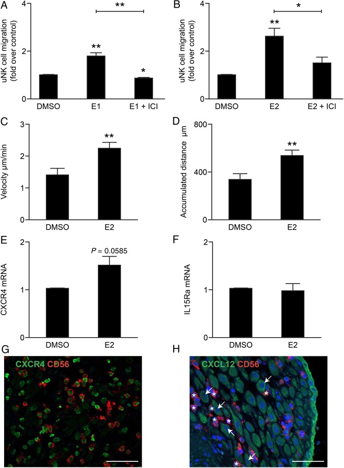
Estrogens promote uNK cell migration and chemokinesis. Uterine natural killer (uNK) cells were treated in duplicate with estrone (E1) or estradiol (E2) alone or in combination with the anti-estrogen I...
Figureu00a02
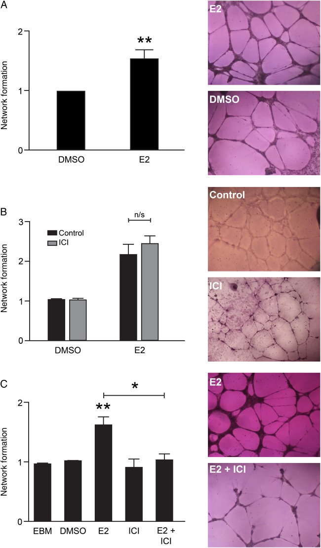
Estradiol promotes uNK-mediated network formation in endometrial endothelial cells via uNK estrogen receptor. Human endometrial endothelial cell (HEEC) network formation was assessed in response to uN...
Figureu00a03
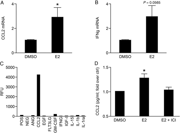
E2-dependent regulation of angiogenic factors in uNK cells. The top two targets identified by PCR array; chemokine (C-C motif) ligand 2 (CCL2) and interferon gamma (IFNu03b3), were selected for follow...
Figureu00a04
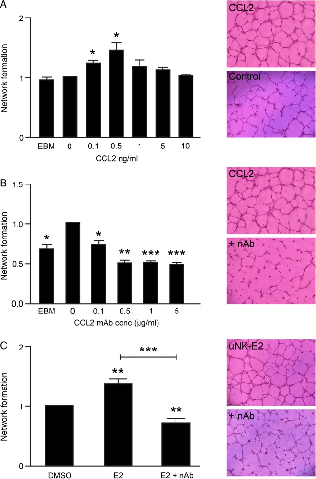
E2-dependent secretion of CCL2 regulates uNK-mediated angiogenesis. HEEC network formation was assessed in response to recombinant human CCL2 (rCCL2). ( A ) Network formation was significantly increas...
Figureu00a05
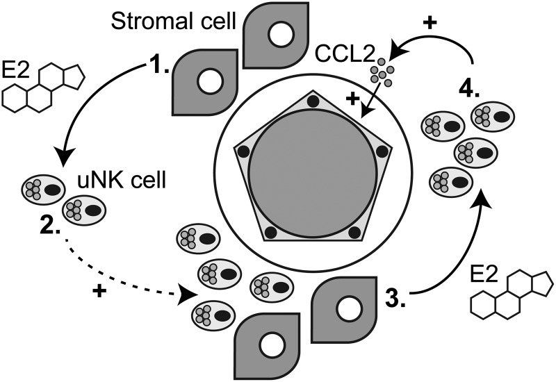
Summary: Regulation of endometrial function in early pregnancy. Stromal cells and uNK cells interact with endometrial arterioles to promote vascular remodelling. 1 . In the decidua, stromal cells secr...
Figure images are served from the NIH/NLM PubMed Central Open Access Subset or Europe PMC; copyright remains with the publishers and authors.
💬 Discussion
0 commentsNo comments yet. Be the first to start a discussion!
Leave a Comment