Abstract
To account for the many functions of phosphatidylinositol 4,5-bisphosphate (PIP(2)), several investigators have proposed that there are separate pools of PIP(2) in the plasma membrane. Recent experiments show the surface concentration of PIP(2) is indeed enhanced in regions where phagocytosis, exocytosis, and cell division occurs. Kinases that produce PIP(2) are also concentrated in these regions. However, how is the PIP(2) produced by these kinases prevented from diffusing rapidly away? First, proteins could act as "fences" around the perimeter of these regions. Second, some factor could markedly decrease the diffusion coefficient, D, of PIP(2) within these regions. We used fluorescence correlation spectroscopy (FCS) and fluorescence recovery after photobleaching (FRAP) to investigate these two possibilities in the forming phagosomes of macrophages injected with fluorescent PIP(2). FCS measurements show that PIP(2) diffuses rapidly (D ~ 1 μm(2)/s) in both the forming phagosomes and unengaged plasma membrane. FRAP measurements show that the fluorescence from PIP(2) does not recover (>100 s) after photobleaching the entire forming phagosome but recovers rapidly (~10 s) in a comparable area of membrane outside the cup. These results (and similar data for a plasma membrane-anchored green fluorescent protein) support the hypothesis that a fence impedes the diffusion of PIP(2) into and out of forming phagosomes.
🔬 Techniques
🔭 Microscopes
✨ Fluorophores
🧪 Sample Preparation
🔬 Cell Lines
🏭 Microscope Brands
🧪 Reagent Suppliers
📷 Detectors
💻 Software Details
🏛️ Research Organizations (ROR)
Affiliated research institutions:
📋 Methods
Materials Bodipy-TMR-PIP 2 was purchased from Echelon Bioscience (Salt Lake City, UT). The chemical structure of this lipid is illustrated in Gambhir et al. (2004) . TopFluor-PI(4,5)P 2 was purchased from Avanti Polar Lipids (Alabaster, AL). DiIC 12 was purchased from Invitrogen (Carlsbad, CA). Arachidoyl Lyso-PC was purchased from Avanti Polar Lipids. Rhodamine B and human IgG were from Sigma-Aldrich (St. Louis, MO). The 8-μm latex beads were from Bangs Labs (Fishers, IN). Glass capillaries, thin walled with filament inner diameter 1.0 mm, were purchased from World Precision Instruments (Sarasota, FL). Needles were pulled on a Flaming Brown micropipette puller, model P.80/PC, Sutter Instrument (Novato, CA). The 35-mm glass-bottom dishes were purchased from MatTek Corporation (Ashland, MA).
Cell culture
J774A.1 macrophages were purchased from the American Type Culture Collection (ATCC; Manassas, VA). Cells were maintained at 37°C with 5% CO 2 in DMEM supplemented with 10% fetal bovine serum (FBS), 50 U/ml penicillin, 50 μg/ml streptomycin sulfate, and sodium pyruvate. J774A.1 cells used in the examination of PIP 2 and PIP 3 levels with GFP-tagged probes were maintained at 37°C with 5% CO 2 in DMEM with 1.5 g/l sodium bicarbonate from Wisent (St. Bruno, QC, Canada) supplemented with 10% FBS. RAW 264.7 cells used for the actin localization and frustrated phagocytosis experiments were from ATCC and were maintained at 37°C with 5% CO 2 in RPMI-1640 from Wisent supplemented with 5% FBS. RAW 264.7 cells were also used for the PM-GFP FCS and FRAP experiments. They were cultured at 37°C in DMEM (Life Technologies, Invitrogen, Carlsbad, CA) supplemented with 10% FBS (Life Technologies), 50 U/ml penicillin, 50 μg/ml streptomycin sulfate, l -glutamine, and 110 mg/ml sodium pyruvate. As recommended by ATCC, CO 2 was maintained at 10% due to the high sodium bicarbonate level of the Life Technologies media used. Sample preparation Bodipy-TMR-PIP 2 and TopFluor-PIP 2 were injected into J774a.1 cells in the form of arachidoyl Lyso-PC/fluorescent-PIP 2 micelles as described previously ( Golebiewska et al. , 2008 ). The composition of micelles was 82% arachidoyl Lyso-PC and 18% fluorescent-PIP 2 . Lipids were suspended in 1 mM EDTA solutions, warmed to 37–42°C, mixed, and diluted with 1 mM EDTA to a concentration of ∼500 μM, and then sonicated in a bath sonicator for 3–5 s to form the mixed micelles. We used mixed micelles rather than PIP 2 micelles because PIP 2 micelles aggregate in the presence of the 1 mM Ca 2+ present in the bathing solution, causing the pipette tips to clog. DiIC 12 was dissolved in ethanol and added to cells by mock microinjections (with the Pi and Pc set to 0). The final concentration of ethanol in the cell was
Show full methods section
Materials Bodipy-TMR-PIP 2 was purchased from Echelon Bioscience (Salt Lake City, UT). The chemical structure of this lipid is illustrated in Gambhir et al. (2004) . TopFluor-PI(4,5)P 2 was purchased from Avanti Polar Lipids (Alabaster, AL). DiIC 12 was purchased from Invitrogen (Carlsbad, CA). Arachidoyl Lyso-PC was purchased from Avanti Polar Lipids. Rhodamine B and human IgG were from Sigma-Aldrich (St. Louis, MO). The 8-μm latex beads were from Bangs Labs (Fishers, IN). Glass capillaries, thin walled with filament inner diameter 1.0 mm, were purchased from World Precision Instruments (Sarasota, FL). Needles were pulled on a Flaming Brown micropipette puller, model P.80/PC, Sutter Instrument (Novato, CA). The 35-mm glass-bottom dishes were purchased from MatTek Corporation (Ashland, MA).
Cell culture
J774A.1 macrophages were purchased from the American Type Culture Collection (ATCC; Manassas, VA). Cells were maintained at 37°C with 5% CO 2 in DMEM supplemented with 10% fetal bovine serum (FBS), 50 U/ml penicillin, 50 μg/ml streptomycin sulfate, and sodium pyruvate. J774A.1 cells used in the examination of PIP 2 and PIP 3 levels with GFP-tagged probes were maintained at 37°C with 5% CO 2 in DMEM with 1.5 g/l sodium bicarbonate from Wisent (St. Bruno, QC, Canada) supplemented with 10% FBS. RAW 264.7 cells used for the actin localization and frustrated phagocytosis experiments were from ATCC and were maintained at 37°C with 5% CO 2 in RPMI-1640 from Wisent supplemented with 5% FBS. RAW 264.7 cells were also used for the PM-GFP FCS and FRAP experiments. They were cultured at 37°C in DMEM (Life Technologies, Invitrogen, Carlsbad, CA) supplemented with 10% FBS (Life Technologies), 50 U/ml penicillin, 50 μg/ml streptomycin sulfate, l -glutamine, and 110 mg/ml sodium pyruvate. As recommended by ATCC, CO 2 was maintained at 10% due to the high sodium bicarbonate level of the Life Technologies media used. Sample preparation Bodipy-TMR-PIP 2 and TopFluor-PIP 2 were injected into J774a.1 cells in the form of arachidoyl Lyso-PC/fluorescent-PIP 2 micelles as described previously ( Golebiewska et al. , 2008 ). The composition of micelles was 82% arachidoyl Lyso-PC and 18% fluorescent-PIP 2 . Lipids were suspended in 1 mM EDTA solutions, warmed to 37–42°C, mixed, and diluted with 1 mM EDTA to a concentration of ∼500 μM, and then sonicated in a bath sonicator for 3–5 s to form the mixed micelles. We used mixed micelles rather than PIP 2 micelles because PIP 2 micelles aggregate in the presence of the 1 mM Ca 2+ present in the bathing solution, causing the pipette tips to clog. DiIC 12 was dissolved in ethanol and added to cells by mock microinjections (with the Pi and Pc set to 0). The final concentration of ethanol in the cell was
📊 Figures
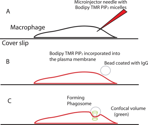
FIGURE 1:
Methods used to study the diffusion of fluorescent PIP 2 in the forming phagosomes of macrophages. (A) Cartoon showing a J774a.1 macrophage and adjacent microinjector needle loaded with micelles conta...
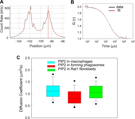
FIGURE 2:
FCS measurements of PIP 2 diffusion in the phagosomal cups of macrophages. (A) Fluorescence intensity scan in the z-direction through the center of the phagosomal cup region of a J774a.1 macrophage. T...
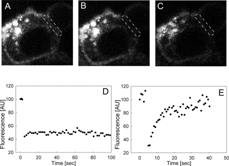
FIGURE 3:
FRAP measurements on fluorescent PIP 2 are consistent with a fence around the forming phagosome. FRAP measurements of TopFluor-PIP 2 in the phagosomal cup of a J774 cell. (A) Cell injected with TopFlu...
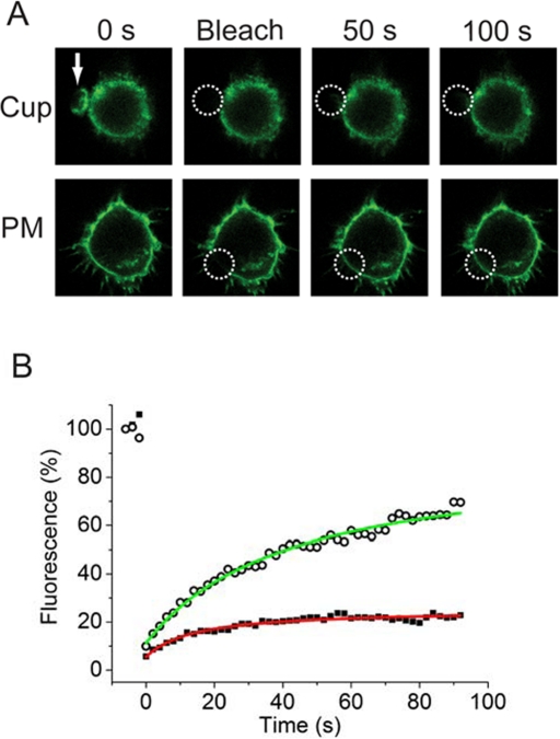
FIGURE 4:
Fluorescence due to PM-GFP within a phagocytic cup does not recover after photobleaching. RAW 264.7 macrophages transfected with a GFP construct that is targeted to the plasma membrane (PM-GFP) were a...
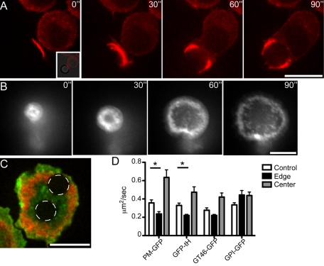
FIGURE 5:
Diffusion of proteins in the actin-rich region at the perimeter of the forming phagosome. (A) RAW macrophages with stable expression of mCherry-actin were allowed to phagocytose 8-u03bcm-diameter late...
Figure images are served from the NIH/NLM PubMed Central Open Access Subset or Europe PMC; copyright remains with the publishers and authors.
💬 Discussion
0 commentsNo comments yet. Be the first to start a discussion!
Leave a Comment