Abstract
Abstract Here we demonstrate that primary cultures of human fetal liver cells (HFLC) reliably support infection with laboratory strains of hepatitis C virus (HCV), although levels of virus replication vary significantly between different donor cell preparations and frequently decline in a manner suggestive of active viral clearance. To investigate possible contributions of the interferon (IFN) system to control HCV infection in HFLC, we exploited the well-characterized ability of paramyxovirus (PMV) V proteins to counteract both IFN induction and antiviral signaling. The V proteins of measles virus (MV) and parainfluenza virus 5 (PIV5) were introduced into HFLC using lentiviral vectors encoding a fluorescent reporter for visualization of HCV-infected cells. V protein-transduced HFLC supported enhanced (10 to 100-fold) levels of HCV infection relative to untransduced or control vector-transduced HFLC. Infection was assessed by measurement of virus-driven luciferase, by assays for infectious HCV and viral RNA, and by direct visualization of HCV-infected hepatocytes. Live cell imaging between 48 and 119 hours postinfection demonstrated little or no spread of infection in the absence of PMV V protein expression. In contrast, V protein-transduced HFLC showed numerous HCV infection events. V protein expression efficiently antagonized the HCV-inhibitory effects of added IFNs in HFLC. In addition, induction of the type III IFN, IL29, following acute HCV infection was inhibited in V protein-transduced cultures. Conclusion: These studies suggest that the cellular IFN response plays a significant role in limiting the spread of HCV infection in primary hepatocyte cultures. Strategies aimed at dampening this response may be key to further development of robust HCV culture systems, enabling studies of virus pathogenicity and the mechanisms by which HCV spreads in its natural host cell population. (Hepatology 2011;)
🔬 Techniques
🔭 Microscopes
✨ Fluorophores
🧪 Sample Preparation
🔬 Cell Lines
🏭 Microscope Brands
🧪 Reagent Suppliers
🏛️ Research Organizations (ROR)
Affiliated research institutions:
📋 Methods
Patients and Methods Human Subjects
All protocols involving human tissue were reviewed and exempted by the Rockefeller University Institutional Review Board.
Isolation and Culture of HFLC
Deidentified fetal livers (16-24 weeks gestation) were procured through Advanced Bioscience Resources (ABR; Alameda, CA) or the Human Fetal Tissue Repository of the Albert Einstein College of Medicine (AECOM; New York, NY). Livers received on ice were washed with hepatocyte wash buffer (HWB) consisting of Williams' E Medium (WEM) plus 10 mM HEPES, 50 μg/mL gentamicin, 100 U/mL penicillin, and 100 μg/mL streptomycin (Invitrogen). Tissue was minced then resuspended in 20-40 mL warm digestion buffer consisting of Hanks Balanced Salt Solution plus 40 mM HEPES, 3.26 mM CaCl 2 , 2 U/mL DNase I Grade II (Roche), and 0.2% Collagenase type IV (Sigma). Tissue was digested for 30 minutes at 37°C, then diluted 1:1 with HWB and gently pushed through 70 μm cell-strainers (BD Biosciences). The suspension was centrifuged at 100 g for 3 minutes and the cell pellet containing large hepatocytes was washed twice by resuspension in 50 mL HWB and centrifugation at 100 g for 4 minutes. Hepatocytes were enriched by 1 g sedimentation in 25 mL HWB for 1 hour at room temperature, followed by additional washing. In some experiments hepatocytes were further enriched by centrifugation through lymphocyte separation medium (Cellgro, Manassas, VA) as described. 33 Hepatocyte yields ranged from 0.5 to 4 × 10 7 cells per tissue and cells were generally >80% viable as assessed by Trypan blue exclusion and collagen attachment. Hepatocytes were plated at ≍1 × 10 5 /cm 2 on 24- or 48-well collagen I-coated plates (BD Biosciences) in WEM containing 10% fetal bovine serum (FBS) (Omega Scientific, Tarzana, CA), 2 mM L-glutamine (Invitrogen), 1X ITS Plus (BD Biosciences) and antibiotics. After overnight incubation, adherent cells were washed with WEM, then maintained in Hepatocyte Defined Medium (HDM; BD Biosciences) plus L-glutamine and antibiotics. The culture medium was aspirated and replaced every 2 days.
Show full methods section
Patients and Methods Human Subjects
All protocols involving human tissue were reviewed and exempted by the Rockefeller University Institutional Review Board.
Isolation and Culture of HFLC
Deidentified fetal livers (16-24 weeks gestation) were procured through Advanced Bioscience Resources (ABR; Alameda, CA) or the Human Fetal Tissue Repository of the Albert Einstein College of Medicine (AECOM; New York, NY). Livers received on ice were washed with hepatocyte wash buffer (HWB) consisting of Williams' E Medium (WEM) plus 10 mM HEPES, 50 μg/mL gentamicin, 100 U/mL penicillin, and 100 μg/mL streptomycin (Invitrogen). Tissue was minced then resuspended in 20-40 mL warm digestion buffer consisting of Hanks Balanced Salt Solution plus 40 mM HEPES, 3.26 mM CaCl 2 , 2 U/mL DNase I Grade II (Roche), and 0.2% Collagenase type IV (Sigma). Tissue was digested for 30 minutes at 37°C, then diluted 1:1 with HWB and gently pushed through 70 μm cell-strainers (BD Biosciences). The suspension was centrifuged at 100 g for 3 minutes and the cell pellet containing large hepatocytes was washed twice by resuspension in 50 mL HWB and centrifugation at 100 g for 4 minutes. Hepatocytes were enriched by 1 g sedimentation in 25 mL HWB for 1 hour at room temperature, followed by additional washing. In some experiments hepatocytes were further enriched by centrifugation through lymphocyte separation medium (Cellgro, Manassas, VA) as described. 33 Hepatocyte yields ranged from 0.5 to 4 × 10 7 cells per tissue and cells were generally >80% viable as assessed by Trypan blue exclusion and collagen attachment. Hepatocytes were plated at ≍1 × 10 5 /cm 2 on 24- or 48-well collagen I-coated plates (BD Biosciences) in WEM containing 10% fetal bovine serum (FBS) (Omega Scientific, Tarzana, CA), 2 mM L-glutamine (Invitrogen), 1X ITS Plus (BD Biosciences) and antibiotics. After overnight incubation, adherent cells were washed with WEM, then maintained in Hepatocyte Defined Medium (HDM; BD Biosciences) plus L-glutamine and antibiotics. The culture medium was aspirated and replaced every 2 days.
Lentiviral Vectors V proteins and control protein firefly luciferase
(Fluc) were expressed from a hybrid albumin promoter in a bi-cistronic lentiviral vector 34 modified to express the HCV-dependent fluorescence relocalization (HDFR) cassette TagRFP-NLS-IPS. 32 In this cassette the fluorescent reporter TagRFP is fused to both a nuclear localization sequence (NLS) and the transmembrane domain of the mitochondrially tethered adapter protein IPS-1. 32 Following HCV infection of HDFR-expressing cells, cleavage of IPS-1 by the viral NS3-4A protease 2 leads to migration of the fluorescent reporter from mitochondria to the nucleus, enabling visualization of HCV-infected cells. 32 Vector construction and source of protein-coding sequences are detailed in the Supporting Information and Supporting Fig. S1A . Transduction with Lentiviral Pseudoparticles (PP) PP were prepared by cotransfection of 293T cells with lentiviral and packaging plasmids as described. 15 , 32 HFLC were transduced 1-3 days postplating by incubation for 3-6 hours with PP stocks diluted 1:3 in HDM plus 20 mM HEPES and 4 μg/mL polybrene, then washed and fed with HDM. HCVcc Inocula HCVcc inocula used were the Gaussia luciferase reporter virus Jc1FLAG2 (p7-nsGluc2A) 35 (JC1G) and J6JFH Clone 2, 36 which lacks a reporter but replicates to higher titer in Huh-7.5 cells. Virus stocks were prepared by electroporation of in vitro transcribed RNA into Huh-7.5 cells as described 35 and virus was collected in serum-free medium, or medium containing 1.5 or 10% FBS. For some experiments virus stocks were concentrated using Amicon Ultracel-100K filters (Millipore). Infectious titers of HCVcc inocula were determined by titration on Huh-7.5 cells as described 12 and are expressed as 50% tissue culture infectious doses (TCID 50 ). HFLC were infected for 3 to 6 hours with HCVcc diluted in HDM, then washed and fed with HDM.
Detection of HCV RNA Total
RNA was extracted from washed HFLC using RNEasy Kits (Qiagen). HCV RNA was detected by quantitative RT-PCR using the Eragen MultiCode-RTx method (Eragen Biosciences, Madison, WI) and primers directed to the 5′ untranslated region of the HCV genome. Quantitation of HCV RNA copy number was achieved using a synthetic RNA standard (Apath, Brooklyn, NY). Cytokines and Drugs Recombinant human cytokines, IFN-beta (IFN-β), IL-28A, and IL-29, were from Peprotech (Rocky Hill, NJ). The HCV NS5B polymerase inhibitor 2′ C-methyl adenosine (2′CMA) was the gift of D. Olsen and S. Carroll (Merck Research Laboratories, West Point, PA). Immunofluorescence Analysis (IFA), Immunoblotting, and Enzyme-Linked Immunosorbent Assay (ELISA) IFA 9 and immunoblotting 14 were carried out as described. Antibodies and reagents are detailed in the Supporting Information. Human IL-29 was detected using ELISA kits from eBioscience (San Diego, CA). ELISA for human albumin is described in the Supporting Information.
Time-Lapse Live Cell Imaging
HFLC were plated on collagen-coated optical dishes and maintained in HDM. Images were acquired on a Zeiss Axiovert-200 inverted microscope equipped with an environmental chamber.
Visualization of Tag-RFP in transduced
HFLC was achieved by laser excitation and emission as described. 32
Supplementary material Additional Supporting Information may be found in the online version of this article. Supporting Video 1: Firefly luciferase (Fluc) Supporting Video 2: Parainfluenza virus 5 V protein (PIV5 V) or (C). Supporting Video 3: Measles virus V protein (MV V).
📊 Figures
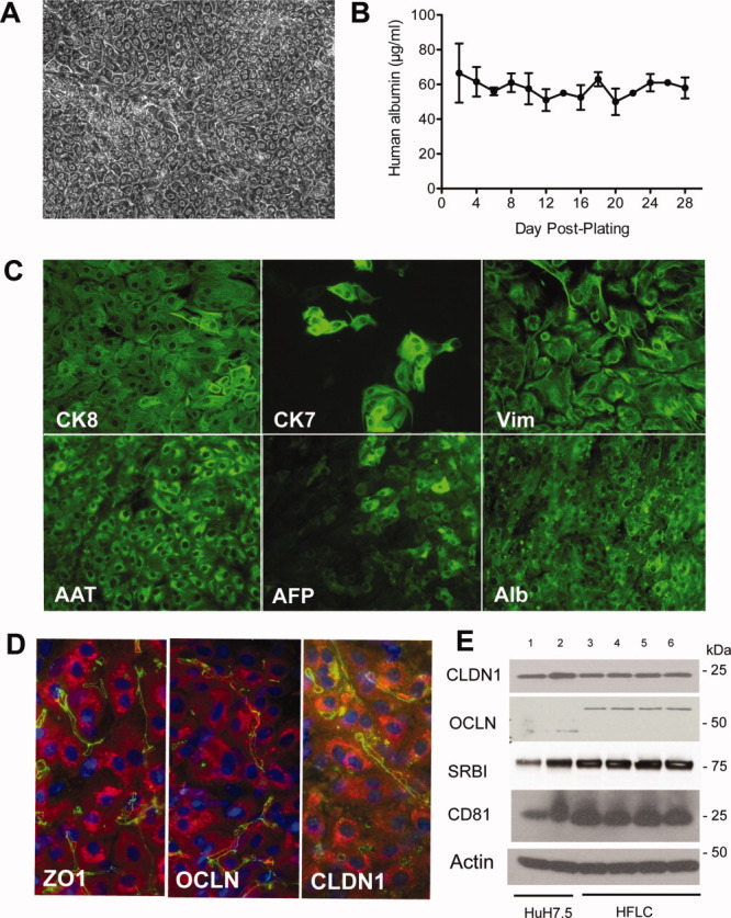
Fig. 1
Cultured HFLC are long-lived, express markers of differentiated hepatocytes, and are positive for the expression of HCV entry factors. (A) Phase contrast microscopy of HFLC 2 weeks postplating. (B) Al...
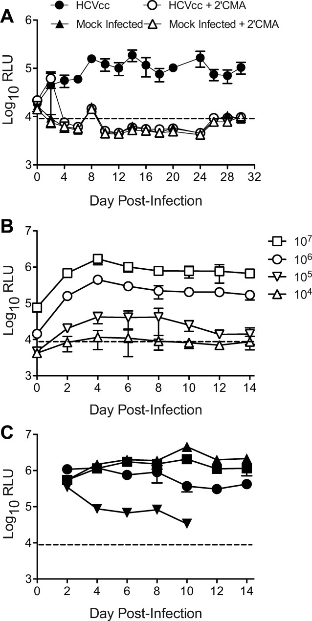
Fig. 2
Cultured HFLC are susceptible to infection with HCVcc. (A) Gaussia luciferase secretion by HFLC (ABR-15168, 23 weeks gestation) following infection with the reporter virus JC1G in the presence or abse...
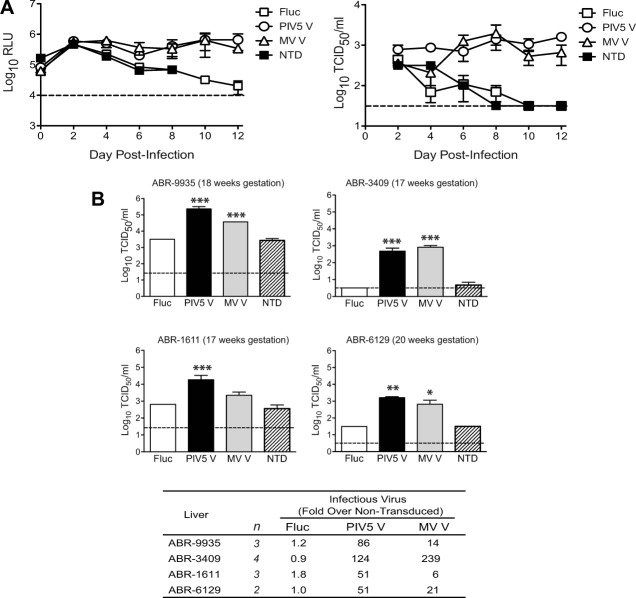
Fig. 3
PMV V protein expression enhances productive infection with JC1G in cultured HFLC. (A) One day postplating, HFLC (ABR-5591, 17 weeks gestation) were transduced with lentiviral vectors encoding firefly...
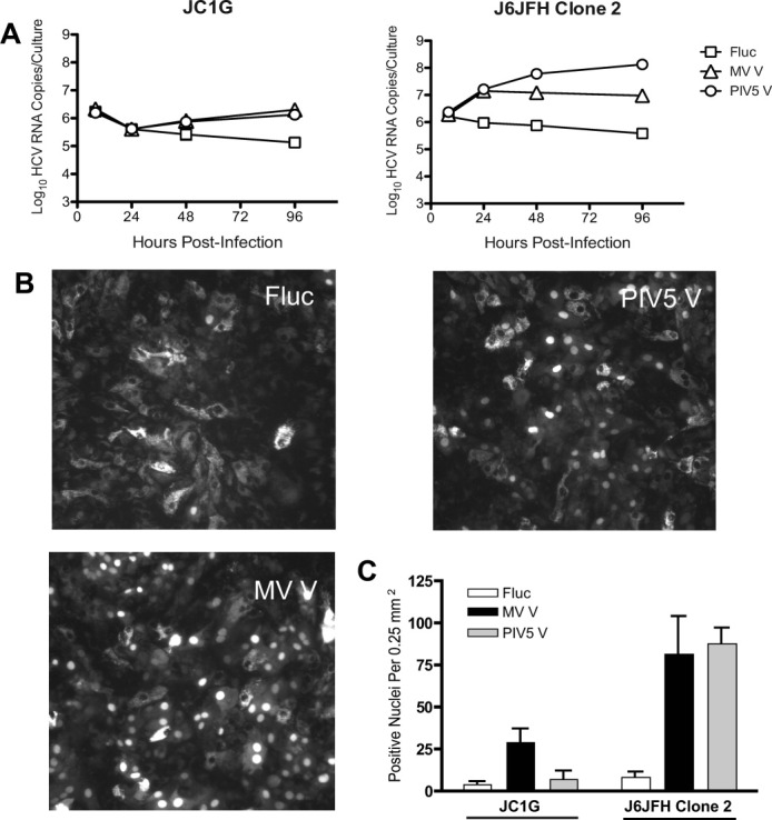
Fig. 4
J6JFH Clone 2 replicates to higher levels in HFLC than JC1G, and replication is enhanced by V protein expression. Four days posttransduction with lentiviral vectors, HFLC (AECOM-063010; gestational ag...
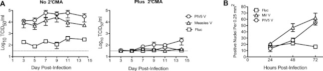
Fig. 5
Kinetics of J6JFH Clone 2 replication in cultured HFLC. (A) Five days posttransduction, HFLC (AECOM-031010, gestational age unavailable) were incubated with 1 u00d7 10 7 TCID5 0 J6JFH Clone 2, washed ...
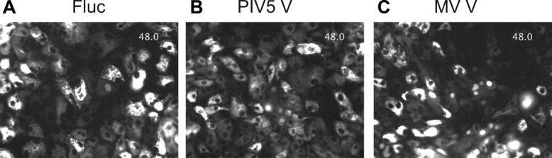
Fig. 6
Live-cell imaging of J6JFH Clone 2 infection in PMV V protein- or Fluc-transduced HFLC. Five days posttransduction with lentiviral vectors, HFLC (ABR-3676; 19 weeks gestation) were infected with 1 u00...
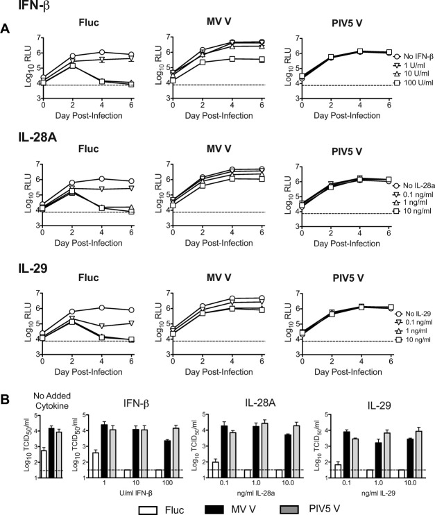
Fig. 7
V protein expression counteracts the HCV-inhibitory effects of added type I and type III interferons in HFLC. Four days posttransduction with lentiviral vectors, HFLC (ABR-1108; 19 weeks gestation) we...
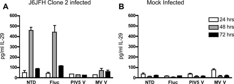
Fig. 8
V protein expression inhibits IL-29 protein induction following HCV infection. Four days posttransduction with lentiviral vectors, cultures of gradient-enriched HFLC (ABR-8338; 20 weeks gestation) wer...
Figure images are served from the NIH/NLM PubMed Central Open Access Subset or Europe PMC; copyright remains with the publishers and authors.
💬 Discussion
0 commentsNo comments yet. Be the first to start a discussion!
Leave a Comment