Abstract
AbstractSensing bacterial products in the cytosol of mammalian cells by NOD-like receptors leads to the activation of caspase-1 inflammasomes, and the production of the pro-inflammatory cytokines interleukin (IL)-18 and IL-1β. In addition, mouse caspase-11 (represented in humans by its orthologs, caspase-4 and caspase-5) detects cytosolic bacterial LPS directly. Activation of caspase-1 and caspase-11 initiates pyroptotic host cell death that releases potentially harmful bacteria from the nutrient-rich host cell cytosol into the extracellular environment. Here we use single cell analysis and time-lapse microscopy to identify a subpopulation of host cells, in which growth of cytosolic Salmonella Typhimurium is inhibited independently or prior to the onset of cell death. The enzymatic activities of caspase-1 and caspase-11 are required for growth inhibition in different cell types. Our results reveal that these proteases have important functions beyond the direct induction of pyroptosis and proinflammatory cytokine secretion in the control of growth and elimination of cytosolic bacteria.
🔬 Techniques
🔭 Microscopes
✨ Fluorophores
🧪 Sample Preparation
🔬 Cell Lines
🏭 Microscope Brands
🧪 Reagent Suppliers
🏛️ Research Organizations (ROR)
Affiliated research institutions:
📋 Methods
Antibodies and reagents
Antibodies for immunoblotting were from Sigma (actin, AC-74 used at 1:5,000 dilution and caspase-11, 17D9 used at 1:1,000 dilution), Cell Signaling (caspase-7, 9492 used at 1:1,000 dilution), Adipogen (caspase-1 p20, AG-20B-0042 used at 1:1,000 dilution), DSHB (tubulin, E7 used at 1:5,000 dilution) and Santa Cruz Biotechnology (ASC sc-22514-R used at 1:1,000 dilution). Propidium iodide was from Life Technologies and Lipofectamine2000 from Invitrogen. siRNAs for caspase-7 and caspase-11 were purchased from Santa Cruz and used at 40 pmol. zVAD-FMK and YVAD-FMK were from R&D systems.
Bacterial infections Salmonella enterica serovar
Typhimurium (strain 12023) was grown overnight in LB. GFP-expressing Salmonella carry plasmid pFPV25.1, mCherry-expressing Salmonella carry plasmid pDiGc (ref. 61 ). prgH mutant Salmonella carry the plasmid pRI203, expressing Yersinia InvA (ref. 62 ). Bacteria (20 μl) were opsonized with 20 μl mouse serum (Sigma) in 170 μl DMEM for 20 min before addition of 600 μl DMEM. Macrophages (in 500 μl media in 24 well plates) were infected with 40 μl of opsonized bacteria (MOI 5–10), centrifuged at 110 g for 5 min and incubated for 25 min at 37 °C. Following two washes with PBS, cells were incubated with 100 μg ml −1 gentamicin for 2 h and then 20 μg ml −1 , or directly incubated with 20 μg ml −1 gentamicin. For SPI-1 T3SS-mediated invasion of 3T3 fibroblasts or MEFs, stationary phase bacterial cultures were sub-cultured (1:33) in fresh LB and grown for 3.5 h at 37 °C before inoculation. Cells in 24 well plates (500 μl media/well) were infected with 7 μl of sub-cultured bacteria for 7 min. After two PBS washes cells were incubated with 100 μg ml −1 gentamicin for 2 h and 20 μg ml −1 gentamicin thereafter. Cell culture C57BL/6 WT, Asc −/− , Casp11 −/− , Casp1/11 −/− , Casp7 −/− , IL-18 −/− , IL-1r −/− and Nlrc4 −/− BMDM were infected with the v-myc/v-raf expressing J2 retrovirus 63 , and differentiated in 20% L929-MCSF supernatant. Cells were then maintained in Dulbecco's modified Eagle medium (DMEM, Sigma), 10% fetal calf serum (FCS), 20% L929-MCSF and 1 mM sodium pyruvate at 37 °C, 5% CO 2 . 3T3 fibroblasts, MEFs, 293ETs and RAW 264.7 macrophages (ATCC) were cultured in DMEM containing 10% FCS. C57BL/6 control and Gsdmd −/− iBMDMs were maintained as described above. Cell lines, tested for mycoplasma, were chosen for ease of Salmonella infection, enabling analysis after both invasive and non-invasive uptake. Primary BMDMs were differentiated in 20% L929-MCSF supernatant for 1 week after isolation. For assays investigating the effect of caspase inhibitors, cells were incubated in DMEM with 10% FCS supplemented with 20 μM zVAD-FMK, 20 μM YVAD-FMK or 20 μM of other peptide inhibitors ( Supplementary Fig. 1D ) or DMSO (1:1000) as vehicle control, for 1 h before infection. When indicated KCl was added at 50 mM, 1 h before infection. Constructs and retroviral transductions Plasmids encoding GFP-tagged galectin-8 and LC3B were kind gifts from Dr Felix Randow and have previously been described 36 . Genes encoding murine caspase-1 or caspase-11 were ligated into a replication-defective retroviral plasmid (m6p) (ref. 64 ). Site directed mutagenesis was used to introduce mutations, which were verified by sequencing. Caspase-1 6D-N comprises 6 Asp to Asn mutations, preventing self-cleavage, while maintaining catalytic activity 42 . For transduction, retroviral particles were packaged into vesicular stomatitis virus pseudotyped virus after co-transfection of 293ET cells. After 48 h, cells were selected in puromycin (2.5 μg ml −1 ) or blasticidin (5 μg ml −1 ) so that all cells within a population expressed the transgene. Where GFP fusions were used, cells were sorted by Fluorescence-Activated Cell Sorting to obtain a 100% GFP-positive population.
Show full methods section
Antibodies and reagents
Antibodies for immunoblotting were from Sigma (actin, AC-74 used at 1:5,000 dilution and caspase-11, 17D9 used at 1:1,000 dilution), Cell Signaling (caspase-7, 9492 used at 1:1,000 dilution), Adipogen (caspase-1 p20, AG-20B-0042 used at 1:1,000 dilution), DSHB (tubulin, E7 used at 1:5,000 dilution) and Santa Cruz Biotechnology (ASC sc-22514-R used at 1:1,000 dilution). Propidium iodide was from Life Technologies and Lipofectamine2000 from Invitrogen. siRNAs for caspase-7 and caspase-11 were purchased from Santa Cruz and used at 40 pmol. zVAD-FMK and YVAD-FMK were from R&D systems.
Bacterial infections Salmonella enterica serovar
Typhimurium (strain 12023) was grown overnight in LB. GFP-expressing Salmonella carry plasmid pFPV25.1, mCherry-expressing Salmonella carry plasmid pDiGc (ref. 61 ). prgH mutant Salmonella carry the plasmid pRI203, expressing Yersinia InvA (ref. 62 ). Bacteria (20 μl) were opsonized with 20 μl mouse serum (Sigma) in 170 μl DMEM for 20 min before addition of 600 μl DMEM. Macrophages (in 500 μl media in 24 well plates) were infected with 40 μl of opsonized bacteria (MOI 5–10), centrifuged at 110 g for 5 min and incubated for 25 min at 37 °C. Following two washes with PBS, cells were incubated with 100 μg ml −1 gentamicin for 2 h and then 20 μg ml −1 , or directly incubated with 20 μg ml −1 gentamicin. For SPI-1 T3SS-mediated invasion of 3T3 fibroblasts or MEFs, stationary phase bacterial cultures were sub-cultured (1:33) in fresh LB and grown for 3.5 h at 37 °C before inoculation. Cells in 24 well plates (500 μl media/well) were infected with 7 μl of sub-cultured bacteria for 7 min. After two PBS washes cells were incubated with 100 μg ml −1 gentamicin for 2 h and 20 μg ml −1 gentamicin thereafter. Cell culture C57BL/6 WT, Asc −/− , Casp11 −/− , Casp1/11 −/− , Casp7 −/− , IL-18 −/− , IL-1r −/− and Nlrc4 −/− BMDM were infected with the v-myc/v-raf expressing J2 retrovirus 63 , and differentiated in 20% L929-MCSF supernatant. Cells were then maintained in Dulbecco's modified Eagle medium (DMEM, Sigma), 10% fetal calf serum (FCS), 20% L929-MCSF and 1 mM sodium pyruvate at 37 °C, 5% CO 2 . 3T3 fibroblasts, MEFs, 293ETs and RAW 264.7 macrophages (ATCC) were cultured in DMEM containing 10% FCS. C57BL/6 control and Gsdmd −/− iBMDMs were maintained as described above. Cell lines, tested for mycoplasma, were chosen for ease of Salmonella infection, enabling analysis after both invasive and non-invasive uptake. Primary BMDMs were differentiated in 20% L929-MCSF supernatant for 1 week after isolation. For assays investigating the effect of caspase inhibitors, cells were incubated in DMEM with 10% FCS supplemented with 20 μM zVAD-FMK, 20 μM YVAD-FMK or 20 μM of other peptide inhibitors ( Supplementary Fig. 1D ) or DMSO (1:1000) as vehicle control, for 1 h before infection. When indicated KCl was added at 50 mM, 1 h before infection. Constructs and retroviral transductions Plasmids encoding GFP-tagged galectin-8 and LC3B were kind gifts from Dr Felix Randow and have previously been described 36 . Genes encoding murine caspase-1 or caspase-11 were ligated into a replication-defective retroviral plasmid (m6p) (ref. 64 ). Site directed mutagenesis was used to introduce mutations, which were verified by sequencing. Caspase-1 6D-N comprises 6 Asp to Asn mutations, preventing self-cleavage, while maintaining catalytic activity 42 . For transduction, retroviral particles were packaged into vesicular stomatitis virus pseudotyped virus after co-transfection of 293ET cells. After 48 h, cells were selected in puromycin (2.5 μg ml −1 ) or blasticidin (5 μg ml −1 ) so that all cells within a population expressed the transgene. Where GFP fusions were used, cells were sorted by Fluorescence-Activated Cell Sorting to obtain a 100% GFP-positive population.
Colony forming unit assay
To enumerate intracellular bacteria, cells from duplicate or triplicate wells of a 24 well plate, infected as above, were lysed in 1 ml of ice cold PBS containing 0.1% Triton X100 for 5 min. Serial dilutions were plated on duplicate LB agar and plates were incubated overnight at 37 °C. Colonies were counted using an Acolyte colony counter. Where CQ treatment was used (Sigma, 250 μM) it was added between 1.5 and 3 h (3T3 fibroblasts) or 2 and 4 h (WT infected iBMDMs) or 6 and 7 h ( ΔsifA Salmonella). For 3T3 fibroblasts the colony counts are represented as the fold growth in vacuolar bacteria (total—CQ resistant) and cytosolic bacteria (CQ resistant). For iBMDMs the fold growth in CQ-resistant bacteria (cytosolic) are shown. ELISA Concentrations of IL-1β in macrophage culture supernatants were measured using mouse IL-1β kits according to manufacturer's recommendations (Affymetrix ebioscience) after uptake of Salmonella Flow cytometry To measure the replication of GFP-expressing Salmonella in intact cells, cells were infected as above and harvested following trypsin treatment, washed and re-suspended in Optimem (Invitrogen) containing 1 μg ml −1 Propidium Iodide (PI). Data, consisting of at least 10,000 events, were acquired on a FACs Calibur and analysed using FlowJo 8.8.6. Data are represented as the fold-change (from 1 or 2 h p.u.) in geometric mean of cells harbouring GFP-expressing bacteria.
Immunoblotting
Proteins in post nuclear supernatants from 1 × 10 6 cells were separated on either 10% or 12% Tris polyacrylamide gels. Proteins were transferred to Nitrocellulose membranes, which were then blocked in 5% milk in TBST (100 mM Tris Cl pH 7.4, 150 mM NaCl, 0.1% Tween20). Membranes were incubated overnight at 4 °C with primary antibodies, washed three times with TBST and then incubated for 2 h with secondary antibodies at room temperature. Visualization was done using ECL+detection regents (GE Healthcare). Uncropped blots are shown in Supplementary Fig. 6 .
LDH cytotoxicity assay
Host cell death was measured as a percentage of total LDH release, according to the recommended protocol (Promega). Medium was used as a blank control to obtain background measurements and supernatants from non-infected samples were subtracted from infected conditions. Total LDH release was measured after cell lysis at −80 °C.
Mice
For primary BMDMs, Caspase-1/11 double knockout mice were from the Swiss Immunological Mouse Repository (SwImMR) and caspase-7 −/− mice were purchased from Jackson Laboratories. Casp11 −/− primary BMDMs had been isolated from previously described mice 65 . C57BL/6 control mice were from Charles Rivers. All animals were bred in accordance with accredited animal facility regulations at Imperial College London. Imperial College Animal Welfare and Ethical Review Body (AWERB) granted approval for all mouse work. iBMDMs, that have been previously described were prepared from Nlrc4 −/− , Asc −/− (ref. 45 ), Casp1/11 −/− (ref. 66 ) and Casp11 −/− (ref. 65 ) mice. For mouse infections, mice (8–10 week old, C57BL/6 or Casp1/11 −/− ) were inoculated intraperitoneal with 1 × 10 5 CFUs with GFP expressing WT or ΔsifA Salmonella. After 48 h, spleens were collected, homogenized and splenic CD11b(+) cells enriched using magnetic beads according to the manufacturer instructions (Miltenyi Biotec). Purified cells were analysed by flow cytometry in Optimem containing PI. Mice showing very poor infection of the spleen were excluded. Randomization and blinding were not used.
Microscopy and digitonin assays
Cells were seeded on glass cover slips one-day prior to infection and fixed in 4% paraformaldehyde for 20 min. Confocal images were taken on a Zeiss 710 microscope with a × 100 objective. For digitonin-mediated permeabilisation of the plasma membrane, live cells were treated with 40 μg ml −1 digitonin for 5 min on ice prior to immunolabelling with anti-CSA1 (1:400, Kirkegaard and Perry Laboratories), anti-GM130 (1:500, BD Transduction laboratories) and anti-PDI (Protein disulfide-isomerase, 1:100, Enzo) for 30 min on ice. Cells were then washed twice in PBS and fixed in 4% paraformaldehyde. After permeabilisation in PBS, 0.1% Triton X100 and 10% horse serum, cover slips were incubated with appropriate AlexaFluor secondary antibodies (Invitrogen) and DAPI (4′,6-Diamidino-2-Phenylindole, Dihydrochloride) for 30 min before mounting onto glass slides. PI uptake PI uptake was used to determine plasma membrane integrity. Macrophages (3 × 10 5 cells per ml) were seeded in white clear-bottomed 96-well plates (Greiner) and infected with opsonised late stationary phase Salmonella (MOI 10:1) for 30 min at 37 °C. Following infection, cells were washed twice with PBS and 200 μl Optimem medium containing 10% FCS, 20 μg ml −1 gentamicin and 1 μg ml −1 PI was added. Triton X-100 (0.1%) was included in Optimem medium in wells used for positive controls. Optimem medium without PI was added to negative control wells. Plates were incubated at 37 °C in 5% CO 2 within a Tecan Infinite M200PRO fluorescent plate reader throughout infection, with PI fluorescence measured every 15 min. Non-infected controls were subtracted from infected samples and then divided by the fluorescence of wells treated with Triton-X100 to give the relative PI uptake. Quantitative reverse transcriptase (RT)-PCR Total RNA was isolated from 1 × 10 6 cells (Qiagen RNAeasy mini kit) and 400 ng was used to synthesize complementary DNA (cDNA) according to manufactures recommendations (QuantiTect RT kit, Qiagen). cDNA (0.5 μl) was used in quantitative RT-PCRs (SybrGreen PCR master mix, Applied biosystems) containing 0.2 μM gene-specific primers. After determining the cycle threshold (Ct) required to reach a significant emission of Sybr Green reporter dye (Rotor-Gene 3000, Corbett Research), relative mRNA was calculated from a titration curve of cDNA. Data represent the relative amounts of mRNA, normalized to rps9 house keeping gene. The following primers were used: Caspase-1 For 5′- ACTGGGACCCTCAAGTTTTG, Rev 5′- CATCTCCAGAGCTGTGAG Caspase-7 For 5′-TGGAAAAGGTGGATTCTTCC, Rev 5′-CTTTGTCGAAGTTCTTGTTG Caspase-11 For 5′-AAACACCCTGACAAACCACT, Rev 5′-TTCCTCCATTTCCAGATTAG Rps9 For 5′- CTGGACGAGGGCAAGATGAAGC-3′ Rev 5′- TGACGTTGGCGGATGAGCACA-3′ Time-lapse microscopy Cells seeded in dishes (Matek) with an embedded glass cover slip were infected as above. Prior to imaging, medium was replaced with Optimem (Invitrogen) containing 10% FCS, 40 μM Hepes (Sigma), 20 μg ml −1 gentamicin and 1 μg ml −1 PI. Cells were maintained at 37 °C in a heated chamber and images were acquired at 20-min intervals using the × 40 objective on a Zeiss 710 confocal microscope. At least 100 infected cells were scored per experiment.
Statistics Either an one-way
ANOVA with Dunnett's multiple comparisons test or one and two-tailed unpaired equal variance Students t -test were used for statistical comparison from three or more independent experiments as indicated. * P
📊 Figures
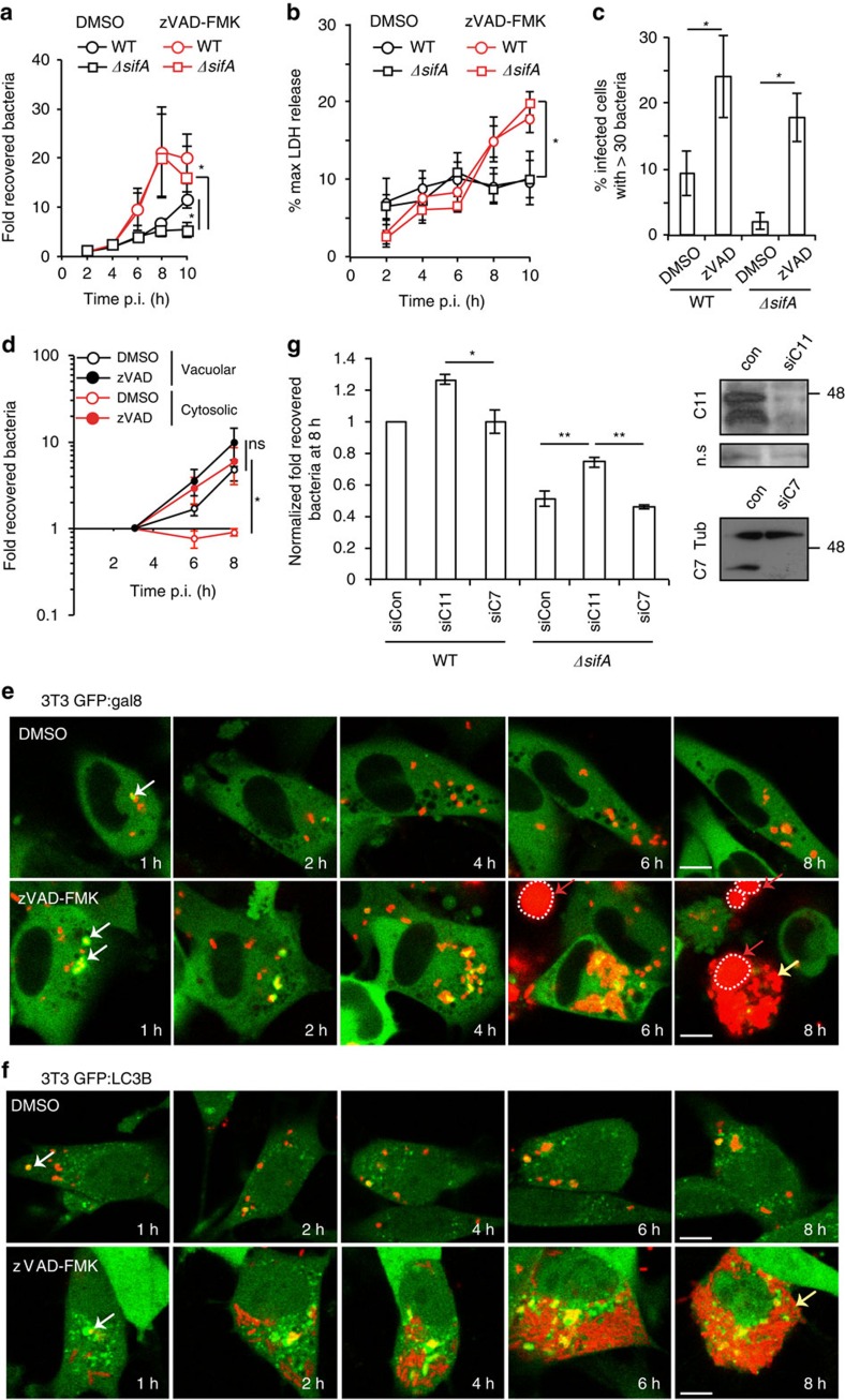
Figure 1
Caspase-dependent inhibition of cytosolic Salmonella in 3T3 fibroblasts.
( a ) Bacterial replication in 3T3 fibroblasts pre-treated with DMSO or zVAD-FMK and infected with WT or u0394sifA Salmonella was determined by enumeration of colony-forming units (CFUs) after cell ly...
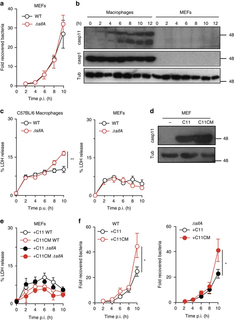
Figure 2
Expression of caspase-11 in MEFs reduces growth of Salmonella.
( a ) MEFs were infected with WT or u0394sifA Salmonella then lysed at the indicated time points. Bacterial CFUs were counted on LB agar and normalized to CFUs at 2u2009h p.i. ( b ) Protein extracts f...
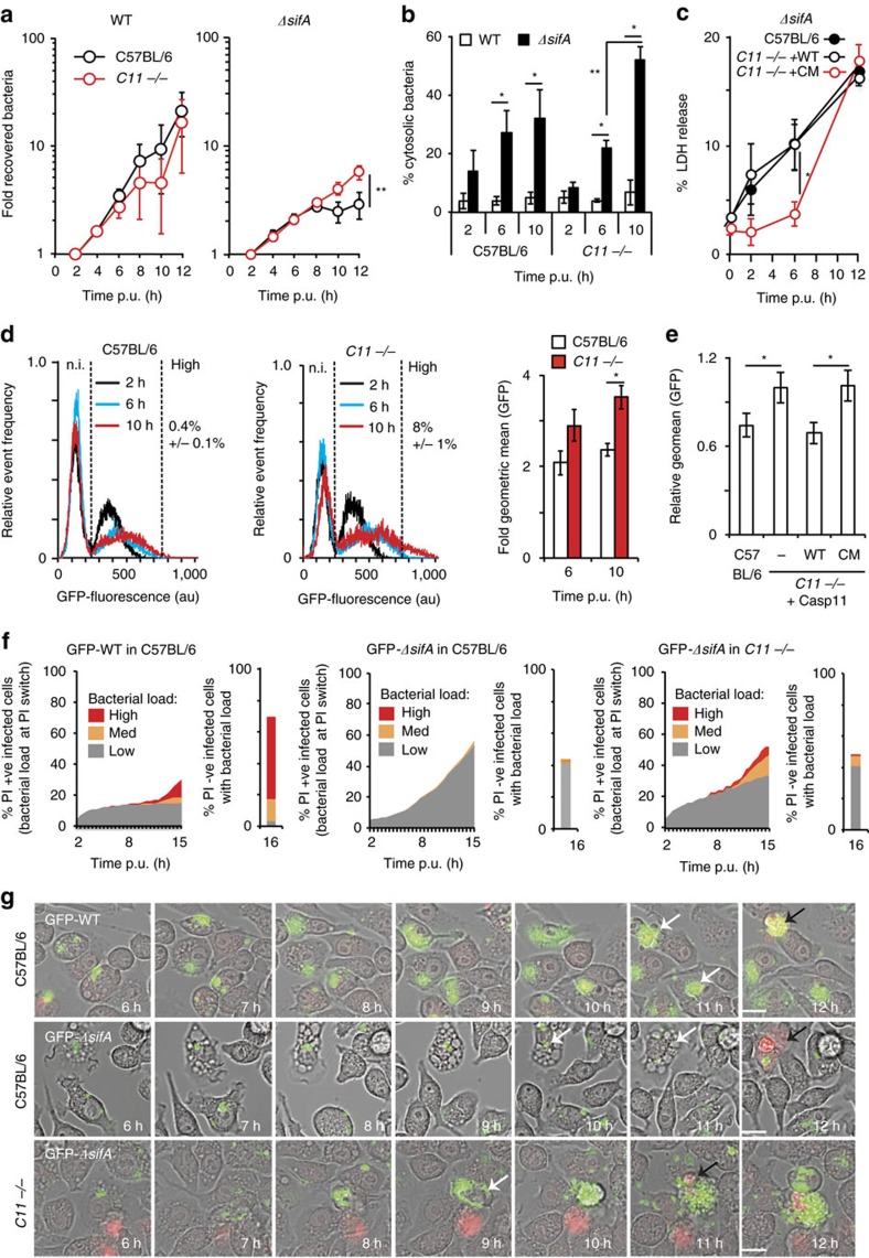
Figure 3
u0394sifA growth within macrophages is partially inhibited by caspase-11.
( a ) Kinetics of intracellular growth of wild-type (WT) or u0394sifA Salmonella after infection of the indicated iBMDMs. Following host-cell lysis at the indicated time-points, intracellular bacteria...
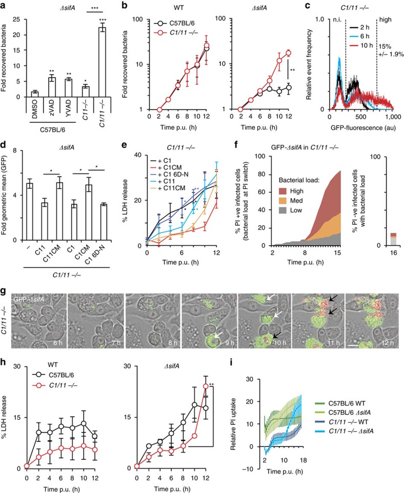
Figure 4
Both caspase-1 and caspase-11 inhibit growth of u0394sifA Salmonella.
( a ) C57BL/6 iBMDMs exposed to DMSO, YVAD-FMK or zVAD-FMK, or Casp11 u2212/u2212 or Casp1/11 u2212/u2212 iBMDMs, were infected with u0394sifA Salmonella for 17u2009h. CFUs were normalized to bacteria...
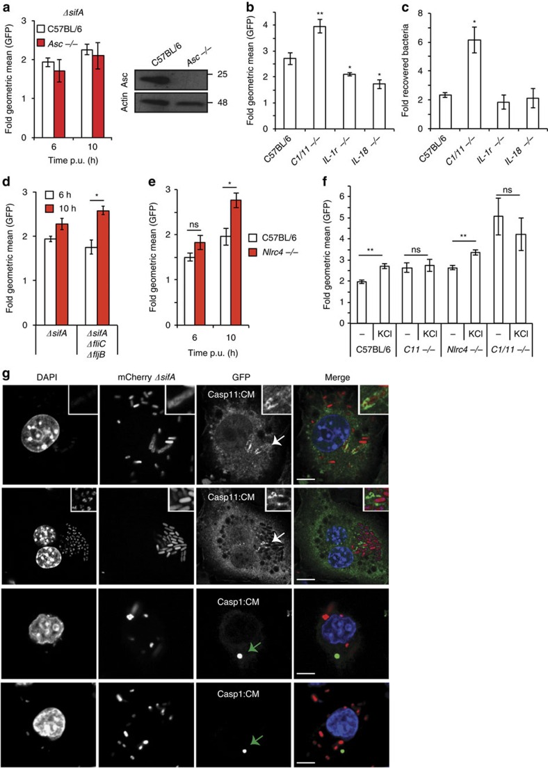
Figure 5
Growth inhibition of u0394sifA does not require cytokine production.
( a , b ) Flow cytometry analysis of GFP fluorescence per PI-negative cell of the indicated genotype after infection with GFP- u0394sifA Salmonella, expressed as fold geometric mean from 2u2009h. Inse...
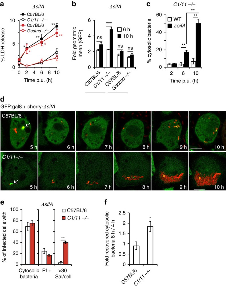
Figure 6
Cytosolic replication of u0394sifA in Casp1/11 u2212/u2212 macrophages.
( a ) Macrophage cytotoxicity was determined by LDH release from the indicated iBMDMs infected with GFP- sifA Salmonella. ( b ) Growth of GFP- sifA Salmonella in Gsdmd u2212/u2212 , Casp1/11 u2212/u22...
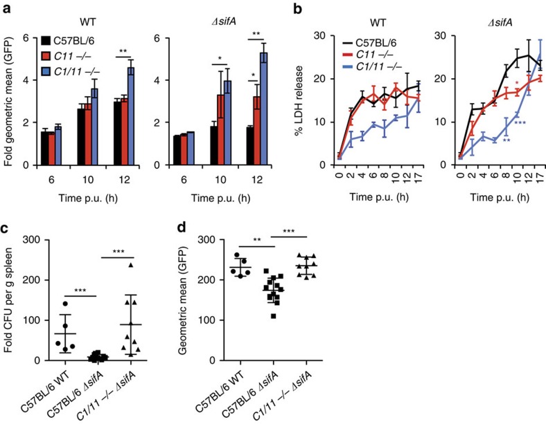
Figure 7
Caspase-mediated inhibition of u0394sifA in primary macrophages.
( a ) Growth of GFP-WT or GFP- sifA Salmonella in primary BMDMs isolated from C57BL/6 mice, caspase-11 knockout mice ( C11 u2212 /u2212 ) or caspase-1 and caspase-11 double knockout mice ( C1/11 u2212...
Figure images are served from the NIH/NLM PubMed Central Open Access Subset or Europe PMC; copyright remains with the publishers and authors.
💬 Discussion
0 commentsNo comments yet. Be the first to start a discussion!
Leave a Comment