Abstract
Myocardium is composed of two main cell populations: cardiomyocytes (CMs) and interstitial cells (e.g. fibroblasts, immunoreactive cells, capillaries). However, very recently we have showed that a novel type of interstitial cell called telocytes (TCs) does exist in epi-, myo- and endocardium. They have very long and thin telopodes (Tp) formed by alternating podomeres and podoms. Heterocellular communication between TCs and CMs it is supposed to occur by shed vesicles and close apposition. If TCs have to play a role in cardiac physiology it is expected to develop direct and unambiguous contacts with CMs. Because a clear membrane-to-membrane junction has not been reported by electron microscopy we have investigated the heterocellular communication in the mouse heart by electron tomography. This advanced technique showed that small dense structures (10-15 nm nanocontacts) directly connect TCs with CMs. More complex and atypical junctions could be observed between TCs and CMs at the level of intercalated discs. This study proves that TCs and CMs are directly connected and might represent a 'functional unit'.
🔬 Techniques
🧪 Sample Preparation
🔬 Cell Lines
🏭 Microscope Brands
📷 Detectors
💻 Software Details
🏛️ Research Organizations (ROR)
Affiliated research institutions:
📋 Methods
This study was performed on mouse heart samples obtained from 8-month-old C57BL/6 mice. The tissue samples were processed according to routine Epon-embedding procedure, as previously described [ 5 , 7 ]. The thin and thick sections were cut with a diamond knife using an RMC ultramicrotome (Boeckeler Instruments, Inc., Tucson, AZ, USA) and double stained with 1% uranyl acetate and Reynolds lead citrate. 1. Transmission electron microscopy was performed on 60-nm-thin sections using a Morgagni 286 transmission electron microscope (FEI Company, Eindhoven, The Netherlands) at 60 kV. Digital electron micrographs were recorded with a MegaView III charged-coupled device (CCD) using iTEM-SIS software (Olympus, Soft Imaging System GmbH, Münster, Germany). 2. ET was performed was performed on 250-nm-thick sections using a Tecnai G2 Spirit BioTwin electron microscope with single-tilt specimen holder (FEI Company) at 100 kV and electron tomographic data sets were recorded with a MegaView G2 CCD camera (Olympus) in ET mode [ 31 , 32 ]. Tomographs were acquired at 1° angular increment from –65° to +65° about an axis perpendicular to the optical axis of the microscope, at 36,000× magnifications (1.64 nm/px). After data alignment, the data sets were reconstructed into 3D volume (data collection, reconstruction and visualization) using Xplore3D Tomography Suite software (FEI Company). Amira 5.0.1 software (Visage Imaging GmbH, Berlin, Germany) was used for 3D imaging.
📊 Figures
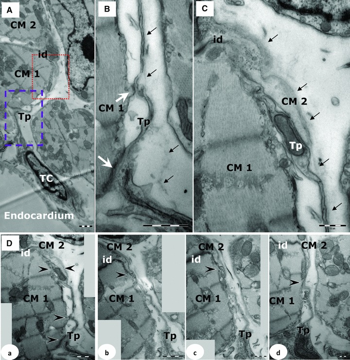
Fig 1
(A)u2013(C) Electron microscopy on 250-nm-thick sections shows a TC extending a Tp beneath basal lamina (black arrows) of the CMs (CM1, CM2). id: intercalated disc. (B) Higher magnification of dashed ...
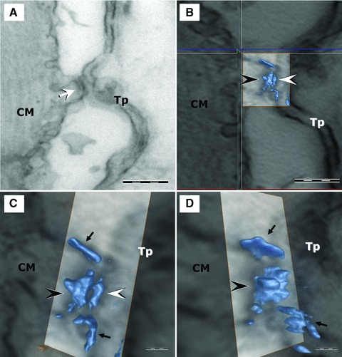
Fig 2
(A) Digital section from tomographic volume shows a junction between the Tp and a discrete projection of the CM. (B) Top view of the 3D isosurface reconstruction of the junction shows a macromolecular...
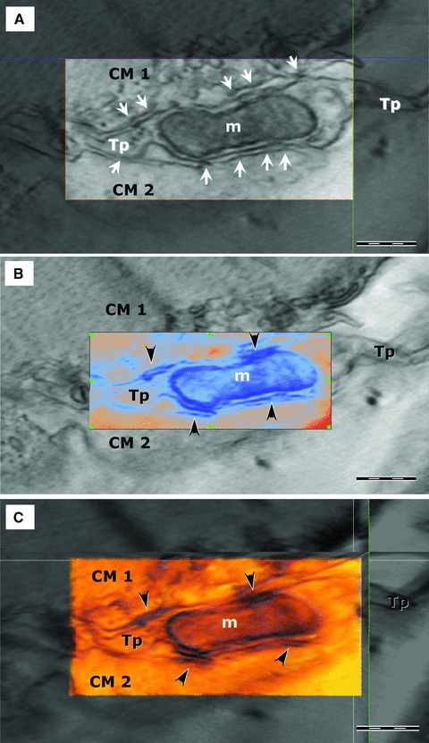
Fig 3
ET shows small electron-dense structures (arrows) connecting the cellular membranes of a TC (Tp) and two CMs (CM1, CM2). A mitochondrion (m) is visible in the dilation of the Tp (podom). (A) A digital...
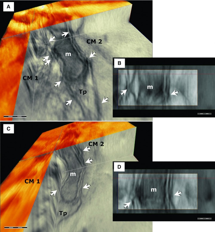
Fig 4
ET shows nanocontacts (arrows) connecting the cellular membranes of the Tp and CMs (CM1, CM2). The mitochondrion (m) is visible in the dilation of Tp. Images in (A) and (C) illustrate nanocontacts at ...
Figure images are served from the NIH/NLM PubMed Central Open Access Subset or Europe PMC; copyright remains with the publishers and authors.
💬 Discussion
0 commentsNo comments yet. Be the first to start a discussion!
Leave a Comment