Abstract
Over the last decade microfluidics has created a versatile platform that has significantly advanced the ways in which micro-scale organisms and objects are controlled, processed and investigated, by improving the cost, compactness and throughput aspects of analysis. Microfluidics has also expanded into optics to create reconfigurable and flexible optical devices such as reconfigurable lenses, lasers, waveguides, switches, and on-chip microscopes. Here we present a new opto-fluidic microscopy modality, i.e., Holographic Opto-fluidic Microscopy (HOM), based on lensless holographic imaging. This imaging modality complements the miniaturization provided by microfluidics and would allow the integration of microscopy into existing on-chip microfluidic devices with various functionalities. Our imaging modality utilizes partially coherent in-line holography and pixel super-resolution to create high-resolution amplitude and phase images of the objects flowing within micro-fluidic channels, which we demonstrate by imaging C. elegans, Giardia lamblia, and Mulberry pollen. HOM does not involve complicated fabrication processes or precise alignment, nor does it require a highly uniform flow of objects within microfluidic channels.
🔬 Techniques
🧬 Organisms
💻 Software
✨ Fluorophores
🧪 Sample Preparation
🏭 Microscope Brands
💻 Software Details
🏛️ Research Organizations (ROR)
Affiliated research institutions:
📊 Figures
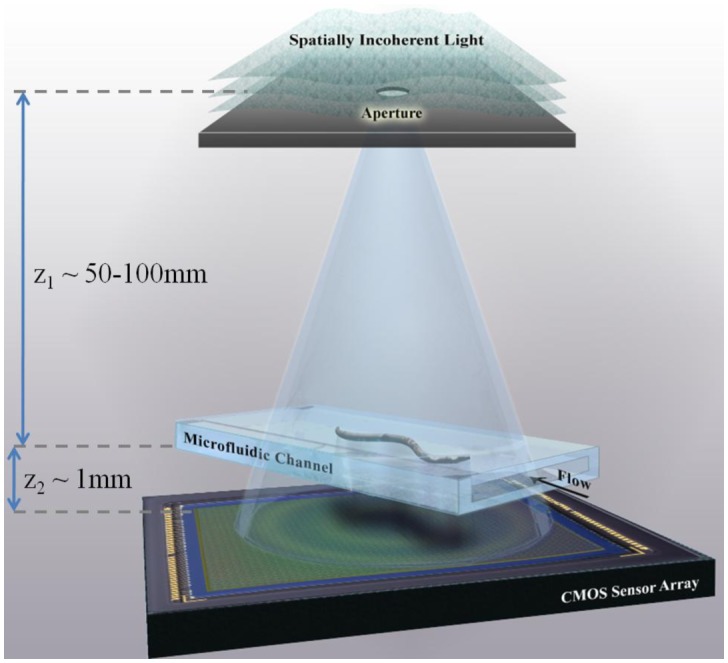
Fig. 1
A schematic of the experimental set-up of Holographic Opto-fluidic Microscopy (HOM). The sample to be imaged flows within a micro-fluidic channel, due to either electro-kinetic or pressure driven moti...
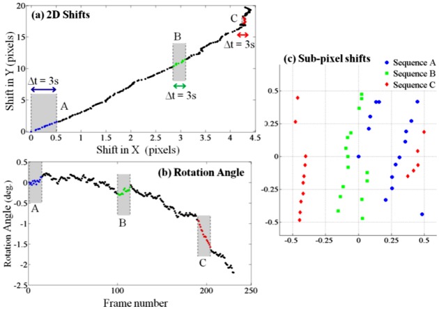
Fig. 2
Electro-kinetic motion: As the object flows through the micro-channel due to electro-kinetic motion, lensfree in-line holograms are continuously captured by the CMOS sensor at a rate of ~5-6 frames/se...
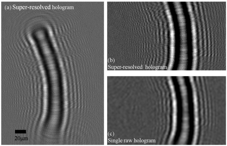
Fig. 3
(a) A super-resolved hologram of a C. elegans sample is shown. This high-resolution hologram is digitally computed using sequence A of Fig. 2 (15 consecutive lensfree raw holograms). (b) An enlarged s...
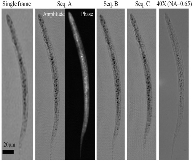
Fig. 4
HOM images of a C. elegans sample. A single raw lensfree hologram gives a lower resolution image of the object after twin image elimination as shown in the far left image. When using 15 consecutive ho...
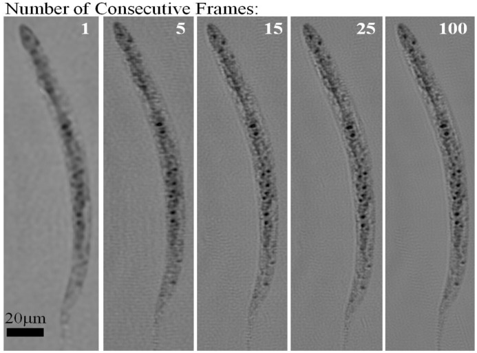
Fig. 5
Electro-kinetic HOM images with different number of consecutive frames used as input to the pixel super-resolution algorithm. The images show progressive enhancement with increasing number of frames, ...
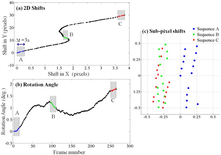
Fig. 6
Pressure flow: By creating a pressure imbalance between the two ends of the micro-fluidic channel, the sample was forced to flow along the channel and frames were continuously captured. Again, only 15...
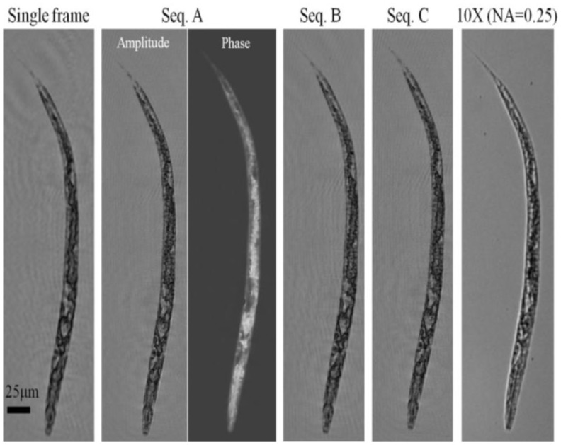
Fig. 7
Pressure driven HOM images of a C. elegans sample. As with the electro-kinetic flow case ( Fig. 4 ), we have used 3 different sequences (A, B and C) of 15 consecutive frames to generate 3 independent ...
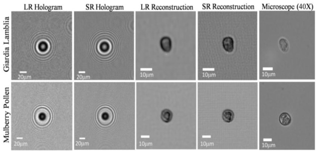
Fig. 8
HOM imaging results are summarized for a Giardia lamblia cyst and a Mulberry pollen. The low-resolution (LR) hologram exhibit aliasing which is resolved in the super-resolved (SR) hologram. Note that ...
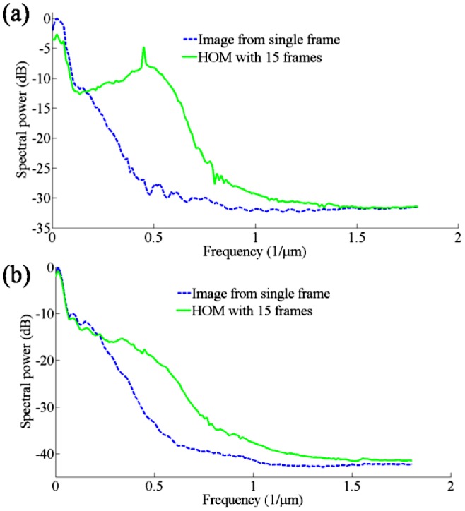
Fig. 9
The radial spectral power of HOM images and images reconstructed from single raw holograms. (a) The radial spectral power of images with electro-kinetic flow ( Fig. 4 ). (b) The radial spectral power ...
Figure images are served from the NIH/NLM PubMed Central Open Access Subset or Europe PMC; copyright remains with the publishers and authors.
💬 Discussion
0 commentsNo comments yet. Be the first to start a discussion!
Leave a Comment