Abstract
BACKGROUND: Interleukin-33 (IL-33) is increasingly being recognized as a key immunomodulatory cytokine in many neurological diseases. METHODS: In the present study, wild-type (WT) and IL-33-/- mice received intracerebroventricular (i.c.v.) injection of lipopolysaccharide (LPS) to induce neuroinflammation. Intravital microscopy was employed to examine leukocyte-endothelial interactions in the brain vasculature. The degree of neutrophil infiltration was determined by myeloperoxidase (MPO) staining. Real-time PCR and western blotting were used to detect endothelial activation. Enzyme-linked immunosorbent assay and quantitative PCR were conducted to detect pro-inflammatory cytokine levels in the brain. RESULTS: In IL-33-/- mice, neutrophil infiltration in the brain cortex and leukocyte-endothelial cell interactions in the cerebral microvessels were significantly decreased as compared to WT mice after LPS injection. In addition, IL-33-/- mice showed reduced activation of microglia and cerebral endothelial cells. In vitro results indicated that IL-33 directly activated cerebral endothelial cells and promoted pro-inflammatory cytokine production in LPS-stimulated microglia. CONCLUSIONS: Our study indicated that IL-33/ST2 signaling plays an important role in the activation of microglia and cerebral endothelial cells and, therefore, is essential in leukocyte recruitment in brain inflammation. The role of IL-33/ST2 in LPS induced neuroinflammation.
🔬 Techniques
🧬 Organisms
✨ Fluorophores
🧪 Sample Preparation
🔬 Cell Lines
🏭 Microscope Brands
🧪 Reagent Suppliers
📷 Detectors
💻 Software Details
🏛️ Research Organizations (ROR)
Affiliated research institutions:
📋 Methods
In the present study, wild-type (WT) and IL-33 −/− mice received intracerebroventricular (i.c.v.) injection of lipopolysaccharide (LPS) to induce neuroinflammation. Intravital microscopy was employed to examine leukocyte–endothelial interactions in the brain vasculature. The degree of neutrophil infiltration was determined by myeloperoxidase (MPO) staining. Real-time PCR and western blotting were used to detect endothelial activation. Enzyme-linked immunosorbent assay and quantitative PCR were conducted to detect pro-inflammatory cytokine levels in the brain.
Methods
Animals and reagents Male
C57BL/6J mice (7−8 weeks old, 20−25 g), used as WT controls, were purchased from the Animal Core Facility of Nanjing Medical University. IL-33-deficient (IL-33 −/− ) mice (C57BL/6J background) were obtained from Dr. Hiroshi Kiyonari (Laboratory for Animal Resources and Genetic Engineering, Center for Developmental Biology, Institute of Physical and Chemical Research, Kobe, Japan). ST2-deficient (ST2 −/− ) mice (C57BL/6J background) were obtained from Dr. Fang Zheng (Department of Immunology, School of Basic Medicine, Tongji Medical College, Huazhong University of Science and Technology, Wuhan, China). All mice were maintained in the Animal Core Facility of Nanjing Medical University under specific pathogen-free conditions with free access to food and water. All animal experimental protocols were reviewed and approved by the Institutional Animal Care and Use Committee of Nanjing Medical University and were in compliance with institutional guidelines. LPS ( Escherichia coli serotype 0111: B4 strain) was purchased from InvivoGen (San Diego, CA, USA). Recombinant mouse IL-33 protein was purchased from R&D Systems (Minneapolis, MN, USA). Antibodies against VCAM-1, P-selectin, E-selectin, IL-33, myeloperoxidase (MPO), and mouse serum albumin were purchased from Abcam (Cambridge, MA, USA). Antibody against ST2 was purchased from Santa Cruz Biotechnology (Santa Cruz, CA, USA). Antibodies against β-actin, ERK, phospho-ERK, p38 mitogen-activated protein kinase (MAPK), phospho-p38 MAPK, JNK, phospho-JNK, NF-κB p65, and phospho-NF-κB p65 were purchased from Cell Signaling Technology (Beverly, MA, USA). The anti-ionized calcium-binding adaptor molecule 1 (Iba-1) antibody was purchased from Wako Pure Chemical (Osaka, Japan).
Show full methods section
In the present study, wild-type (WT) and IL-33 −/− mice received intracerebroventricular (i.c.v.) injection of lipopolysaccharide (LPS) to induce neuroinflammation. Intravital microscopy was employed to examine leukocyte–endothelial interactions in the brain vasculature. The degree of neutrophil infiltration was determined by myeloperoxidase (MPO) staining. Real-time PCR and western blotting were used to detect endothelial activation. Enzyme-linked immunosorbent assay and quantitative PCR were conducted to detect pro-inflammatory cytokine levels in the brain.
Methods
Animals and reagents Male
C57BL/6J mice (7−8 weeks old, 20−25 g), used as WT controls, were purchased from the Animal Core Facility of Nanjing Medical University. IL-33-deficient (IL-33 −/− ) mice (C57BL/6J background) were obtained from Dr. Hiroshi Kiyonari (Laboratory for Animal Resources and Genetic Engineering, Center for Developmental Biology, Institute of Physical and Chemical Research, Kobe, Japan). ST2-deficient (ST2 −/− ) mice (C57BL/6J background) were obtained from Dr. Fang Zheng (Department of Immunology, School of Basic Medicine, Tongji Medical College, Huazhong University of Science and Technology, Wuhan, China). All mice were maintained in the Animal Core Facility of Nanjing Medical University under specific pathogen-free conditions with free access to food and water. All animal experimental protocols were reviewed and approved by the Institutional Animal Care and Use Committee of Nanjing Medical University and were in compliance with institutional guidelines. LPS ( Escherichia coli serotype 0111: B4 strain) was purchased from InvivoGen (San Diego, CA, USA). Recombinant mouse IL-33 protein was purchased from R&D Systems (Minneapolis, MN, USA). Antibodies against VCAM-1, P-selectin, E-selectin, IL-33, myeloperoxidase (MPO), and mouse serum albumin were purchased from Abcam (Cambridge, MA, USA). Antibody against ST2 was purchased from Santa Cruz Biotechnology (Santa Cruz, CA, USA). Antibodies against β-actin, ERK, phospho-ERK, p38 mitogen-activated protein kinase (MAPK), phospho-p38 MAPK, JNK, phospho-JNK, NF-κB p65, and phospho-NF-κB p65 were purchased from Cell Signaling Technology (Beverly, MA, USA). The anti-ionized calcium-binding adaptor molecule 1 (Iba-1) antibody was purchased from Wako Pure Chemical (Osaka, Japan).
Cell culture
The murine cerebral microvascular endothelial cell line bEND.3 and the murine microglial cell line BV2 were purchased from the American Type Culture Collection (Manassas, VA, USA). Cells were cultured in high-glucose Dulbecco’s modified Eagle’s medium (DMEM; GE Healthcare Hyclone, Logan, UT, USA) containing 10% fetal bovine serum (FBS; Gibco, Gaithersburg, MD, USA), at 37 °C in a 5% CO 2 incubator. The cells were serum-starved for 12 h before they were stimulated with IL-33 for western blotting.
Intracerebroventricular LPS injection
Mice were administered an intracerebroventricular (i.c.v.) LPS injection as previously described [ 10 ]. In brief, the mice were anesthetized via intraperitoneal injection of 200 mg/kg ketamine and 10 mg/kg xylazine. The mice were placed onto a rodent stereotaxic frame (David Kopf Instruments, Tujunga, CA, USA). Then, 2 μg of LPS in 2 μl saline was injected into the left ventricle using a Hamilton microsyringe over a 5-min period. Control animals received an i.c.v. injection of an equal volume of saline. After i.c.v. injection, the animals were maintained at 36 ± 1 °C on a thermostatic heating system (Harvard Apparatus, MA, USA) throughout the experiment.
Intravital microscopy
Intravital microscopy was performed as previously described [ 10 ]. After anesthetization, a craniotomy was performed in the right parietal bone using a high-speed drill and the dura was carefully removed to expose the brain microvessels. The mice were given an intravenous injection of rhodamine 6G (Sigma-Aldrich, St. Louis, MD, USA) (0.5 mg/kg body weight) to label leukocytes. Leukocyte–endothelial interactions in the brain microvasculature were photographed using a sCMOS camera (ORCA-Flash 4.0; Hamamatsu, Japan) mounted on Nikon FN1 microscope. Three different microvessels with diameters of 30−60 μm were visualized and imaged. Rolling leukocytes were defined as cells moving at a velocity less than that of erythrocytes. Cells were considered adherent when they remained stationary for 30 s. Enzyme-linked immunosorbent assay (ELISA) The mice were anesthetized after i.c.v. LPS injection and subsequently perfused through the heart with 20−30 ml of ice-cold PBS to clear blood cells and proteins from the circulation. The brains were rapidly removed and subsequently homogenized in 1 ml of ice-cold PBS, followed by centrifugation at 12,000× g for 5 min at 4 °C. The supernatants were assayed for TNF-α, IL-6, IL-1β, and MCP-1 concentrations using commercial ELISA kits (for TNF-α, IL-6, and IL-1β: BD Biosciences, San Diego, CA, USA; for MCP-1: R&D Systems) following the manufacturers’ instructions. RNA isolation and quantitative reverse transcription (qRT)-PCR After perfusion of the heart with ice-cold PBS, the mouse brain was collected and homogenized in 1 ml of TRIzol (Takara Bio, Shiga, Japan) on ice, and RNA was extracted using TRIzol reagent following the protocols supplied by the manufacturer. One microgram of total RNA was reverse-transcribed into cDNA using PrimeScript RT Master Mix (Takara Bio). Then, real-time PCR was conducted using SYBR® Green (Bio-Rad Laboratories, Hercules, CA, USA) following the manufacturer’s instructions. The following primer sets were used: TNF-α fwd, 5′-ACGGCATGGATCTCAAAGAC-3′, and TNF-α rev, 3′-AGATAGCAAATCGGCTGACG-5′; IL-6 fwd, 5′-ACAACCACGGCCTTCCCTAC-3′, and IL-6 rev, 3′-AGATAGCAAATCGGCTGACG-5′; IL-1β fwd, 5′-TGTCTTGGCCGAGGACTAAGG-3′, and IL-1β rev, 3′-TGGGCTGGACTGTTTCTAATGC-5′; P-selectin fwd, 5′-TCCAGGAAGCTCTGACGTACTTG-3′, and P-selectin rev, 3′-GCAGCGTTAGTGAAGACTCCGTAT-5′; E-selectin fwd, 5′-TGAACTGAAGGGATCAAGAAGACT-3′, and E-selectin rev, 3′-GCCGAGGGACATCATCACAT-5′; ICAM-1 fwd, 5′-CCTGTTTCCTGCCTCTGAA-3′, and ICAM-1 rev, 3′-GTCTGCTGAGACCCCTCTTG-5′; VCAM-1 fwd, 5′-TGACAAGTCCCCATCGTTGA-3′, and VCAM-1 rev, 3′-ACCTCGCGACGGCATAATT-5′; IL-33 fwd 5′-TCCAACTCCAAGATTTCCCCG-3′, and IL-33 rev, 3′-AAGACGGTACAGATGACGTAC-5′; and GAPDH fwd, 5′-TGCAGTGGCAAAGTGGAGATT-3′, and GAPDH rev, 3′-TCGCTCCTGGAAGATGGTGAT-5′. The housekeeping gene GAPDH was used for normalization. qPCR were conducted in triplicate for each sample, and target mRNA levels were quantified using the 2 –ΔΔCt method.
Western blotting
Mice were anesthetized and perfused through the heart with ice-cold PBS to clear blood cells and proteins from the circulation. The mouse brains were rapidly removed and homogenized in 1 ml of ice-cold PBS and then centrifuged at 13,800× g for 5 min at 4 °C. For cell-based assays, cells were treated with radioimmunoprecipitation assay lysis buffer for 30 min at 4 °C and centrifuged at 13,800× g for 10 min at 4 °C. The supernatants of brain homogenates or cell lysates were diluted in loading buffer and boiled at 100 °C for 10 min. The samples were subjected to 10% sodium dodecyl sulfate-polyacrylamide gel electrophoresis and the proteins were transferred to a polyvinylidene difluoride membrane (Millipore, Billerica, MA, USA). The membranes were blocked with 5% bovine serum albumin in PBS for 2 h at room temperature and then incubated with primary antibody overnight at 4 °C. The membranes were washed with PBST (0.05% Tween-20 in PBS) three times and then incubated with species-appropriate HRP-conjugated secondary antibody for 1.5−2 h at room temperature. Then, the membranes were washed with PBST three times and subjected to immunodetection with enhanced chemiluminescence reagents (PerkinElmer, Waltham, MA, USA). Primary culture of purified microglia and astrocytes The brains of newborn mice were harvested and the cerebella, white matter, and leptomeninges were separated from the cerebral cortices. Then, the cerebral cortices were trypsinized for 5 min at 37 °C and filtrated through a 70-μm pore-size filter (Millipore, Billerica, MA, USA). Cells from six cerebra were seeded in a 75-cm 2 culture flask containing 15 ml of DMEM/F12 with 10% FBS and incubated in 5% CO 2 at 37 °C. After 24 h, the entire medium was replaced, and then half of the medium was exchanged every 3−4 days. On days 13−14, microglia were isolated from the mixed glial culture by shaking the flask at 200 rpm for 1 h at 37 °C [ 31 ]. The microglia were centrifuged and seeded in 24-well plates for further stimulation. The mixed glial cells were passaged two to three times and shaken at 220 rpm for 6 h; the supernatants were discarded and the remaining adherent cells were collected as astrocytes. The purity of the isolated microglia and astrocytes was 95 and 90%, respectively.
Isolation and culture of murine cerebral endothelial cells
Seven- to 8-week-old mice were sacrificed, and their brains were collected. The cerebella, striata, optic nerves, and white matter were removed, and cerebral cortices were collected. The tissue samples were digested in 15 ml of 0.1% collagen B (Roche, Indianapolis, IN, USA) supplemented with 30 U/ml DNase I (Sigma, St. Louis, MO, USA) for 1.5 h at 37 °C and shaken every half hour. The microvessel pellets were resuspended in medium supplemented with 3 ng/ml of bovine fibroblast growth factor (Peprotech, Rocky Hill, NJ, USA), 30% FBS, 10 U/ml of heparin, 100 U/ml of penicillin, and 100 μg/ml of streptomycin. The microvessel suspensions were placed in six-well plates pre-coated with rat-tail collagen I (Sigma-Aldrich) and incubated in 5% CO 2 at 37 °C. The medium was changed within the first 24 h and then every 2 days. The endothelial cells grew to confluence within 7–10 days.
Immunohistochemical analysis
Brain blocks were embedded in paraffin after fixation in 4% paraformaldehyde and then sliced into 4-μm sections. After blocking with bovine serum albumin, for the detection of infiltrating neutrophils, the sections were incubated with anti-MPO antibodies at 4 °C overnight. This was followed by a 1-h incubation with horseradish peroxidase (HRP)-labeled secondary antibodies at room temperature. Different fields of view were analyzed at magnifications of ×100 and ×400, and cells stained positive for primary antibody were counted under a Nikon E100 microscope.
Immunofluorescence
Mice were perfused with ice-cold PBS and 4% paraformaldehyde, and the brains were collected, conserved in paraformaldehyde, and dehydrated in a 30% sucrose solution. Then, 25-μm cortex sections were incubated overnight with anti-Iba1. Iba-1 is known to be the activation marker of microglia. The cortex was observed in four fields of view at a primary magnification of ×200 in every brain section. Images were captured using a fluorescence microscope (Nikon Eclipse TE2000-S, Nikon, Tokyo, Japan) at a magnification of ×200.
Statistical analysis
Statistical analysis was performed using GraphPad Prism 5 software. The data are shown as the mean ± standard error of the mean (SEM). Means were compared using Student’s t test for two groups or one-way ANOVA for multiple groups. P < 0.05 was considered significant.
Availability of data and materials The datasets supporting the conclusions of this study are included in this article.
📊 Figures
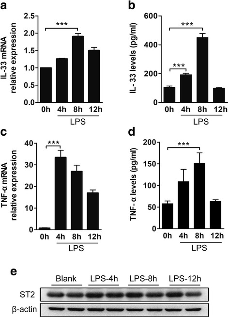
Fig. 1
IL-33 and ST2 expression in the brain after LPS stimulation. I.c.v. LPS injection, used as a standard neuroinflammation model, resulted in a significant increase in inflammatory cytokines in WT mice. ...
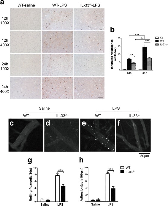
Fig. 2
IL-33 u2212/u2212 mice exhibit decreased neutrophil recruitment and leukocyteu2013endothelial interactions in brain microvessels after i.c.v. LPS administration. WT and IL-33 u2212/u2212 mice received...
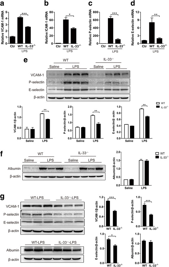
Fig. 3
Role of IL-33 in brain endothelial adhesion molecule expression and bloodu2013brain barrier (BBB) integrity changes. Four hours after i.c.v. LPS injection, the mRNA levels of adhesion molecules in WT ...
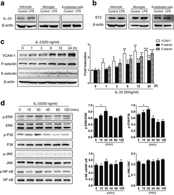
Fig. 4
IL-33 stimulates endothelial cell activation and adhesion molecule expression. Primary astrocytes, microglia, and endothelial cells were isolated from WT mice and treated with saline or LPS (100u00a0n...
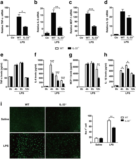
Fig. 5
Pro-inflammatory cytokine levels and microglial activation in the brains of WT and IL-33 u2212/u2212 mice after i.c.v. LPS injection. WT mice and IL-33 u2212/u2212 mice received i.c.v. injection of sa...
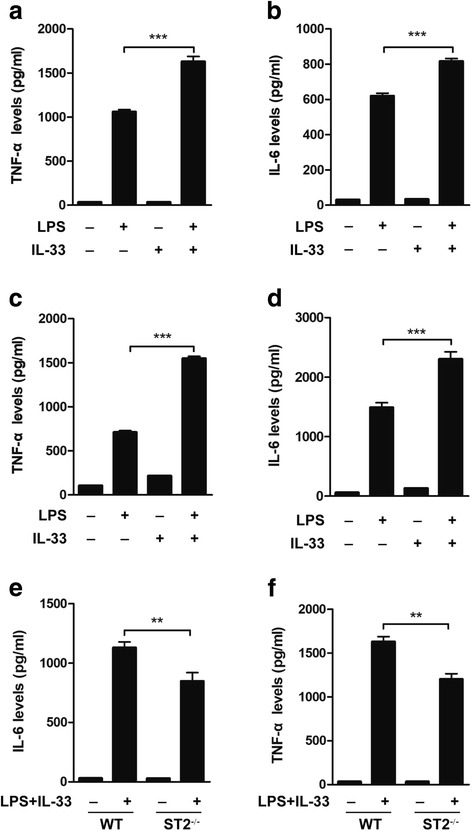
Fig. 6
IL-33 promotes LPS-induced pro-inflammatory cytokine production by microglia. Primary microglia from WT mice were stimulated with LPS (100u00a0ng/ml), IL-33 (50u00a0ng/ml), or LPS (100u00a0ng/ml) plus...
Figure images are served from the NIH/NLM PubMed Central Open Access Subset or Europe PMC; copyright remains with the publishers and authors.
💬 Discussion
0 commentsNo comments yet. Be the first to start a discussion!
Leave a Comment