Abstract
SummaryMalaria parasites undergo a population expansion inside the host liver before disease onset. Developmental arrest inside host hepatocytes elicits protective immune responses. Therefore, elucidation of the molecular mechanisms leading to mature hepatic merozoites, which initiate the pathogenic blood phase, also informs anti‐malaria vaccine strategies. Using targeted gene deletion in the rodent model malaria parasite Plasmodium berghei, we show that a Plasmodium‐specific Apicoplast protein plays an important role for Liver Merozoite formation (PALM). While the resulting knockout mutants develop normally for most of the life cycle, merozoite release into the blood stream and the ability to establish an infection are severely impaired. Presence of a signature blood‐stage antigen, merozoite surface protein 1 and normal apicoplast morphology indicate that the inability to finalize merozoite segregation is a direct consequence of loss of PALM function. Experimental immunization of mice with as few as two doses of palm‐ sporozoites can elicit sterile protection up to 110 days after final immunization. Our data establish that a tailor‐made arrest in the final steps of hepatic merozoite formation can induce strong protective immune responses and that malaria parasites employ a distinct apicoplast protein for efficient formation of pre‐erythrocytic merozoites.
🔬 Techniques
🧬 Organisms
✨ Fluorophores
🧪 Sample Preparation
🏭 Microscope Brands
🧪 Reagent Suppliers
🎨 Filters
🏛️ Research Organizations (ROR)
Affiliated research institutions:
📋 Methods
Impaired liver-stage development leads to reduced incidence of experimental cerebral malaria (ECM) We first followed the clinical symptoms of population ‘+’, i.e. those mice that became palm - blood-stage-positive ( Fig. 6C ). Only few palm - -infected mice developed symptoms of ECM, a fatal outcome of an acute Plasmodium infection, as compared with ANKA-GFP-infected animals. Signature signs of ECM are sudden onset of ataxia, paralysis, convulsion or coma ( de Souza et al ., 2010 ). Symptoms of ECM typically develop on the fourth day after patency in sporozoite-induced infections. Of note, none of the mice that were infected by bite of palm - -infected mosquitoes developed ECM. It is important to exclude any impairment in parasite virulence due to the genetic manipulation. We previously detected normal blood-stage parasite growth ( Fig. S1A ). To confirm that palm - parasites are as virulent as WT parasites; we inoculated 1000 erythrocytes infected with either palm - ANKA-GFP or, as controls, ANKA-GFP parasites via blood transfusion, thus bypassing the liver merozoite phase ( Fig. 6C ). All animals developed the signature ECM symptoms and had to be carefully culled to avoid unnecessary suffering. Therefore, the reduced ECM incidence of palm - -infected mice can be entirely attributed to the altered pre-erythrocytic development of these parasites. We finally determined the integrity of the blood–brain barrier in sporozoite-injected animals by Evan's blue stain at the peak of clinical symptoms in WT-infected animals, typically day 4 after patency ( Fig. 6D ). This analysis further corroborated the strongly reduced cases and severity of ECM-related pathology in the palm - -positive mice.
Show full methods section
Impaired liver-stage development leads to reduced incidence of experimental cerebral malaria (ECM) We first followed the clinical symptoms of population ‘+’, i.e. those mice that became palm - blood-stage-positive ( Fig. 6C ). Only few palm - -infected mice developed symptoms of ECM, a fatal outcome of an acute Plasmodium infection, as compared with ANKA-GFP-infected animals. Signature signs of ECM are sudden onset of ataxia, paralysis, convulsion or coma ( de Souza et al ., 2010 ). Symptoms of ECM typically develop on the fourth day after patency in sporozoite-induced infections. Of note, none of the mice that were infected by bite of palm - -infected mosquitoes developed ECM. It is important to exclude any impairment in parasite virulence due to the genetic manipulation. We previously detected normal blood-stage parasite growth ( Fig. S1A ). To confirm that palm - parasites are as virulent as WT parasites; we inoculated 1000 erythrocytes infected with either palm - ANKA-GFP or, as controls, ANKA-GFP parasites via blood transfusion, thus bypassing the liver merozoite phase ( Fig. 6C ). All animals developed the signature ECM symptoms and had to be carefully culled to avoid unnecessary suffering. Therefore, the reduced ECM incidence of palm - -infected mice can be entirely attributed to the altered pre-erythrocytic development of these parasites. We finally determined the integrity of the blood–brain barrier in sporozoite-injected animals by Evan's blue stain at the peak of clinical symptoms in WT-infected animals, typically day 4 after patency ( Fig. 6D ). This analysis further corroborated the strongly reduced cases and severity of ECM-related pathology in the palm - -positive mice.
Experimental procedures
Experimental animals
This study was carried out in strict accordance with the German ‘Tierschutzgesetz in der Fassung vom 22. Juli 2009’ and the Directive 2010/63/EU of the European Parliament and Council ‘On the protection of animals used for scientific purposes’. The protocol was approved by the ethics committee of the Berlin state authority (‘Landesamt für Gesundheit und Soziales Berlin’, permit number G0469/09). C57BL/6 mice were used for sporozoite challenges and analysis of ECM. All other parasite infections were conducted in NMRI mice, unless otherwise indicated.
Generation of PALM-mCherry-myc and palm - parasites
Mutant parasites were generated as described previously ( Janse et al ., 2006 ). See supporting experimental procedures and Table S2 for further details on transfection vector construction and confirmation of successful transfection. Plasmodium mosquito stage development Anopheles stephensi mosquitoes were raised under a 14 h light/10 h dark cycle at 28°C and 75% humidity. Blood-feeding and mosquito dissection were performed as described previously ( Vanderberg, 1975 ). In order to determine infectivity, and quantify midgut and salivary gland associated sporozoites, infected mosquitoes were dissected at days 10, 14 and 17 after feeding respectively. Midguts of PALM-mCherry-myc infected mosquitoes were dissected at day 10 and fixed with 4% paraformaldehyde with 0.0075% glutaraldehyde. Subsequently, midguts were permeabilized with 0.5% Triton X-100 and blocked with 3% bovine serum albumin. Incubation with mouse anti-myc antibodies (1:200 dilution, Santa Cruz Biotechnology) was done overnight at 4°C. Bound antibodies were detected using donkey anti-mouse IgG Alexa Fluor 488 conjugated antibodies (1:2000 dilution, Invitrogen). Nuclei were visualized with the DNA-dye DRAQ5 (1:1000 dilution, Axxora) and coverslips were mounted with Fluoromount-G (Southern Biotech). Salivary glands of PALM-mCherry-myc infected mosquitoes were dissected and liberated sporozoites were settled in RPMI medium containing 3% bovine serum albumin and fixed with 4% paraformaldehyde. Subsequently, sporozoites were permeabilized with 0.1% Triton X-100 in PBS and blocked with 3% bovine serum albumin. Incubation with mouse anti-myc antibodies (1:100 dilution, Santa Cruz Biotechnology) was done overnight at 4°C. Bound antibodies were detected using donkey anti-mouse IgG Alexa Fluor 546 conjugated antibodies (1:1000 dilution, Invitrogen). Coverslips were mounted with Fluoromount-G (Southern Biotech). To determine sporozoite infectivity to mice, sporozoites collected from mosquito salivary glands were injected intravenously at the numbers indicated into C57BL/6 mice. Patency was determined by examination of daily Giemsa-stained thin blood smears. Phenotyping of Plasmodium life cycle progression Plasmodium berghei mosquito stages were maintained and analysed using standard techniques ( Vanderberg, 1975 ). During all experiments, mice were monitored for the development of behavioural and functional abnormalities ( Lackner et al ., 2006 ), as indicators of ECM development. Mice were classified as suffering from ECM when they were diagnosed with at least three behavioural and functional abnormalities (e.g. positional passivity, body position, limb grasping, toe pinch, etc.). Mice were immediately sacrificed upon showing sudden onset of signature symptoms of ECM, such as ataxia, paralysis, convulsions or coma. To test the integrity of the blood–brain barrier as a further indication for the susceptibility to ECM, we injected 100 µl of 2% Evans Blue in saline i.v. into naïve C57BL/6 mice, or mice infected with 10 000 or 100 000 palm - , or 10 000 ANKA-GFP sporozoites at day 4 after patency. After 1 h, mice were sacrificed, brains prepared, and digital images taken using standardized lighting, exposure and white balance settings.
Plasmodium liver-stage development in cultured hepatoma cells
Plasmodium berghei in vitro liver stages were cultured and analysed using standard techniques ( Silvie et al ., 2008b ). In short, 30 000 hepatoma (HuH7) cells per well were plated in eight-well chamber slides (Nalge Nunc International). After 24 h the cells were incubated with 10 000 sporozoites, first for 60 min at room temperature, then at 37°C for 90–120 min. Non-invaded sporozoites were washed off and medium was changed daily. At the time points indicated, infected hepatoma cultures were fixed for 10 min with ice-cold methanol and blocked with PBS/10% FCS. For confirmation of expression, liver-stage PALM-mCherry-myc parasites were fixed and incubated with mouse anti-myc antibodies (1:1000 dilution, Santa Cruz Biotechnology). To confirm apicoplast targeting of PALM, liver-stage parasites were co-stained with rabbit anti- P. berghei ACP peptide antiserum (1:750 dilution; Friesen et al ., 2010 ). Completion of liver merozoite formation was analysed using rabbit anti- P. berghei upregulated in infectious sporozoites protein 4 (UIS4) peptide antiserum (1:2000 dilution; kindly provided by G. Montagna, MPI-IB, Berlin) and a monoclonal mouse anti- P. yoelii merozoite surface protein 1 (MSP1) antibody against a 90 kDa N-terminal fragment of the protein that shares 74% identity with P. berghei MSP1 (1:2000 dilution; kindly provided by T. Holder, National Institute for Medical Research, London, UK). Liver-stage parasites were visualized and quantified using monoclonal mouse anti- P. berghei heat shock protein 70 (HSP70) antibodies (1:300 dilution; Tsuji et al ., 1994 ). Bound antibodies were detected using donkey anti-rabbit/mouse IgG Alexa Fluor 488/546 conjugated antibodies (1:3000 dilution, Invitrogen). Nuclei were visualized with DNA-dyes Hoechst 33342 (Invitrogen) and DRAQ5 (Axxora; both 1:1000 dilution) and coverslips were mounted with Fluoromount-G (Southern Biotech). Images were recorded using a Leica TCS SP-1 confocal microscope. Total numbers of parasites were counted using a Leica DM2500 epifluorescence microscope. To confirm the apicoplast localization of PALM, we used 1 µM azithromycin (Pfizer) treatment of sporozoite-infected hepatoma cells as described previously ( Friesen et al ., 2010 ). Merosome formation was followed using two different methods, either by (i) seeding of 100 000 to 150 000 hepatoma cells per well in 24-well plates and inoculation with 100 000 sporozoites per well 24 h later, or by (ii) seeding of 30 000 hepatoma cells per well in eight-well chamber slides (Nalge Nunc International) and inoculation with 10 000 sporozoites 24 h later. Thereafter, standard procedures were used ( Silvie et al ., 2008b ). Merosomes were harvested and counted in a Neubauer chamber 72 h after infection. Infectivity of in vitro cultured ANKA-GFP and palm - parasites was tested by injection of the complete merosome containing liver-stage culture supernatants in naïve NMRI mice. All animals were monitored for parasitaemia by daily Giemsa-stained thin blood smears. Immunization and parasite challenge experiments Age-matched female C57BL/6 mice were immunized with two doses of 1000 or 10 000 palm - sporozoites extracted from salivary glands of infected mosquitoes. Sporozoites were injected intravenously in a volume of 100 µl. For the prime/boost protocol, animals that remained malaria-free after the first immunization were given a second dose 5–7 weeks after the first immunization. Only animals that remained blood-stage parasite-negative after the first immunization and subsequent boost were used for the challenge experiments at 4–6 weeks after the last immunization. Mice were challenged with five ANKA-GFP-infected mosquitoes, 10 000 intravenously injected ANKA-GFP sporozoites, or 10 intravenously injected ANKA-GFP blood-stage parasites. The number of ANKA-GFP salivary gland sporozoites per mosquito used for the challenge by bite ranged from 20 000 to 70 000. At least three age-matched naïve animals were included to verify infectivity of sporozoites during all challenge experiments. Parasitaemia was monitored by daily Giemsa-stained thin blood smears, starting from day 3 after immunization or ANKA-GFP challenge until at least day 17.
Experimental animals
This study was carried out in strict accordance with the German ‘Tierschutzgesetz in der Fassung vom 22. Juli 2009’ and the Directive 2010/63/EU of the European Parliament and Council ‘On the protection of animals used for scientific purposes’. The protocol was approved by the ethics committee of the Berlin state authority (‘Landesamt für Gesundheit und Soziales Berlin’, permit number G0469/09). C57BL/6 mice were used for sporozoite challenges and analysis of ECM. All other parasite infections were conducted in NMRI mice, unless otherwise indicated.
Supporting information Additional supporting information may be found in the online version of this article. Please note: Wiley-Blackwell are not responsible for the content or functionality of any supporting materials supplied by the authors. Any queries (other than missing material) should be directed to the corresponding author for the article.
📊 Figures
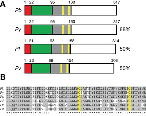
Fig. 1
The Plasmodium -specific apicoplast protein important for liver merozoite formation (PALM). A. Primary structure of Plasmodium PALM proteins. Shown are the overall sequence structures and amino acid s...
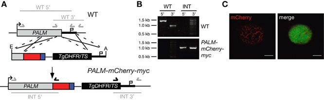
Fig. 2
Live cell imaging of PALM in infected hepatoma cells. A. Generation of PALM-mCherry-myc parasites. The PbPALM genomic locus was targeted with a replacement plasmid containing the C-terminal PALM fragm...
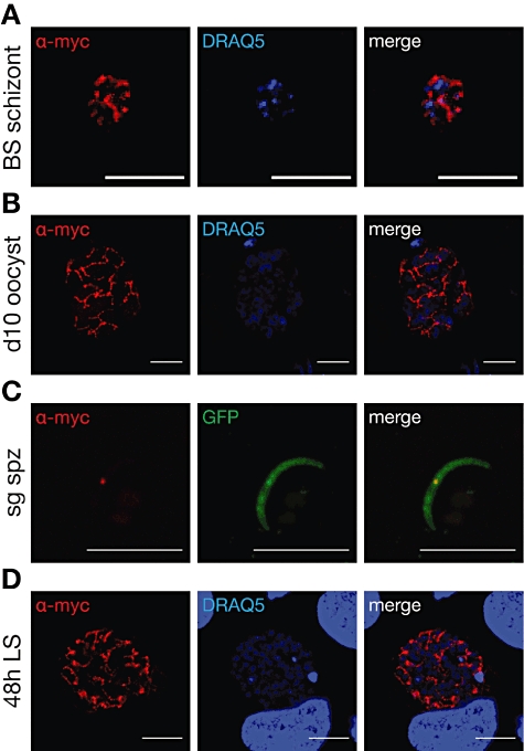
Fig. 3
Expression of PALM during the Plasmodium berghei life cycle. PALM-mCherry-myc parasites were used to infect mice and Anopheles stephensi mosquitoes. Intra- and extracellular parasite stages were fixed...
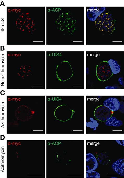
Fig. 4
Apicoplast localization of PALM. A. Co-staining of fixed, PALM-mCherry-myc parasite-infected hepatoma cells 48 h after infection using anti-myc and anti-ACP antibodies. Note the substantial overlap be...
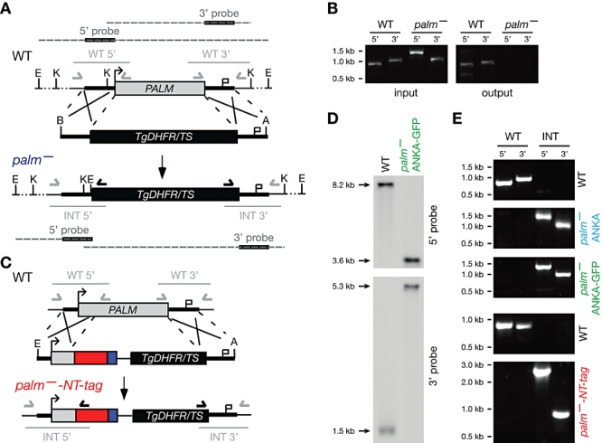
Fig. 5
Generation of palm - parasites. A. The PbPALM genomic locus was targeted with a replacement plasmid containing PALM 5u2032 and 3u2032 fragments (thick black lines) and the DHFR/TS positive selectable ...
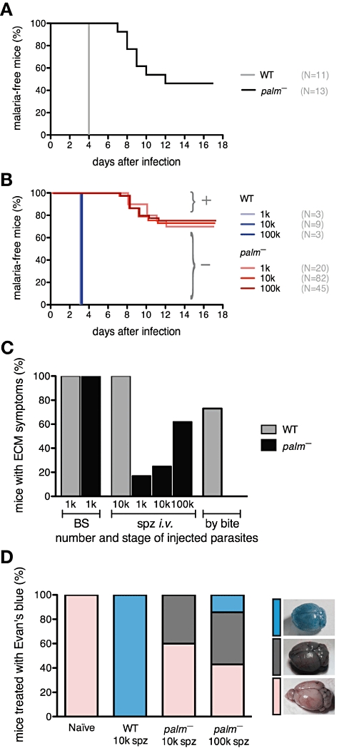
Fig. 6
Infection with palm - sporozoites leads to attenuated liver-stage development in vivo . A. Kaplanu2013Meier analysis of time to malaria blood-stage infection. C57BL/6 mice were infected by natural bit...
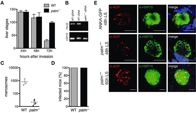
Fig. 7
palm - parasites display a defect in liver-stage maturation. A. Quantification of liver stages in cultured hepatoma cells at 24, 48 and 72 h after infection with 10 000 P. berghei ANKA-GFP and palm - ...
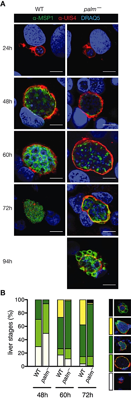
Fig. 8
palm - parasites cannot finalize liver merozoite formation efficiently. A. Defect in liver-stage merozoite segregation in palm - parasites. In vitro cultured P. berghei ANKA-GFP and palm - ANKA-GFP li...
Figure images are served from the NIH/NLM PubMed Central Open Access Subset or Europe PMC; copyright remains with the publishers and authors.
💬 Discussion
0 commentsNo comments yet. Be the first to start a discussion!
Leave a Comment