Abstract
BACKGROUND: Extracellular high mobility group box 1 protein (HMGB1) serves a central role in inflammation as a transporter protein, which binds other immune-activating molecules that are endocytosed via the receptor for advanced glycation end-products (RAGE). These pro-inflammatory complexes are targeted to the endolysosomal compartment, where HMGB1 permeabilizes the lysosomes. This enables HMGB1-partner molecules to avoid degradation, to leak into the cytosol, and to reach cognate immune-activating sensors. Lipopolysaccharide (LPS) requires this pathway to generate pyroptosis by accessing its key cytosolic receptors, murine caspase 11, or the human caspases 4 and 5. This lytic, pro-inflammatory cell death plays a fundamental pathogenic role in gram-negative sepsis. The aim of the study was to identify molecules inhibiting HMGB1 or HMGB1/LPS cellular internalization. METHODS: Endocytosis was studied in cultured macrophages using Alexa Fluor-labeled HMGB1 or complexes of HMGB1 and Alexa Fluor-labeled LPS in the presence of an anti-HMGB1 monoclonal antibody (mAb), recombinant HMGB1 box A protein, acetylcholine, the nicotinic acetylcholine receptor subtype alpha 7 (α7 nAChR) agonist GTS-21, or a dynamin-specific inhibitor of endocytosis. Images were obtained by fluorescence microscopy and quantified by the ImageJ processing program (NIH). Data were analyzed using student's t test or one-way ANOVA followed by the least significant difference or Tukey's tests. RESULTS: Anti-HMGB1 mAb, recombinant HMGB1 antagonist box A protein, acetylcholine, GTS-21, and the dynamin-specific inhibitor of endocytosis inhibited internalization of HMGB1 or HMGB1-LPS complexes in cultured macrophages. These agents prevented macrophage activation in response to HMGB1 and/or HMGB1-LPS complexes. CONCLUSION: These results demonstrate that therapies based on HMGB1 antagonists and the cholinergic anti-inflammatory pathway share a previously unrecognized molecular mechanism of substantial clinical relevance.
🔬 Techniques
🧬 Organisms
✨ Fluorophores
🧪 Sample Preparation
🔬 Cell Lines
🏭 Microscope Brands
🧪 Reagent Suppliers
💻 Software Details
🏛️ Research Organizations (ROR)
Affiliated research institutions:
📋 Methods
Endocytosis was studied in cultured macrophages using Alexa Fluor-labeled HMGB1 or complexes of HMGB1 and Alexa Fluor-labeled LPS in the presence of an anti-HMGB1 monoclonal antibody (mAb), recombinant HMGB1 box A protein, acetylcholine, the nicotinic acetylcholine receptor subtype alpha 7 (α7 nAChR) agonist GTS-21, or a dynamin-specific inhibitor of endocytosis. Images were obtained by fluorescence microscopy and quantified by the ImageJ processing program (NIH). Data were analyzed using student’s t test or one-way ANOVA followed by the least significant difference or Tukey’s tests.
Materials and methods Materials Recombinant mouse TNF, RAGE-Fc chimera protein and enzyme-linked immunosorbent assay (ELISA) kits were obtained from R & D System Inc. (Minneapolis, MN). Triton X-114, Lipopolysaccharide (LPS, E. coli. 0111:B4), peptidoglycan, pyridostigmine bromide, human macrophage-colony stimulating factor (M-CSF), acetylcholine chloride, GTS-21, 3, 3′, 5, 5′-Tetramethylbenzidine (TMB) substrate solution, lactate dyhydrogenase (LDH) cytotoxicity assay kit, non-immune rabbit IgG (Cat# I5006) and mouse IgG (Cat# I5381) were purchased from Sigma-Aldrich (St. Louis, MO). Ultrapure E. coli LPS (Cat # tlrl-pelps), poly I:C, and type B CpG oligonucleotide were obtained from InvivoGen (San Diego, CA). Thioglycollate medium was purchased from Becton Dickinson Co., (Sparks, MD). Fluorescent labeling kits were purchased from Molecular Probes (Eugene, OR). Alexa 568 labeled LPS was obtained from Invitrogen (Waltham, MA). Dynasore was purchased from Tocris Bioscience (Bristol, UK). Microscope cover glasses were obtained from Fisher Scientific (Cat# 12–545-82, Waltham, MA). Dako Fluorescence mounting medium was purchased from Agilent (Santa Clara, CA).
Show full methods section
Endocytosis was studied in cultured macrophages using Alexa Fluor-labeled HMGB1 or complexes of HMGB1 and Alexa Fluor-labeled LPS in the presence of an anti-HMGB1 monoclonal antibody (mAb), recombinant HMGB1 box A protein, acetylcholine, the nicotinic acetylcholine receptor subtype alpha 7 (α7 nAChR) agonist GTS-21, or a dynamin-specific inhibitor of endocytosis. Images were obtained by fluorescence microscopy and quantified by the ImageJ processing program (NIH). Data were analyzed using student’s t test or one-way ANOVA followed by the least significant difference or Tukey’s tests.
Materials and methods Materials Recombinant mouse TNF, RAGE-Fc chimera protein and enzyme-linked immunosorbent assay (ELISA) kits were obtained from R & D System Inc. (Minneapolis, MN). Triton X-114, Lipopolysaccharide (LPS, E. coli. 0111:B4), peptidoglycan, pyridostigmine bromide, human macrophage-colony stimulating factor (M-CSF), acetylcholine chloride, GTS-21, 3, 3′, 5, 5′-Tetramethylbenzidine (TMB) substrate solution, lactate dyhydrogenase (LDH) cytotoxicity assay kit, non-immune rabbit IgG (Cat# I5006) and mouse IgG (Cat# I5381) were purchased from Sigma-Aldrich (St. Louis, MO). Ultrapure E. coli LPS (Cat # tlrl-pelps), poly I:C, and type B CpG oligonucleotide were obtained from InvivoGen (San Diego, CA). Thioglycollate medium was purchased from Becton Dickinson Co., (Sparks, MD). Fluorescent labeling kits were purchased from Molecular Probes (Eugene, OR). Alexa 568 labeled LPS was obtained from Invitrogen (Waltham, MA). Dynasore was purchased from Tocris Bioscience (Bristol, UK). Microscope cover glasses were obtained from Fisher Scientific (Cat# 12–545-82, Waltham, MA). Dako Fluorescence mounting medium was purchased from Agilent (Santa Clara, CA).
Cell culture
Murine macrophage-like RAW 264.7 cells were obtained from American Type Culture Collection, (ATCC, Rockville, MD). Thioglycollate-elicited peritoneal macrophages were isolated from BALB/c mice (male, 8–12 weeks old) using a previously described method (Li et al. 2007 ). Cells were plated in 24-well plate, and treatment was carried out in serum-free Opti-MEM I medium (Life Technologies, Waltham, MA). Human primary monocytes were purified by density gradient centrifugation through Ficoll from blood donated to the Long Island Blood Bank by healthy individuals (New York Blood Center, Melville, NY) (Yu et al. 2006 ). Cells were allowed to differentiate into macrophages for 7 days in complete DMEM medium containing M-CSF (1 ng/ml) in 24-well culture plate with microscope cover glasses. For cytokine measurements, macrophages were plated in 96-well plates and treatment was carried out in serum-free Opti-MEM I medium. Cells were incubated with stimuli (HMGB1, alone or with LPS; TLR3 agonist Poly I:C, TLR2 agonist PGN, and TLR9 agonist CpG DNA), plus increasing amounts of m2G7, box A, acetylcholine or GTS-21 (or IgG control) as indicated in the text for 16 h. Cell culture supernatants were collected for cytokine measurements.
Expression and isolation of recombinant HMGB1 and box
A, generation of HMGB1 redox isoforms, anti-HMGB1 antibodies and HMGB1/TLR4 antagonist K883 Recombinant HMGB1 (disulfide isoform) and box A were expressed in E. coli and purified to homogeneity as previously described (Yang et al. 2004 ; Li et al. 2004 ; Antoine et al. 2014 ). The integrity of HMGB1 proteins was verified by SDS-PAGE with Coomassie Blue staining, with the purity consistently over 85%. HMGB1 isoforms (disulfide, fully reduced and sulfonyl) were generated as previously described (Yang et al. 2012 ). LPS content was typically less than 1 pg/μg recombinant protein or un-detectable as measured by the Limulus assay. Polyclonal antibodies against HMGB1 B box were raised in rabbits, the titer was determined by immuno-blotting and the antibodies were affinity purified using cyanogen bromide activated Sepharose beads following standard procedure (Cocalico Biological, Inc. Reamstown, PA). Neutralizing activity of anti-HMGB1 was confirmed in macrophage cultures exposed to recombinant HMGB1 and assayed for ability to inhibit TNF release (Yang et al. 2004 ). Generation of neutralizing monoclonal antibodies to HMGB1 (m2G7) and epitope mapping were reported previously (Qin et al. 2006 ; Lundback et al. 2016 ). Clone m2G7 was found to bind a region between amino acids 53–63 in the box A region of HMGB1. Non-immune mouse IgG was used as isotype control in experiments where anti-HMGB1 antibody was used. HMGB1 antagonist K883 is a peptidomimetic analog of the tetra peptide P5779 (Yang et al. 2015a ). K883 peptides were generated by Dr. Yousef Al-Abed (Feinstein Institute). The peptides were purified to 90% purity as determined by HPLC. Endotoxin was not detectable in the synthetic peptide preparations as measured by the Limulus assay. Peptides were first dissolved in DMSO and further diluted in PBS, and prepared freshly before each use.
LPS measurements
Limulus assay, measuring LPS content, was performed following manufacturer’s instructions (BioWhittaker, Inc., Walkersville, MD). Contaminating LPS from protein preparations was removed by Triton X-114 extraction (Li et al. 2004 ).
Quantitative PCR for mRNA measurements
For mRNA measurement, human primary macrophages were plated in 60 mm plates and treated in Opti-MEM I medium containing LPS +/− monoclonal anti-HMGB1 antibodies for 60 min. Cells were scraped off the plate and total RNA was isolated by Trizol method, in accordance with the manufacturer’s instructions (Tel-Test “B”, Inc., Friendswood, TX). Total RNA (1–4 μg) was reverse-transcribed to cDNA as previously described (Chaung et al. 2008 ). Primer pairs for human TNF and GAPDH were obtained from R&D systems (Cat # RDP-10 and RDP-39). Real time PCR was performed using 7300 Real-Time PCR system (Applied Biosystems, Foster City, CA) with SYBR green as detection dye. The gene expression is presented as fold change from the GAPDH level.
Fluorescent microscopy for measurements of macrophage endocytic uptake
The labeling of HMGB1 with Alexa Fluor was performed according to the manufacturer’s instructions (Molecular Probes). To examine the uptake process of HMGB1, LPS, or complex of HMGB1 and LPS, RAW 264.7 cells, primary mouse or human macrophages seeded on glass coverslips were incubated with Alexa Fluor-labeled HMGB1, LPS or LPS plus HMGB1 in the presence or absence of increasing amounts of m2G7, box A, acetylcholine and GTS-21 simultaneously for 2 h at 37 °C. Acetylcholine esterase inhibitor pyridostigmine bromide was added (1 μM final concentration) whenever acetylcholine was used. As a positive control of endocytosis, cells were pre-incubated for 30 min with endocytosis inhibitor Dynasore (8 μM) (Saenz et al. 2014 ) before the addition of Alexa Fluor-labeled HMGB1 or LPS in some experiments. After incubation, cells were rinsed with phosphate buffered saline and fixed by using 4% PFA (paraformaldehyde) for 30 min at room temperature. Cells were mounted using permanent mounting medium containing 4′,6- diamidino-2-phenylinodole (DAPI) (Vecta mount from Vector laboratories, Burlingame, CA). Images were taken by Carl Zeiss fluorescence microscope with a 40 x objective and quantified using the Image J program (NIH, Gov).
Cytokine measurements
Levels of TNF and IL-6 released in the cell culture supernatants or mice sera were measured by commercially obtained ELISA kits (Cat # DY410 for mouse TNF, DY401 for mouse IL-1β and DY406 for mouse IL-6, DY210 for human TNF and DY206 for human IL-6) according to the instructions of the manufacturer (R & D System). ELISA demonstrating box A binding to RAGE Human RAGE–Fc chimera protein (0.25 nM) was incubated with or without box A (10 μM) in PBS containing 0.05% Tween 20 at room temperature for 30 min. The mixture was then added to HMGB1 coated plates (60 nM solution on Maxisorp plate from Nunc, (Thermos Fisher)) and incubated for an additional 100 min. After washing the plate with PBS containing 0.05% Tween 20, anti-human HRP antibody diluted in 1% BSA in PBS was added (1:2000 dilution, Dako, Agilent Technologies). After incubation at room temperature for 90 min, the plate was washed and TMB substrate solution was added. The reaction was stopped by adding 2 N H 2 SO 4 and the plate was subjected to reading at 450 nm.
Animals
BALB/c mice (male 8–12 weeks old) were purchased from Taconic Laboratories (Hudson, NY). Mice were allowed to acclimate for 7 days before experiments. All animal procedures were approved by the Feinstein Institute for Medical Research Institutional Animal Care and Use Committee (IACUC). Mice were housed in the Center for Comparative Physiology of the Feinstein Institute for Medical Research under standard temperature, light and dark cycle conditions. LPS toxicity BALB/c mice (male, n = 5–10 in each group) were given intraperitoneal (IP) injection of 500 μg/mouse polyclonal rabbit anti-HMGB1 antibody at various time points, and were challenged with 7.5 mg/kg LPS injected IP. Two hours later, mice were euthanized, serum was collected, and TNF was measured by ELISA. In some experiments, mice were given an LD 75 dose of LPS (7.5 mg/kg) injected IP and treated with anti-HMGB1 monoclonal antibodies m2G7 (50 μg/mouse) administered IP either simultaneously or prior to LPS injection at the times indicated in the text.
Statistical analysis
Data are presented as means + SEM. Differences between treatment groups were determined by Student’s t test. One-way ANOVA was used for multiple comparisons. For post-hoc analysis, we used the least significance test for groups less than 4 and the Tukey test for groups more than 4. P < 0.05 was considered statistically significant.
Materials Recombinant mouse TNF, RAGE-Fc chimera protein and enzyme-linked immunosorbent assay (ELISA) kits were obtained from R & D System Inc. (Minneapolis, MN). Triton X-114, Lipopolysaccharide (LPS, E. coli. 0111:B4), peptidoglycan, pyridostigmine bromide, human macrophage-colony stimulating factor (M-CSF), acetylcholine chloride, GTS-21, 3, 3′, 5, 5′-Tetramethylbenzidine (TMB) substrate solution, lactate dyhydrogenase (LDH) cytotoxicity assay kit, non-immune rabbit IgG (Cat# I5006) and mouse IgG (Cat# I5381) were purchased from Sigma-Aldrich (St. Louis, MO). Ultrapure E. coli LPS (Cat # tlrl-pelps), poly I:C, and type B CpG oligonucleotide were obtained from InvivoGen (San Diego, CA). Thioglycollate medium was purchased from Becton Dickinson Co., (Sparks, MD). Fluorescent labeling kits were purchased from Molecular Probes (Eugene, OR). Alexa 568 labeled LPS was obtained from Invitrogen (Waltham, MA). Dynasore was purchased from Tocris Bioscience (Bristol, UK). Microscope cover glasses were obtained from Fisher Scientific (Cat# 12–545-82, Waltham, MA). Dako Fluorescence mounting medium was purchased from Agilent (Santa Clara, CA).
Availability of data and materials Agreed.
📊 Figures
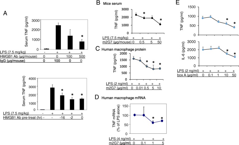
Fig. 1
Anti-HMGB1 antibodies attenuated LPS-induced TNF release in vitro and in vivo . a . Upper: Male BALB/c mice received IP injection of polyclonal rabbit anti-HMGB1 antibody or rabbit control IgG as indi...
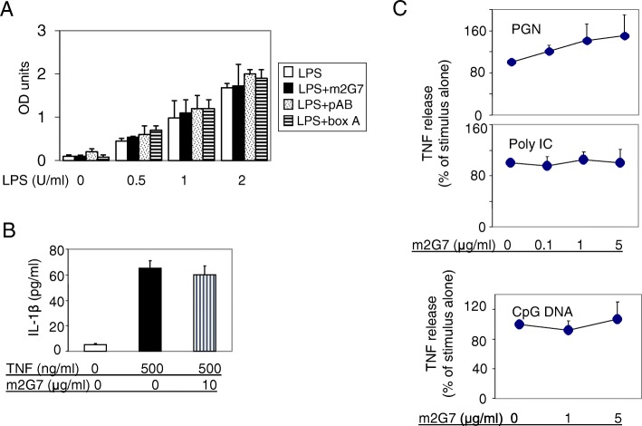
Fig. 2
Neither anti-HMGB1 antibodies nor box A suppressed LPS activity in the Limulus assay; m2G7 did not suppress pro-inflammatory cytokine release induced by TNF, peptidoglycan (PEG), Poly I: C or CpG DNA ...
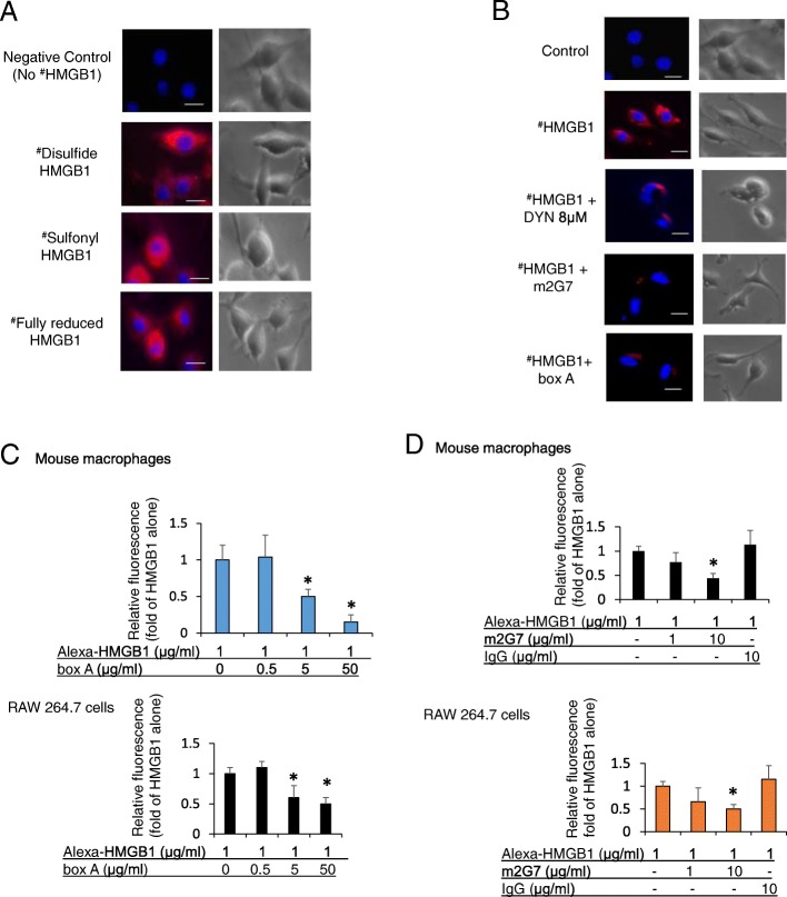
Fig. 3
Anti-HMGB1 m2G7 and box A inhibited macrophages endocytosis of HMGB1. a . Murine macrophage-like RAW 264.7 cells seeded on 24-well culture plate on cover slips (1u2009u00d7u200910 4 /well) were incuba...
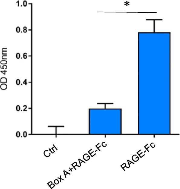
Fig. 4
ELISA demonstrating box A binding to RAGE. Soluble RAGE-Fc chimera (0.25u2009nM) was incubated with an excess amount of box A (10u2009u03bcM) at room temperature for 30u2009min. The mixture was then a...
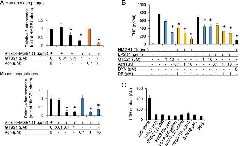
Fig. 5
Cholinergic agonists acetylcholine and GTS-21 inhibited HMGB1 endocytosis, reduced HMGB1 and LPS-induced TNF release in macrophages. a . Primary human macrophages (upper) or thioglycollate-elicited mo...
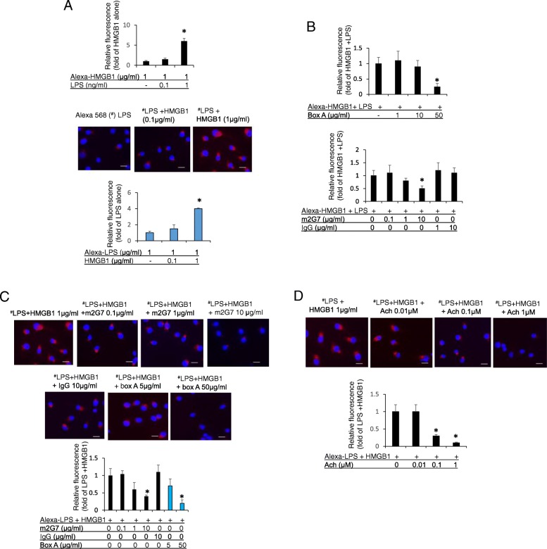
Fig. 6
Macrophage endocytosis of LPS-HMGB1 complexes was inhibited by m2G7, box A and acetylcholine. a . Upper: RAW 264.7 cells, seeded on 24-well culture plate on cover slips (1u2009u00d7u200910 4 /well), w...
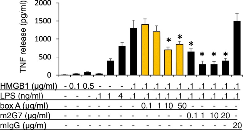
Fig. 7
TNF release induced by LPS-HMGB1 complexes in RAW 264.7 cells was reduced by box A and m2G7. RAW 264.7 cells in 96-well culture plates were used at 90% confluence. Cells were stimulated with HMGB1 or ...
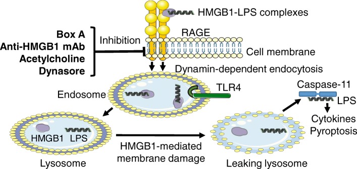
Fig. 8
Molecules inhibiting RAGE-mediated endocytosis of LPS-HMGB1 complexes. The pathogenesis of endotoxemia is caused by extracellular LPS getting attaching to extracellular HMGB1 and forming a complex tha...
Figure images are served from the NIH/NLM PubMed Central Open Access Subset or Europe PMC; copyright remains with the publishers and authors.
💬 Discussion
0 commentsNo comments yet. Be the first to start a discussion!
Leave a Comment