Abstract
Recent studies have been successful at utilizing ectopic expression of transcription factors to generate induced cardiomyocytes (iCMs) from fibroblasts, albeit at a low frequency in vitro. This work investigates the influence of small molecules that have been previously reported to improve differentiation to cardiomyocytes as well as reprogramming to iPSCs in conjunction with ectopic expression of the transcription factors Hand2, Nkx2.5, Gata4, Mef2C, and Tbx5 on the conversion to functional iCMs. We utilized a reporter system in which the calcium indicator GCaMP is driven by the cardiac Troponin T promoter to quantify iCM yield. The TGFβ inhibitor, SB431542 (SB), was identified as a small molecule capable of increasing the conversion of both mouse embryonic fibroblasts and adult cardiac fibroblasts to iCMs up to ∼5 fold. Further characterization revealed that inhibition of TGFβ by SB early in the reprogramming process led to the greatest increase in conversion of fibroblasts to iCMs in a dose-responsive manner. Global transcriptional analysis at Day 3 post-induction of the transcription factors revealed an increased expression of genes associated with the development of cardiac muscle in the presence of SB compared to the vehicle control. Incorporation of SB in the reprogramming process increases the efficiency of iCM generation, one of the major goals necessary to enable the use of iCMs for discovery-based applications and for the clinic.
💻 Software
✨ Fluorophores
🧪 Sample Preparation
🔬 Cell Lines
🏭 Microscope Brands
🧪 Reagent Suppliers
💻 Software Details
💾 Data Repositories
🏛️ Research Organizations (ROR)
Affiliated research institutions:
📋 Methods
Ethics Statement
All animal work was conducted under a protocol (804335) approved by the University of Pennsylvania Institutional Animal Care and Use Committee.
Primary Cell Isolation
Mouse embryonic fibroblasts
(MEFs, isolated at E14.5) were prepared as previously described [5] . Briefly, embryos were harvested from mice of mixed background at 14.5 dpc followed by decapitation and removal of internal visceral organs, including the heart. The tissue was minced and digested with trypsin and trituration. Cells were resuspended in MEF medium (10% FBS and 2 mM L-Glutamine) and plated onto one 10 cm dish per embryo. After 24 hours, cells were passaged at 1∶3 (passage 1). MEFs were used at passages 3–5 for all reprogramming experiments. Adult mouse cardiac fibroblasts were prepared as previously described [5] . Hearts were removed from mice (8–12 weeks in age) and minced in cold PBS. The tissue was digested in 4 mg mL −1 collagenase IV (Sigma) and 10 U mL −1 deoxyribonuclease I (Worthington Biochemical Corporation) and agitation at 37°C for 10 minutes. Samples were spun down and resuspended in TrypLE (Invitrogen) at 37°C with agitation. After 5 minutes, medium (DMEM supplemented with 15% FBS, 1% NEAA) was added and the resulting solution was plated onto gelatin coated 6-well plates. When confluent, the cells were passaged after filtration through a 40 µM filter at 1∶1 to a gelatin coated 10 cm dishes (passage 1). Cells were then passaged 1∶5 and frozen when confluent. Cardiac fibroblasts were used at passage 3 for reprogramming experiments. Plasmid Information All plasmids were constructed as previously described [5] and can be found on Addgene using the following catalog numbers: Troponin T-GCaMP5-Zeo (46027), tetO-Hand2 (46028), tetO-NKX2.5 (46029), tetO-GATA4 (46030), tetO-MEF2C (46031), tetO-TBX5 (46032). Plasmids that were used from Addgene also include: FUdeltaGW-rtTA (19780), psPAX2 (12260), pMD2.G (12259), and PGK-H2B-mCherry (21217). All plasmids were amplified in STBL3 bacteria (Invitrogen) and prepared with Qiagen MidiPrep Kits.
Show full methods section
Ethics Statement
All animal work was conducted under a protocol (804335) approved by the University of Pennsylvania Institutional Animal Care and Use Committee.
Primary Cell Isolation
Mouse embryonic fibroblasts
(MEFs, isolated at E14.5) were prepared as previously described [5] . Briefly, embryos were harvested from mice of mixed background at 14.5 dpc followed by decapitation and removal of internal visceral organs, including the heart. The tissue was minced and digested with trypsin and trituration. Cells were resuspended in MEF medium (10% FBS and 2 mM L-Glutamine) and plated onto one 10 cm dish per embryo. After 24 hours, cells were passaged at 1∶3 (passage 1). MEFs were used at passages 3–5 for all reprogramming experiments. Adult mouse cardiac fibroblasts were prepared as previously described [5] . Hearts were removed from mice (8–12 weeks in age) and minced in cold PBS. The tissue was digested in 4 mg mL −1 collagenase IV (Sigma) and 10 U mL −1 deoxyribonuclease I (Worthington Biochemical Corporation) and agitation at 37°C for 10 minutes. Samples were spun down and resuspended in TrypLE (Invitrogen) at 37°C with agitation. After 5 minutes, medium (DMEM supplemented with 15% FBS, 1% NEAA) was added and the resulting solution was plated onto gelatin coated 6-well plates. When confluent, the cells were passaged after filtration through a 40 µM filter at 1∶1 to a gelatin coated 10 cm dishes (passage 1). Cells were then passaged 1∶5 and frozen when confluent. Cardiac fibroblasts were used at passage 3 for reprogramming experiments. Plasmid Information All plasmids were constructed as previously described [5] and can be found on Addgene using the following catalog numbers: Troponin T-GCaMP5-Zeo (46027), tetO-Hand2 (46028), tetO-NKX2.5 (46029), tetO-GATA4 (46030), tetO-MEF2C (46031), tetO-TBX5 (46032). Plasmids that were used from Addgene also include: FUdeltaGW-rtTA (19780), psPAX2 (12260), pMD2.G (12259), and PGK-H2B-mCherry (21217). All plasmids were amplified in STBL3 bacteria (Invitrogen) and prepared with Qiagen MidiPrep Kits.
Lentivirus Preparation
Lentiviral vectors were packaged into Lenti-X 293T cells (Clontech) using Lipofectamine 2000 (Invitrogen) to deliver 12 µg of the lentiviral backbone plasmid, 7.7 µg psPAX2, and 4.3 µg pMD2.G in 3 mL OPTI-MEM (Invitrogen) to ∼90% confluent 10 cm plates of 293T cells with 10 mL of fresh MEF medium. Viral supernatant was collected at 24 and 48 hours post-transfection (total ∼23 mL), filtered using a 0.45 µM filter (Millipore), aliquots were prepared and frozen at −80°C until use. Viral titer was determined using Lenti-X GoStix (Clontech) and only lentiviruses with a minimum titer of 5×10 5 IFU mL −1 were used for reprogramming experiments. Direct Conversion of Fibroblasts to iCMs Direct conversion of MEFs and cardiac fibroblasts was completed using a protocol similar to that previously described [5] , as shown in Figure 1A . Briefly, glass bottom 12-well plates (MatTek) were coated with poly-L-Lysine solution overnight followed by incubation with MEF medium for 1 hour prior to seeding. At Day -2 cells were dissociated using TrypLE and plated at 30k (∼7.5 k cm −2 ) per well with 250 µL of each of the FUdeltaGW-rtTA and Troponin T-GCaMP5-ZEO reporter lentivirus as well as 500 µL MEF medium (1 mL total per well). On Day-1, the culture medium was replaced with 250 µL MEF medium and 250 µL of each tetO-transcription factor lentivirus (1.5 mL total) as well as any candidate small molecules or vehicle controls (see Table 1 ) where indicated. After another 24 hours (Day 0), the media was changed to Reprogramming medium with doxycycline (2 µg mL −1 ) and the PGK-H2B-mCherry lentivirus. Reprogramming Medium consists of AGM (Lonza, CC-3186) without EGF and supplemented with 2 µg mL −1 doxycycline (Sigma). Cells were also transduced with 200 µL of PGK-H2B-mCherry lentivirus for constitutive expression of nuclear-localized mCherry expression. Reprogramming medium was changed every 2–3 days. For microarray analysis, cells were plated into standard tissue-culture treated 12 well plates (BD Falcon) using the same volumes and protocol described above. For immunocytochemistry, the identical protocol was followed, except cells were plated onto 12 mm diameter poly-L-Lysine coated glass coverslips in 24 well plates (1 coverslip per well) and using half of the volumes described above for all steps. 10.1371/journal.pone.0089678.g001 Figure 1 Direct conversion of mouse embryonic fibroblasts to iCMs can be influenced by treatment with small molecules. Schematic of direct reprogramming strategy of MEFs and timeline (A). Percentage of total cells with flashing Troponin T-GCaMP activity for control and small molecule treated MEFs (B). Representative immunocytochemistry images for HNGMT +DMSO (C), or +BIX (D), +CHIR+XAV (E), and +SB (F) treatments for cardiac Troponin T (green) and nuclei (red). * indicates p
📊 Figures
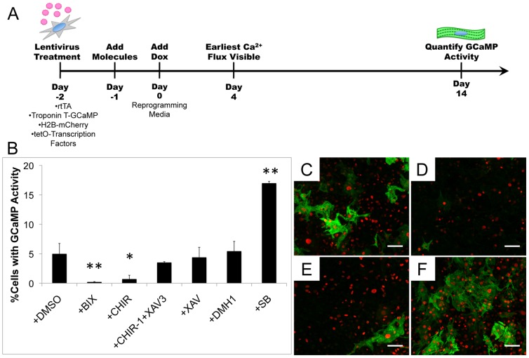
Figure 1
Direct conversion of mouse embryonic fibroblasts to iCMs can be influenced by treatment with small molecules.
Schematic of direct reprogramming strategy of MEFs and timeline (A). Percentage of total cells with flashing Troponin T-GCaMP activity for control and small molecule treated MEFs (B). Representative i...
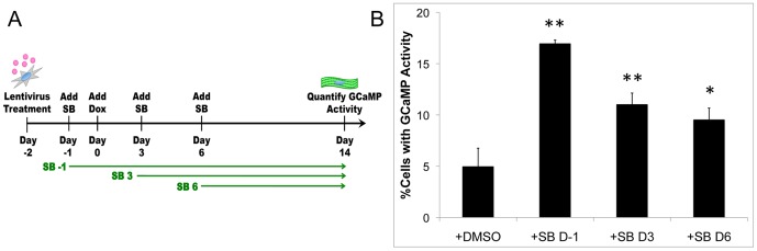
Figure 2
Inhibition of TGFu03b2 early in the conversion process leads to the greatest increase in iCM yield.
Schematic depicting the addition of SB at different time points following induction of the transcription factors (A). Quantification at Day 14 of the number of cells with Troponin T-GCaMP activity upo...
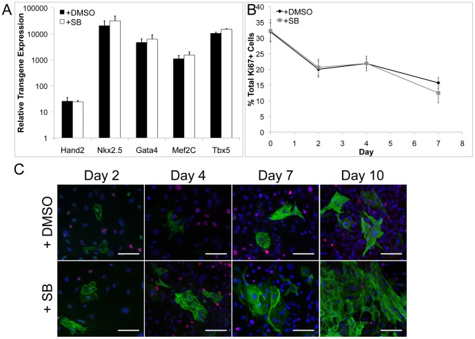
Figure 3
The increase in iCM number with SB treatment is not due to increased transgene expression or changes in cell proliferation.
Evaluation of gene expression of the transgenes at Day 2 via qPCR is not significantly different between +DMSO (black) control and +SB treatment groups (white, A). Evaluation of overall cell prolifera...
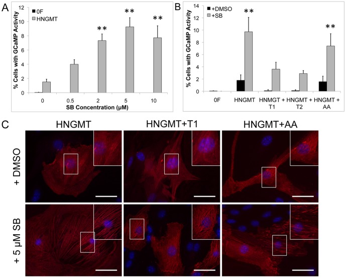
Figure 4
Direct conversion of adult mouse cardiac fibroblasts (CFs) to iCMs.
Conversion efficiency to iCMs increases in yield with increasing concentration of SB (A). Direct conversion of CFs to iCMs is reduced with addition of TGFu03b21 (black, HNGMT+T1) and TGFu03b22 (black,...
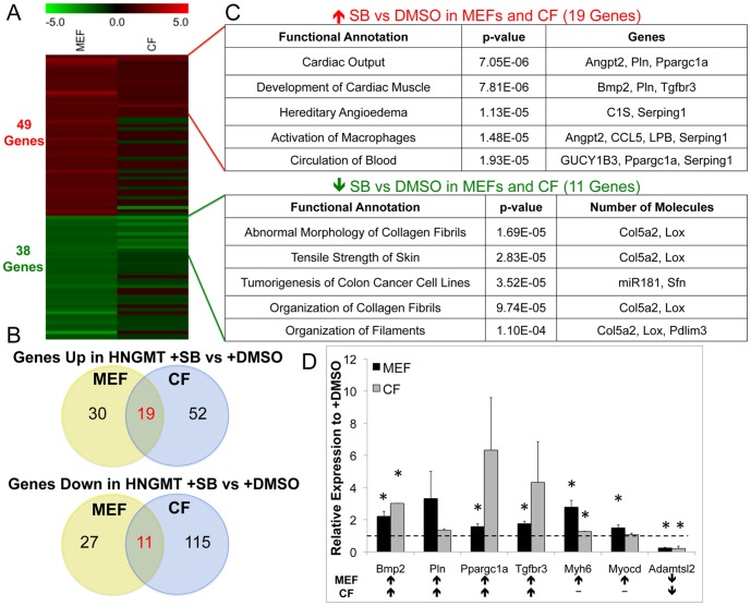
Figure 5
Evaluation of gene expression changes in MEFs and CFs during conversion with and without SB.
Heatmap depicting the 49 and 38 genes up-regulated and down-regulated, respecively exclusively in MEFs for HNGMT + SB versus HNGMT+DMSO as well as the expression of the same genes in CFs (A). Venn dia...
Figure images are served from the NIH/NLM PubMed Central Open Access Subset or Europe PMC; copyright remains with the publishers and authors.
💬 Discussion
0 commentsNo comments yet. Be the first to start a discussion!
Leave a Comment