Abstract
After endocytosis, membrane proteins can recycle to the cell membrane or be degraded in lysosomes. Cargo ubiquitylation favors their lysosomal targeting and can be regulated by external signals, but the mechanism is ill-defined. Here, we studied the post-endocytic trafficking of Jen1, a yeast monocarboxylate transporter, using microfluidics-assisted live-cell imaging. We show that the ubiquitin ligase Rsp5 and the glucose-regulated arrestin-related trafficking adaptors (ART) protein Rod1, involved in the glucose-induced internalization of Jen1, are also required for the post-endocytic sorting of Jen1 to the yeast lysosome. This new step takes place at the trans-Golgi network (TGN), where Rod1 localizes dynamically upon triggering endocytosis. Indeed, transporter trafficking to the TGN after internalization is required for their degradation. Glucose removal promotes Rod1 relocalization to the cytosol and Jen1 deubiquitylation, allowing transporter recycling when the signal is only transient. Therefore, nutrient availability regulates transporter fate through the localization of the ART/Rsp5 ubiquitylation complex at the TGN.
🔬 Techniques
🔭 Microscopes
🧬 Organisms
💻 Software
✨ Fluorophores
🧪 Sample Preparation
🏭 Microscope Brands
🧪 Reagent Suppliers
📷 Detectors
🔎 Objectives
💻 Software Details
📋 Protocols
💾 Data Repositories
🏛️ Research Organizations (ROR)
Affiliated research institutions:
📋 Methods
Yeast strains, transformation and growth conditions Strains are listed and detailed in Supplementary file 1 . All strains are derivatives of the BY4741/2 strains, except for the gga1Δ GGA2-HA and gga1Δ gga2Δ strains, which were kindly provided by Prof. R Piper, University of Iowa, Iowa City, USA ( Scott et al., 2004 ) and the gga1 Δ gga2 Δ and gga1 Δ gga2 Δ ypt6 Δ strains (23344c background) that were kindly provided by Prof. B André, Université Libre de Bruxelles, Belgium ( Lauwers et al., 2009 ). The 9-arrestin mutant was kindly provided by Dr H Pelham (MRC Laboratory of Molecular Biology, Cambridge, UK) ( Nikko and Pelham, 2009 ). Yeast was transformed by standard lithium acetate/polyethylene glycol procedure. Cells were grown in yeast extract/peptone/glucose (YPD) rich medium, or in synthetic complete (SC) medium containing 2% (wt/vol) Glc, or 0.5% (vol/vol) Na-lactate (pH 5.0) (Sigma-Aldrich, Lyon, France). For lactate inductions, cells were grown overnight in SC-Glc, harvested in early exponential phase ( A 600 = 0.3), resuspended in the same volume of SC-lactate and grown for 4 hr ( A 600 = 0.5), before the addition of glucose (2% wt/vol, final concentration). For the observation of Stl1-GFP, cells were grown for 2 hr in lactate medium, and glycerol was then added (3% vol/vol) for 2 hr to induce Stl1-GFP expression and targeting to the plasma membrane. For galactose induction, cells were precultured in SC-Glc medium, and grown overnight to early exponential phase ( A 600 = 0.3) in SC medium containing 2% raffinose (wt/vol) and 0.02% Glc (wt/vol) to initiate growth. Galactose was then added at a final concentration of 2% (wt/vol) and cells were grown for the indicated times. Chase/endocytosis was started by adding glucose to a final concentration of 2% (wt/vol) and incubating for the indicated times. Latrunculin A (Sigma) was used a final concentration of 0.2 mM.
Show full methods section
Yeast strains, transformation and growth conditions Strains are listed and detailed in Supplementary file 1 . All strains are derivatives of the BY4741/2 strains, except for the gga1Δ GGA2-HA and gga1Δ gga2Δ strains, which were kindly provided by Prof. R Piper, University of Iowa, Iowa City, USA ( Scott et al., 2004 ) and the gga1 Δ gga2 Δ and gga1 Δ gga2 Δ ypt6 Δ strains (23344c background) that were kindly provided by Prof. B André, Université Libre de Bruxelles, Belgium ( Lauwers et al., 2009 ). The 9-arrestin mutant was kindly provided by Dr H Pelham (MRC Laboratory of Molecular Biology, Cambridge, UK) ( Nikko and Pelham, 2009 ). Yeast was transformed by standard lithium acetate/polyethylene glycol procedure. Cells were grown in yeast extract/peptone/glucose (YPD) rich medium, or in synthetic complete (SC) medium containing 2% (wt/vol) Glc, or 0.5% (vol/vol) Na-lactate (pH 5.0) (Sigma-Aldrich, Lyon, France). For lactate inductions, cells were grown overnight in SC-Glc, harvested in early exponential phase ( A 600 = 0.3), resuspended in the same volume of SC-lactate and grown for 4 hr ( A 600 = 0.5), before the addition of glucose (2% wt/vol, final concentration). For the observation of Stl1-GFP, cells were grown for 2 hr in lactate medium, and glycerol was then added (3% vol/vol) for 2 hr to induce Stl1-GFP expression and targeting to the plasma membrane. For galactose induction, cells were precultured in SC-Glc medium, and grown overnight to early exponential phase ( A 600 = 0.3) in SC medium containing 2% raffinose (wt/vol) and 0.02% Glc (wt/vol) to initiate growth. Galactose was then added at a final concentration of 2% (wt/vol) and cells were grown for the indicated times. Chase/endocytosis was started by adding glucose to a final concentration of 2% (wt/vol) and incubating for the indicated times. Latrunculin A (Sigma) was used a final concentration of 0.2 mM.
Plasmids and constructs
For the Jen1-KR-GFP mutagenesis, the JEN1 ORF and its promoter were first amplified by PCR from BY4741 genomic DNA (using oligonucleotides oSL337/oSL338), the fragment was digested with SacI/SpeI, cloned at SacI/SpeI sites into a pRS416-based vector containing GFP (pRHT140, lab collection), and sequenced (pSL161). A synthetic gene encoding the Jen1-KR mutant was generated (Eurofins MWG Operon, Ebersberg, Germany), amplified by PCR (oSL371/oSL394), cloned by gap-repair in yeast into pSL161 digested with HindIII/SpeI, and sequenced (pSL163). The galactose-inducible version was cloned similarly: the synthetic gene was amplified by PCR (oSL476/oSL477), and cloned by gap-repair in yeast into pRHT373 ( Becuwe et al., 2012b ) (pSL184). The plasmid encoding Rod1-GFP ( Figures 2D,4G ) was described previously (pSL93) ( Becuwe et al., 2012b ). The plasmid encoding Rod1-KR-GFP ( Figure 6F ) was generated by cloning a SacI/XmaI fragment from pSL143 ( Becuwe et al., 2012b ) into pSL93. The plasmid encoding mTag-BFP2-Rsp5 (pSL303, Figure 5D,E ) was constructed by PCR amplification of the mTagBFP2 sequence (TagBFP2-N, Evrogen JSC, Moscow, Russia) (oSL652/oSL653), digestion and cloning at XbaI/NotI sites in place of GFP in pSL19 (p415-pADH-GFP-Rsp5, Leon et al., 2008 ).
Total protein extracts and phosphatase treatment
For total protein extracts, trichloroacetic acid (TCA; Sigma-Aldrich) was added directly in the culture to a final concentration of 10% (vol/vol), and cells were precipitated on ice for 10 min. Cells were then harvested by centrifugation for 1 min at room temperature at 16,000× g , then lysed with glass beads in a 100 µl of TCA (10%, vol/vol) for 10 min at 4°C. Beads were removed, the lysate was centrifuged for 1 min at room temperature at 16,000× g , and the resulting pellet was resuspended in TCA-sample buffer (Tris–HCl 50 mM pH 6.8, dithiothreitol 100 mM, SDS 2%, bromphenol blue 0.1%, glycerol 10%, containing 200 mM of unbuffered Tris solution) at a concentration of 50 µl/initial OD unit, before being denatured at 37°C for 10 min. Phosphatase treatment was performed as previously described ( Becuwe et al., 2012b ).
Antibodies and immunoblotting
We used monoclonal antibodies raised against GFP (clones 7.1 and 13.1; Roche Diagnostics, Meylan, France), HA (clone F7; Santa Cruz Biotechnology, Dallas, TX), anti-ubiquitin antibody coupled to horseradish peroxidase (clone P4D1; Santa Cruz Biotechnology), and polyclonal antibodies against 3-phosphoglycerate kinase (PGK) (clone 22CS; Life Technologies, Saint Aubin, France). Immunoblots were acquired with the LAS-4000 imaging system (Fuji, Tokyo, Japan). Quantification was performed using ImageJ (NIH) on non-saturated blots.
Fluorescence microscopy
Cells were mounted in synthetic complete medium with the appropriate carbon source and observed with a motorized Olympus BX-61 fluorescence microscope equipped with an Olympus PlanApo 100× oil-immersion objective (1.40 NA), a Spot 4.05 charge-coupled device camera and the MetaVue acquisition software (Molecular Devices; Sunnyvale, CA). Cells were mounted in SD medium and imaged at room temperature. GFP-tagged proteins were visualized using a Chroma GFP II filter (excitation wavelength 440–470 nm). mCh-tagged proteins were visualized using an HcRed I filter (excitation wavelength 525–575 nm). Images were processed in ImageJ (NIH) and Photoshop (Adobe, San Jose, CA) for levels. Vacuolar staining was obtained by incubating cells with 100 µM CMAC (Life Technologies) for 10 min under agitation at 30°C, then cells were then washed twice with water before observations with a confocal microscope (see references below) equipped with a DAPI filter (450QM60). For the microfluidics experiments, cells growing in exponential phase (DO = 0.3–0.6) were injected in a CellASIC microfluidics chamber (ref. YO4C, Merck-Millipore, Darmstadt, Germany), using the Microfluidic Perfusion Platform (ONIX), driven with the interface software ONIX-FG-SW (Merck-Millipore). Cells were trapped and maintained in a uniform plane. Normal growth conditions were reproduced by adjusting the ambient temperature at 30°C with a thermostated chamber, and by flowing cells with the indicated culture medium at 3 psi. The microfluidics device was coupled to a DMI6000 (Leica, Buffalo Grove, IL) microscope, equipped with an oil immersion plan apochromat 100× objective NA 1,4, a QuantEM cooled EMCCD camera (Photometrics, Tucson, AZ), and a spinning-disk confocal system CSU22 (Yokogawa, Tokyo, Japan). Image resolution was 1 pixel = 149 nm. GFP-tagged proteins, mCh-tagged proteins and CMAC staining were visualized with a GFP Filter 535AF45, RFP Filter 590DF35, and DAPI Filter 450QM60 respectively. Images were acquired with MetaMorph 7 software (Molecular Devices, Sunnyvale, CA), and denoised ( Figure 1A,C,D–F ; Figure 3 ; Figure 3A,B,F ; Figure 6A,C,D ; Figure 7B ; and all videos), with the Image J plugin Safir Filter ( Kervrann and Boulanger, 2006 ). For Figure 4—figure supplement 3 , images were acquired with a Revolution xD TuCam system (Andor) equipped with a confocal scanner unit CSU-X1 (Yokogawa) and a Ti microscope with a 100x/1.4 NA objective (Nikon, Tokyo, Japan) and piloted by MetaMorph software (Molecular Devices). Simultaneous acquisitions of two channels were done using the TuCam device (Andor, Belfast, UK) equipped with 580 dicroic filter and 525/50 (green), 616/73 (red) emission filters. Detectors on the TuCam are iXon3 EMCCD camera (Andor) with 8 µm well size. Image stacks are acquired with a Pifoc (Physik Intrumente, Karlsruhe, Germany) with a z-step of 0.2 µm.
Quantification of co-localizations and trafficking delay
Quantifications to evaluate the delayed internalization of Jen1-GFP in rod1Δ vs WT cells were performed manually ( Figure 1G ). The number of Jen1-containing vesicles was quantified over time in each strain by three manual counting on 30 cells, taken from three independent experiments. The total number of Jen1-GFP-labeled structures were counted for the 30 cells and divided by 30. STDEV was also calculated for each time point. Manual quantifications were also performed for co-localization event between Jen1-GFP with markers of internal compartments (Sec7-mCh and Vps17-mCh) ( Figure 4C,I ). Counting was performed three times on 20 cells, taken from three independent experiments. Over time, the percentage of Jen1-GFP-containing vesicles co-localizing with the mCherry marker (Sec7 or Vps17) was calculated for each cell and divided by the mean number of vesicles per cell. STDEV was also calculated for each time point.
Additional files 10.7554/eLife.03307.033 Supplementary file 1. A table listing yeast strains used in this study is provided in Supplementary file 1 . DOI: http://dx.doi.org/10.7554/eLife.03307.033
📊 Figures
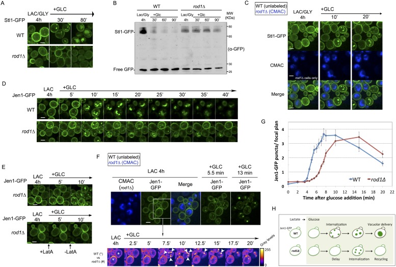
Figure 1.
Dual function of Rod1 in transporter internalization and post-endocytic sorting.
( A ) Rod1 is required for the glucose-induced endocytosis of Stl1, the glycerol/proton symporter, from the plasma membrane to the vacuole. WT (ySL1146) and rod1 u0394 (ySL1153) cells were grown in la...
Video 1.
Rod1 is required for the glucose-induced internalization of the glycerol/proton symporter Stl1.
WT and rod1u0394 (CMAC-positive) cells expressing Stl1-GFP were grown in lactate/glycerol medium and simultaneously observed for 20 min after glucose addition. See also Figure 1C . DOI: http://dx.doi....
Video 2.
Jen1-GFP is internalized upon glucose treatment even in the absence of Rod1.
WT cells (left) and in rod1u0394 cells (right) expressing Jen1-GFP were grown in lactate medium and observed for 45 min after glucose addition. See also Figure 1D . DOI: http://dx.doi.org/10.7554/eLif...
Video 3.
rod1 u0394 cells display a kinetic delay in Jen1 internalization.
WT and rod1u0394 (CMAC positive) cells expressing Jen1-GFP were visualized simultaneously during 20 min after glucose addition (left). Images of the same video were treated in ImageJ using the u2018Fi...
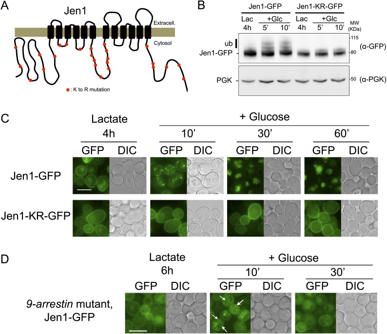
Figure 2.
Jen1 ubiquitylation is required for its glucose-induced endocytosis.
( A ) Schematic of the lysine-to-arginine mutations introduced in the cytosolic loops of Jen1 to generate the Jen1-KR construct. ( B ) Jen1-KR-GFP is not ubiquitylated in response to glucose. WT cells...
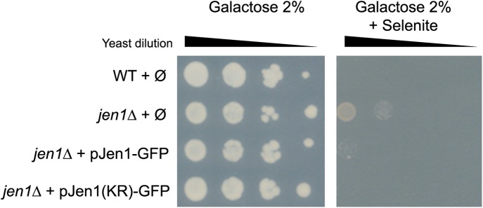
Figure 2u2014figure supplement 1.
Jen1-KR-GFP is a functional protein.
Jen1 transports selenite ( McDermott et al., 2010 ), which is used here as a readout for Jen1 activity. WT and jen1 u0394 strains carrying either an empty vector (u00d8), a plasmid-encoded Jen1-GFP (p...
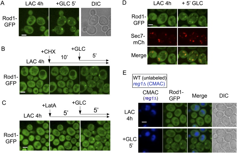
Figure 3.
Rod1 is dynamically recruited to the trans-Golgi network when endocytosis is triggered.
( A ) Rod1-GFP re-localizes from the cytosol to punctate structures in response to glucose. Lactate-grown cells (ySL542) expressing Rod1-GFP were injected into a microfluidics device in lactate medium...
Video 4.
Rod1-GFP relocalizes from the cytosol to punctate structures in response to glucose.
WT cells expressing Rod1-GFP were grown in lactate medium and visualized for 60 min after glucose addition. See also Figure 3A . DOI: http://dx.doi.org/10.7554/eLife.03307.010
Video 5.
Rod1 co-localizes with the trans-Golgi network marker, Sec7-mCherry, in response to glucose.
WT cells expressing both Rod1-GFP and Sec7-mCh were grown during 4 hr in lactate medium and visualized during 45 min after glucose addition. Merge of Rod1-GFP fluorescence (left panel) and Sec7-mCh fl...
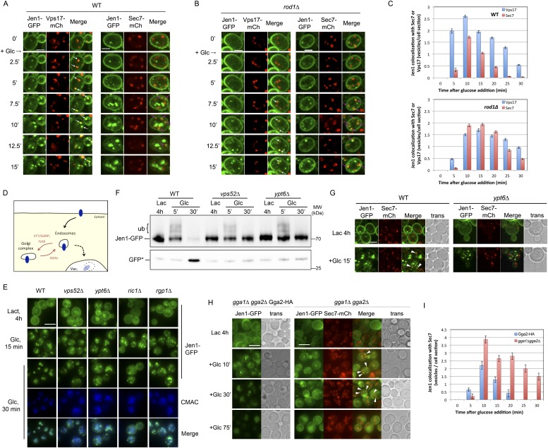
Figure 4.
Jen1 transits through the TGN during its endocytosis and requires Rod1 for exit from the TGN to the vacuole.
( A ) Jen1 co-localizes with the TGN marker, Sec7-mCh, during its trafficking to the vacuole in wild-type cells. WT cells expressing Jen1-GFP and either Vps17-mCh (ySL1168), a marker of the early endo...
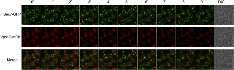
Figure 4u2014figure supplement 1.
Sec7 and Vps17 localize to distinct compartments.
WT cells expressing Sec7-GFP and Vps17-mCh (ySL1531) were injected in the microfluidic device and observed every minute during 9 min. Sec7-GFP-positive vesicles never co-localize with Vps17-mCh-positi...
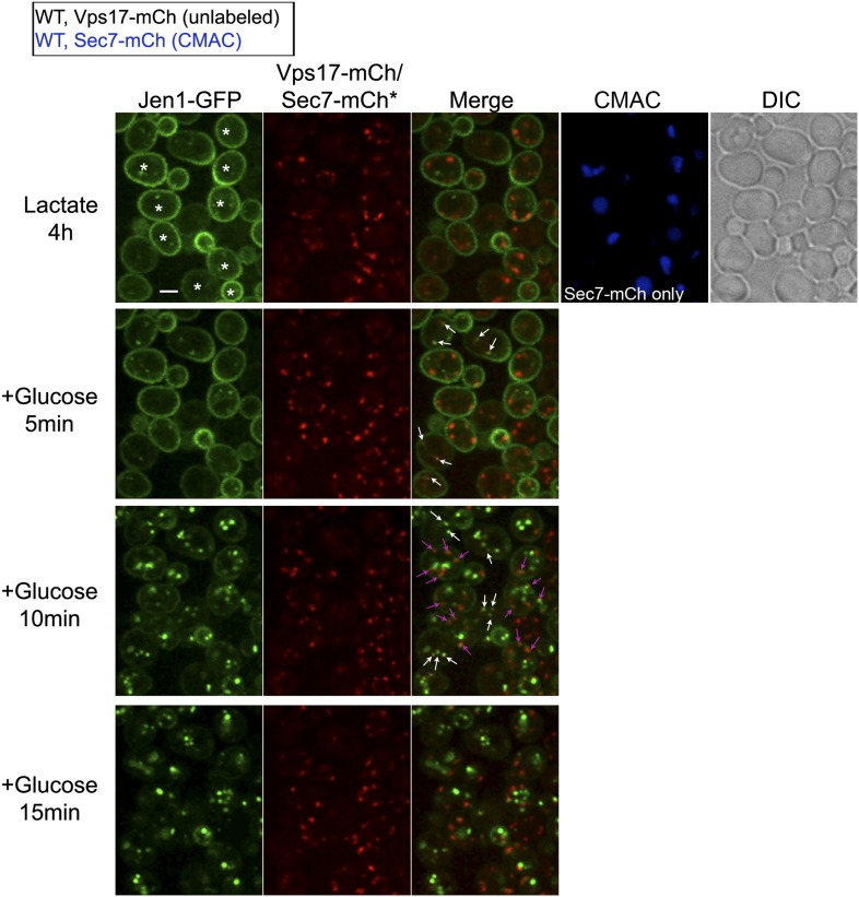
Figure 4u2014figure supplement 2.
Jen1 traffics through the TGN in the course of its endocytosis in wild-type cells.
Uncropped pictures corresponding to the panel presented in Figure 3A . See corresponding legend for details. Cells expressing Sec7-mCh were marked with an asterisk. Co-localization events between Jen1...
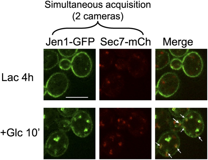
Figure 4u2014figure supplement 3.
Jen1-GFP and Sec7-mCh co-localize to the same compartment when observed simultaneously.
WT cells expressing Jen1-GFP and Sec7-mCh (ySL1165) were grown on lactate medium, and were injected in the microfluidics device in lactate medium, before glucose was added. Cells were imaged at the in...
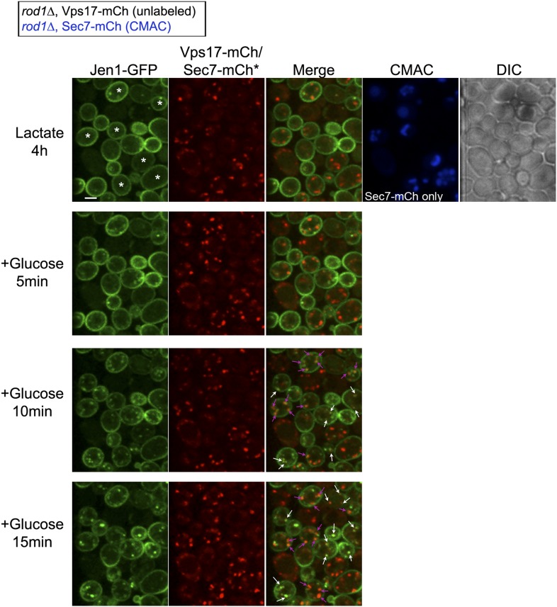
Figure 4u2014figure supplement 4.
Jen1 also co-localizes to the TGN in rod1u0394 mutant cells.
Uncropped pictures corresponding to the panel presented in Figure 3B . See corresponding legend for details. Cells expressing Sec7-mCh were marked with an asterisk. Co-localization events between Jen1...
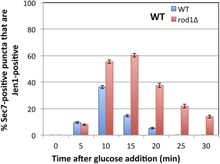
Figure 4u2014figure supplement 5.
Quantification of Sec7-mCh puncta that are also Jen1-GFP positive in WT and rod1u0394 cells.
This quantification was based on the data presented in Figure 4A,B . Jen1 co-localizes more robustly with Sec7-mCh in the rod1u0394 mutant. DOI: http://dx.doi.org/10.7554/eLife.03307.017
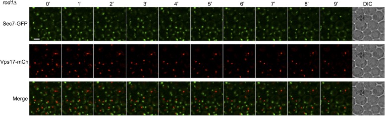
Figure 4u2014figure supplement 6.
Sec7 and Vps17 localize to distinct compartments in rod1u0394 cells.
rod1u0394 cells expressing Sec7-GFP and Vps17-mCh (ySL1602) were injected in the microfluidic device and observed every minute during 9 min. Sec7-GFP-positive vesicles never co-localize with Vps17-mCh...
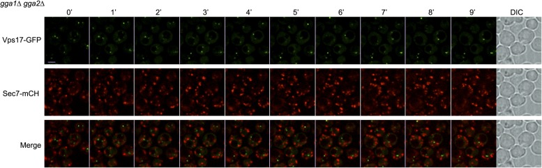
Figure 4u2014figure supplement 7.
Sec7 and Vps17 localize to distinct compartments in gga1u0394gga2u0394 cells.
gga1 u0394 gga2 u0394 cells expressing Vps17-GFP and Sec7-mCh (ySL1615) were injected in the microfluidic device and observed every minute during 9 min. Vps17-GFP-positive vesicles never co-localize w...
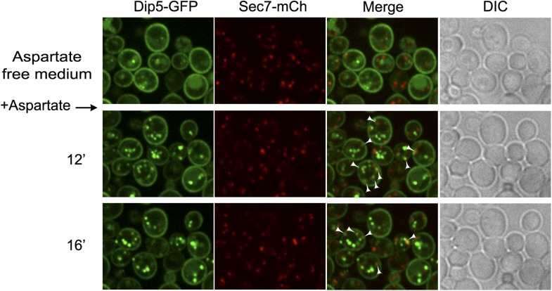
Figure 4u2014figure supplement 8.
Dip5-GFP traffics through the TGN in the course of its endocytosis in WT cells.
WT cells expressing Dip5-GFP and Sec7-mCh (ySL956) were grown on aspartate-free medium, and injected into the microfluidics device in the same medium. Aspartic acid was then added to the medium (200 u...
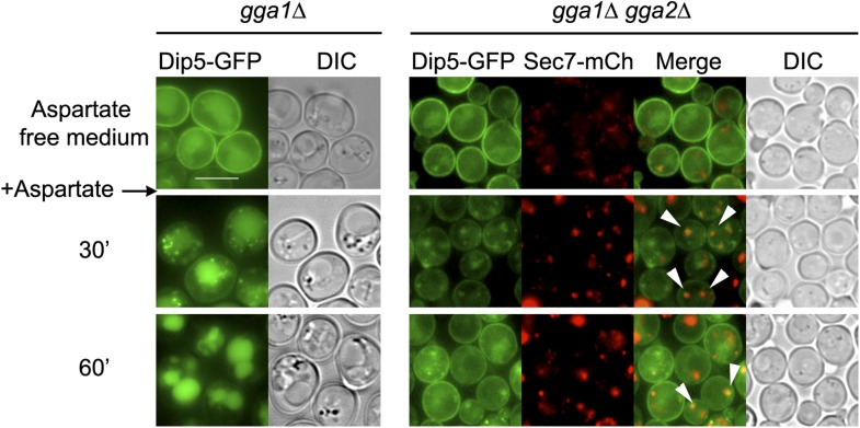
Figure 4u2014figure supplement 9.
Deletions of GGA1 and GGA2 , encoding redundant Golgi-localized clathrin adaptor proteins, alter Dip5 trafficking to the vacuole after endocytosis.
Strains gga1u0394gga2u0394 (ySL1323) and gga1u0394gga2u0394 expressing Gga2-HA (ySL1322), used as a positive control, both expressing Dip5-GFP genomically tagged at its endogenous locus were grown on ...
Video 6.
Uncropped video corresponding to Figure 4A .
Jen1-GFP co-localizes with the endosomal marker Vps17-mCh and the TGN marker Sec7-mCh during its trafficking to the vacuole in wild-type cells. WT cells expressing either both Jen1-GFP and Sec7-mCh (i...
Video 7.
Uncropped video corresponding to Figure 4B .
Jen1-GFP co-localizes with the endosomal marker Vps17-mCh and the TGN marker Sec7-mCh during its trafficking to the vacuole in rod1u0394 cells. rod1u0394 cells expressing either both Jen1-GFP and Sec7...
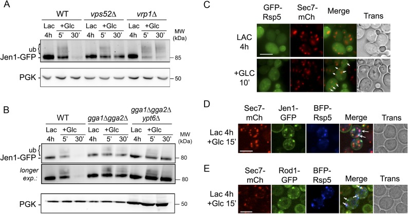
Figure 5.
The prolonged presence of glucose is required for the full endocytosis of Jen1.
( A ) Jen1 is deubiquitylated after endocytosis. WT (ySL1150), vps52 u0394 (ySL1369) and vrp1 u0394 (ySL1610) cells expressing Jen1-GFP were grown in lactate medium and treated with glucose. Crude ext...
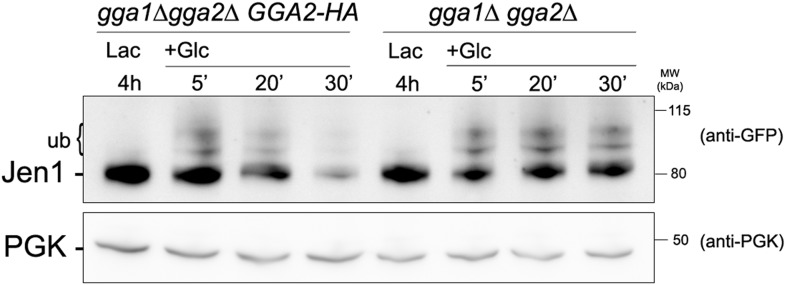
Figure 5u2014figure supplement 1.
Jen1 accumulates in an ubiquitylated form in the gga1u0394 gga2u0394 mutant.
Strains gga1u0394gga2u0394 (ySL1307) or gga1 u0394 gga2u0394 expressing Gga2-HA (ySL1308), used as a positive control, both expressing Jen1-GFP were grown on lactate medium, and crude extracts were pr...
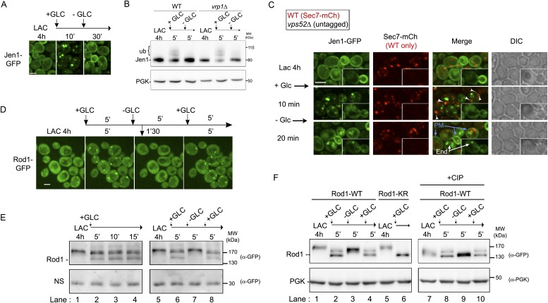
Figure 6.
The control of Jen1 trafficking at the TGN allows the recycling of internalized transporters back to the cell membrane.
( A ) Jen1 endocytosis is reversible upon glucose removal. Lactate-grown WT cells expressing Jen1-GFP (ySL1150) were injected into a microfluidics device in lactate medium. Cells were then imaged over...
Video 8.
Jen1 recycles back to the plasma membrane via the TGN.
WT cells expressing both Jen1-GFP and Sec7-mCh, and vps52u0394 cells expressing only Jen1-GFP were grown 4 hr in lactate medium and observed simultaneously for 10 min of glucose addition and 20 min af...
Video 9.
Jen1 recycling correlates with the loss of Rod1-localization to the TGN.
WT cells expressing Rod1-GFP were grown for 4 hr in lactate medium and observed during 3 cycles of glucose addition/removal (5-min pulses). See also Figure 6D . DOI: http://dx.doi.org/10.7554/eLife.03...
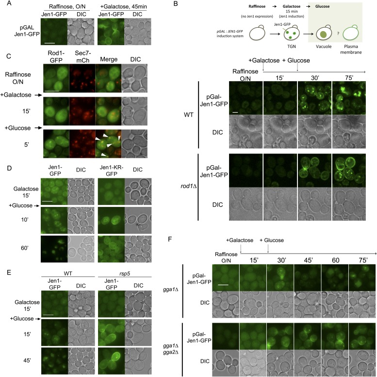
Figure 7.
Rod1 promotes Jen1 exit from the secretory pathway to the vacuole.
( A ) A galactose-inducible Jen1-GFP is targeted to the plasma membrane in galactose medium. WT cells (ySL1083) expressing a galactose-inducible Jen1-GFP were grown in raffinose medium overnight, and ...
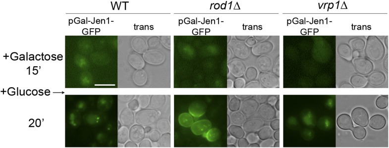
Figure 7u2014figure supplement 1.
The sorting of neosynthesized Jen1 to the vacuole in response to glucose does not require targeting to the plasma membrane and endocytosis.
WT (ySL1083), rod1u0394 (ySL781) and vrp1u0394 (ySL1650) cells both expressing a galactose-inducible Jen1-GFP were grown overnight on raffinose medium. After 15 min of galactose induction, glucose was...
Video 10.
Rod1 is required for the glucose-induced retargeting of Jen1 from the secretory pathway to the vacuole.
WT cells (left panel) and rod1u0394 cells (right panel) expressing Jen1-GFP under a galactose-inducible promoter were grown in raffinose medium overnight and simultaneously observed for 15 min during ...
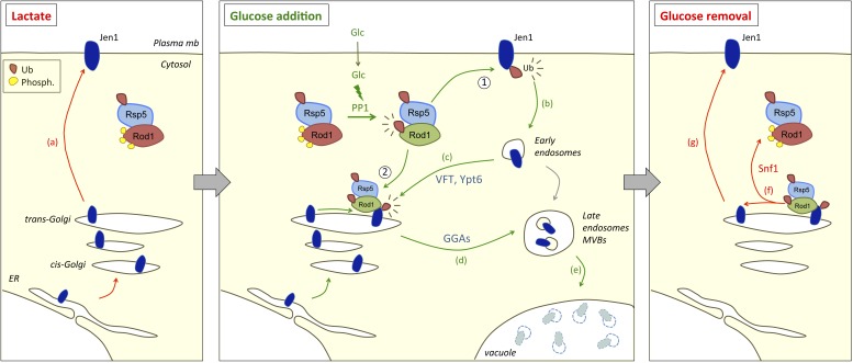
Figure 8.
Working model for the dual function of Rod1 in the regulation of transporter endocytosis and recycling.
Left, in lactate medium, Jen1 is synthesized and targeted to the plasma membrane. Although Rod1 interacts with Rsp5 ( Becuwe et al., 2012b ), it is inactive (phosphorylated) and cytosolic. Middle, In ...
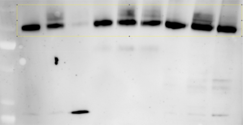
Author response image 1.
Figure images are served from the NIH/NLM PubMed Central Open Access Subset or Europe PMC; copyright remains with the publishers and authors.
💬 Discussion
0 commentsNo comments yet. Be the first to start a discussion!
Leave a Comment