Abstract
Extracellular ATP is a signaling molecule exploited by the immune cells for both autocrine regulation and paracrine communication. By performing live calcium imaging experiments, we show that triggered mouse macrophages are able to propagate calcium signals to resting bystander cells by releasing ATP. ATP-based intercellular communication is mediated by P2X4 and P2X7 receptors and is a feature of pro-inflammatory macrophages. In terms of functional significance, ATP signaling is required for efficient phagocytosis of pathogen-derived molecules and apoptotic cells and may represent a target for macrophage regulation by CD39-expressing cells. These results highlight a cell-to-cell communication mechanism tuning innate immunity.
🔬 Techniques
🔭 Microscopes
🧬 Organisms
✨ Fluorophores
🧪 Sample Preparation
🔬 Cell Lines
🏭 Microscope Brands
🧪 Reagent Suppliers
🔎 Objectives
💻 Software Details
🏷️ Research Resource Identifiers (RRIDs)
Verified research resources used in this paper:
🏛️ Research Organizations (ROR)
Affiliated research institutions:
📋 Methods
Key Resources Table REAGENT or RESOURCE SOURCE IDENTIFIER Antibodies
Rat anti-mouse anti-CD169-AlexaFluor647 (clone 3D6.112) Biorad Cat# MCA884; RRID: AB_322416 Rabbit anti-mouse anti-P2X4R (extracellular) Alomone labs Cat# Apr-024; RRID: AB_2341050 Rabbit anti-mouse anti-P2X7R (extracellular) Alomone labs Cat# Apr-008; RRID: AB_2040065 Rat anti-mouse anti-CD39 (clone Duha-59) Biolegend Cat# 143801; RRID: AB_11203887 Rat anti-mouse anti-CD63 (clone R5G2) MBL International Cat# D263-3; RRID: AB_1278815 Biological Samples Mesenchymal Stem Cell-derived Extracellular Vesicles This lab N/A Chemicals, Peptides, and Recombinant Proteins iso-Ins(1,4,5)P 3 /PM (caged) Enzo Life Sciences Cat# ALX-307-071-C100 Fluo-4-AM ThermoFisher Scientific Cat# F14201 Apyrase from potatoes Sigma Aldrich Cat# A6535 A-740003 Tocris Cat# 3701 5-BDBD Tocris Cat# 3579 2′(3′)-O-(4-Benzoylbenzoyl)adenosine 5′triphosphate triethylammonium salt (Bz-ATP) Sigma Aldrich Cat# B6396 BAPTA, AM, cell permeant chelator ThermoFisher Scientific Cat# B6769 Critical Commercial Assays pHrodo Green E. coli BioParticles Conjugate for Phagocytosis ThermoFisher Scientific Cat# P35366 pHrodo Green Zymosan Bioparticles Conjugate for Phagocytosis ThermoFisher Scientific Cat# P35365 pHrodo Green S. aureus Bioparticles Conjugate for Phagocytosis ThermoFisher Scientific Cat# P35367 Cell Line V Nucleofector Amaxa kit Lonza Cat# VACA-1003 Experimental Models: Cell Lines Raw 264.7 ATCC Cat# TIB-71 B16F10 ATCC Cat# CRL-6475 Experimental Models: Organisms/Strains C57BL/6J mice Internal breeding N/A Oligonucleotides Silencer Select Pre-designed siRNAs for P2X4R Ambion Cat# S71184; s71185 Silencer Select Pre-designed siRNAs for P2X7R Ambion Cat# S71187; s71189 Silencer Select Negative Control No. 1 siRNA Ambion Cat# 4390843 Primers for real-time PCR (see table in Methods ) This paper N/A Software and Algorithms ImageJ (for image analysis) Schneider et al., 2012 N/A FlowJo FlowJo, LLC https://www.flowjo.com/ Other Amicon® Ultra 15 mL Filters Merck Millipore Cat# UFC910096 Contact for Reagent and Resource Sharing Further information and requests for resources and reagents should be directed to and will be fulfilled by the Lead Contact, Sara Zumerle ( sara.zumerle@unipd.it ).
Show full methods section
Key Resources Table REAGENT or RESOURCE SOURCE IDENTIFIER Antibodies
Rat anti-mouse anti-CD169-AlexaFluor647 (clone 3D6.112) Biorad Cat# MCA884; RRID: AB_322416 Rabbit anti-mouse anti-P2X4R (extracellular) Alomone labs Cat# Apr-024; RRID: AB_2341050 Rabbit anti-mouse anti-P2X7R (extracellular) Alomone labs Cat# Apr-008; RRID: AB_2040065 Rat anti-mouse anti-CD39 (clone Duha-59) Biolegend Cat# 143801; RRID: AB_11203887 Rat anti-mouse anti-CD63 (clone R5G2) MBL International Cat# D263-3; RRID: AB_1278815 Biological Samples Mesenchymal Stem Cell-derived Extracellular Vesicles This lab N/A Chemicals, Peptides, and Recombinant Proteins iso-Ins(1,4,5)P 3 /PM (caged) Enzo Life Sciences Cat# ALX-307-071-C100 Fluo-4-AM ThermoFisher Scientific Cat# F14201 Apyrase from potatoes Sigma Aldrich Cat# A6535 A-740003 Tocris Cat# 3701 5-BDBD Tocris Cat# 3579 2′(3′)-O-(4-Benzoylbenzoyl)adenosine 5′triphosphate triethylammonium salt (Bz-ATP) Sigma Aldrich Cat# B6396 BAPTA, AM, cell permeant chelator ThermoFisher Scientific Cat# B6769 Critical Commercial Assays pHrodo Green E. coli BioParticles Conjugate for Phagocytosis ThermoFisher Scientific Cat# P35366 pHrodo Green Zymosan Bioparticles Conjugate for Phagocytosis ThermoFisher Scientific Cat# P35365 pHrodo Green S. aureus Bioparticles Conjugate for Phagocytosis ThermoFisher Scientific Cat# P35367 Cell Line V Nucleofector Amaxa kit Lonza Cat# VACA-1003 Experimental Models: Cell Lines Raw 264.7 ATCC Cat# TIB-71 B16F10 ATCC Cat# CRL-6475 Experimental Models: Organisms/Strains C57BL/6J mice Internal breeding N/A Oligonucleotides Silencer Select Pre-designed siRNAs for P2X4R Ambion Cat# S71184; s71185 Silencer Select Pre-designed siRNAs for P2X7R Ambion Cat# S71187; s71189 Silencer Select Negative Control No. 1 siRNA Ambion Cat# 4390843 Primers for real-time PCR (see table in Methods ) This paper N/A Software and Algorithms ImageJ (for image analysis) Schneider et al., 2012 N/A FlowJo FlowJo, LLC https://www.flowjo.com/ Other Amicon® Ultra 15 mL Filters Merck Millipore Cat# UFC910096 Contact for Reagent and Resource Sharing Further information and requests for resources and reagents should be directed to and will be fulfilled by the Lead Contact, Sara Zumerle ( sara.zumerle@unipd.it ).
Experimental Model and Subject Details Cells
The murine RAW 264.7 cell line was cultured in DMEM 4,5 g/L glucose supplemented with 2 mM L-glutamine, 1 mM sodium pyruvate, 100 U/ml penicillin, 100 U/ml streptomycin (Lonza), and 10% Fetal Bovine Serum (FBS, GIBCO). The murine melanoma B16F10 cell line was cultured in RPMI supplemented with 2mM L-glutamine, 1 mM sodium pyruvate, 100 U/ml penicillin, 100 U/ml streptomycin (Lonza) 0.1% β-mercaptoethanol and 10% Fetal Bovine Serum (FBS, GIBCO). Bone marrow-derived macrophages (BMDMs) were obtained from bone marrow precursor cells. Briefly, femurs and tibiae were collected from 8-12 week old C57BL/6J mice. Bone marrow was flushed with IMDM supplemented with 100 U/ml penicillin, 100 U/ml streptomycin (Lonza), and 10% FBS Superior (Millipore) and red blood cells were removed using ACK lysis buffer (Lonza). 50000 cells/cm 2 were seeded on low-adhesion plates in complete medium supplemented with 40 ng/ml murine M-CSF (Miltenyi Biotech). M-CSF was replenished after 4 days in culture. Experiments were performed at day 7-8. In some experiments, at day 6-7 the cell culture medium was replaced with fresh medium containing 10 ng/ml mIFNγ (Miltenyi) or 20 ng/ml mIL-4 (Miltenyi) to induce the polarization into M1 or M2 status, respectively.
Murine Mesenchymal Stem Cells
(MSCs) were isolated as previously described ( Zanotti et al., 2013 ). Briefly, femurs and tibiae were collected from 8 week-old, C57BL/6 female mice; the bone marrow was flushed and cultured in 25 cm 2 tissue culture flasks at a concentration of 2x10 6 cells/cm 2 using complete Dulbecco modified Eagle medium low glucose (DMEM, Lonza) supplemented with 20% heat-inactivated fetal bovine serum (Biosera), 2 mM glutamine (Lonza), 100 U/ml penicillin/streptomycin (Lonza). After 48 hours, the non-adherent cells were removed. After reaching 70%–80% confluence, the adherent cells were harvested and expanded in larger flasks.
Mice
C57BL/6J mice were kept in regular light and dark cycles, with unrestricted access to water and food. Mice were kept under Italian national and EU directives (2010/63/EU) for animal research with protocols approved by institute Ethical Committee and the Italian Ministry of Health (1045/2016-PR approved on 26/10/2016).
Method Details Confocal calcium imaging
Murine macrophages previously plated on glass coverslips were loaded with the cell membrane permeable ester of caged-IP 3 -PM (1 μM, Enzo Life Science) and the calcium indicator Fluo-4-AM (5 μM, Molecular Probes), Pluronic F-127 (0.1%, w/v, Life Technologies), sulphinpyrazone (250 μM) in unsupplemented culture medium for 30 minutes at 37°C. Live calcium imaging was performed in imaging buffer (Hank’s Balanced Salt Solution supplemented with Ca 2+ 2 mM, Lonza) using a Zeiss LSM700 laser scanning confocal microscope. Time-lapse images were acquired for 1 minute (0.6 frame/sec), using a 40X water immersion objective (Zeiss W Plan-Apochromat 40x/1.0 DIC M27). Fluo-4-AM was excited with a 488-nm laser (0.5% power). Regions of interest (ROIs) were drawn on one distinct cell per field; the UV laser (405 nm, 100% power, 10 ms pulse) was used to release active IP 3 within the ROI. Images were analyzed using ImageJ software ( Schneider et al., 2012 ). Fluo-4 traces were generated by averaging pixel signals within the ROIs, and normalized on the baseline fluorescence of the first 5 frames (ΔF/F 0 ). Control experiments were performed using murine macrophages loaded with calcium indicator alone. In some experiments, the imaging solution was supplemented with 5 U/ml apyrase (Sigma Aldrich), 100 μM A740003 (Sigma Aldrich), 100 μM 5BDBD (Tocris), 100 μM BzATP (Sigma Aldrich) or in Ca 2+ -free HBSS supplemented with 2 mM EGTA (Sigma Aldrich).
Ex vivo lymph node preparation and calcium imaging
C57BL/6J mice were injected with 1 μg of Alexa647-conjugated anti-CD169 antibody (Biorad) in the footpad. After 45 minutes, mice were sacrificed and popliteal lymph nodes were collected and embedded in 4% low-melt agarose (Sigma Aldrich). 200 μm slices were cut with a vibratome in a saline solution containing 150 mM NaCl, 10 mM HEPES–NaOH, 5 mM D–glucose, 5 mM KCl, 2 mM CaCl 2 , 2 mM sodium pyruvate, 1 mM MgCl 2 (pH 7.2, 310 mOsm). The slices were loaded with caged-IP 3 -PM (1 μM, Enzo Life Science), Fluo-4-AM (5 μM, Molecular Probes), Pluronic F-127 (0.1%, w/v, Life Technologies), sulphinpyrazone (250 μM) in unsupplemented phenol red-free IMDM (GIBCO) for 30 minutes at 37°C. Live calcium imaging was performed in phenol red-free IMDM using a Zeiss LSM700 laser scanning confocal microscope. Time-lapse images were acquired for at least 1 minute (0.6 frame/sec), using a 40X water immersion objective (Zeiss W Plan-Apochromat 40x/1.0 DIC M27). Fluo-4-AM was excited with a 488-nm laser (0.5% power). Regions of interest (ROIs) were drawn on one distinct CD169-positive macrophage per field; the UV laser (405 nm, 100% power, 20 ms pulse) was used to release active IP 3 within the ROI. Apyrase (5 U/ml, Sigma) was added to the imaging buffer in some of the experiments. We have observed calcium waves in C57BL/6J murine LNs despite the known DNA polymorphism in the p2x7 gene that reduces receptor function. LN slides loaded with Fluo-4-AM only (without caged-IP 3 ) were negative controls for calcium wave.
Image analysis
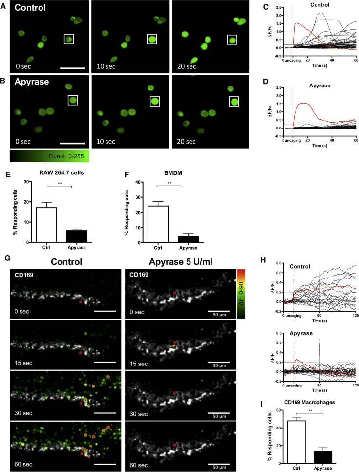
For the analysis of calcium signaling events, one ROI per cell was designed and single cell Fluo-4 traces were generated by averaging pixel signals within the ROIs, and normalized on the baseline fluorescence of the first 5 frames (ΔF/F 0 ) using ImageJ software. A cell was considered as “Responding cell” when its fluorescence variation increased more than 20% over the baseline (ΔF/F 0 ≥ 0.2) in the first 40 s after the uncaging of one single cell. The % of responding cells was calculated as the number of cells showing a ΔF/F 0 value ≥ 0.2 over the total number of cells in each field. Calcium signaling in viable lymph node slices was analyzed using the same approach; ROIs were designed only on CD169 + cells.
RNA and Real-Time PCR Total
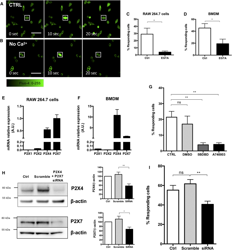
RNA was extracted from macrophages using TriZol reagent (Thermo Fisher Scientific), and cDNA synthetized from 200 ng of RNA using the High Capacity RT kit (Applied Biosystems), according to the manufacturers’ instructions. Real-Time PCR was performed using Sybr Green I (Applied Biosystems) on a 7900HT Fast Real-Time PCR System (Applied Biosystems), with the primers indicated in the table. Gene expression was normalized on RPLP0 expression. Name Forward primer 5′-3′ Reverse primer 5′-3′ Rplp0 GGGCATCACCACGAAAATCTC CTGCCGTTGTCAAACACCT P2X1R ATCTTTGGCTGGTGTCCTGTAG TGACCTTGAAGCGTGGAAAG P2X2R AGGACGCTGTGTACCCTATTAC TTTCAGAAGTCCCATCCTCCAC P2X4R GCAGAAAACTTCACCCTCTTGG AGGTAGGAGGTGGTAATGTTGG P2X7R GCAGGGGAACTCATTCTTTGTC TCCACCCCTTTTTACAACGC Gene Silencing P2X4 and P2X7 gene expression was transiently knocked down transfecting RAW 264.7 cells with Silencer Select Pre-designed siRNAs (Ambion) specific for P2X4 (s71184; s71185) and/or P2X7 (s71187; s71189). Silencer Select Negative Control No. 1 siRNA (Ambion) was adopted as scramble. Transfection was performed with Cell Line V Nucleofector Amaxa kit (Lonza), according to the manufacturer’s instructions. All siRNAs were used at the final concentration of 50 nM. The silencing efficiency was controlled by western blot.
Protein extraction and western blotting
Total protein lysates from macrophages were obtained using RIPA buffer (1mM EDTA, 150 mM NaCl, 1% NP-40, 0.1% SDS, 0.5% Sodium deoxycholate, 50 mM Tris HCl, Protease inhibitors cocktail, pH 8). The protein concentration was quantified with the BCA assay (Euroclone), according to the manufacturer’s instructions. 30 μg of total lysates were resuspended in NuPAGE LDS Loading Buffer (Thermo Fisher Scientific) supplemented with 50 mM DTT and boiled at 99°C for 5 minutes. Samples were loaded in NuPAGE 4%–12% Bis-Tris Gels (Thermo Fisher Scientific) and Western Blot analysis was performed using standard methods. Anti-P2X4R (Alomone Labs), anti-P2X7R (Alomone Labs) and anti-β-actin (Abcam) primary antibodies were used. Immunoblots were acquired with the ImageQuantLS camera and analyzed with ImageJ Software.
Flow cytometry
Macrophages were detached using PBS supplemented with 2mM EDTA and resuspended in the staining solution (PBS supplemented with 2% FBS). For lymph node cell analysis, single cell suspensions were obtained from murine lymph nodes by passing cells through 40-μm cell strainers. After counting, cells were incubated with anti-CD16/CD32 (BD PharMingen), and subsequently stained with the appropriate combinations of the following antibodies: anti-Cd11b-PerCP Cy5.5 (M1/70, BD Biosciences), anti-CD169-AlexaFluor647 (MOMA-1, Biorad), anti-P2X7R-extracellular-FITC (Alomone Labs), purified anti-P2X4R-extracellular (Alomone Labs) followed by incubation with the secondary anti-rabbit-FITC antibody (Thermo Fisher). When indicated, appropriate isotype control antibodies were used to discriminate for antibody specificity. Stained cells were analyzed with a FACSCanto II instrument (BD Biosciences). FlowJo software was used for data analysis. In vitro phagocytosis assay of pHrodo bioparticles In vitro phagocytosis was assessed with Alexa Fluor 488-Zymosan, E. coli or S. aureus PhRodo bioparticles (Molecular Probes). Bone marrow-derived macrophages were starved in suspension in IMDM supplemented with 0.2% Bovine Serum Albumin (Sigma Aldrich), for 40 minutes at 37°C. After 15 minutes on ice, cells were incubated with bioparticle suspension (100 ng bioparticles/100000 cells) at 37°C. Cells pretreated for 30 minutes with 20 μM Cytochalasin D (Calbiochem) were used as negative control. For some experiments, 5 U/ml apyrase, 100 μM A740003, 100 μM 5BDBD, 5 mM EGTA, 200 μM ARL-67516 were added during the experiment. For intracellular calcium chelation, macrophages were loaded with 10 μM BAPTA-AM (Thermo Fisher Scientific) in starvation medium at 37°C for 30 minutes. Phagocytosis was stopped after 15 or 30 minutes by placing cells on ice. The fluorescence of non-internalized beads was quenched using Trypan blue and the samples were analyzed by flow cytometry with a FACSCanto II flow cytometer (BD Bioscience). The phagocytic index was scored as percentage of Alexa Fluor 488-positive macrophages multiplied by their mean of fluorescence (MFI) and normalized on the Cytochalasin-treated samples. In vitro phagocytosis assay of apoptotic cells For induction of apoptosis, murine B16F10 cells were detached using 0.25% Trypsin-2mM EDTA and loaded with 5 μM Calcein green (ThermoFisher) for 30 min at 37°C in serum free medium. Cells were then washed and heated at 56°C for 10 min. After heat-shock, 2 mM staurosporine was added and cells were kept in incubator for at least 1 hour. Before apoptotic cells (AC) phagocytosis, apoptosis was assessed using APC-Annexin V (eBioscience). Apoptosis was generally over the 95%. Bone marrow-derived macrophages were starved in suspension in IMDM supplemented with 0.2% Bovine Serum Albumin (Sigma Aldrich), for 40 minutes at 37°C. During starvation, macrophages were loaded with 5 μM DDAO-1 (ThemoFisher). After 15 minutes on ice, cells were incubated with apoptotic cells at the ratio of 10 apoptotic cells to 1 macrophage at 37°C. Apyrase was added at the concentrations of 5 or 10 U/ml. Cells pretreated for 30 minutes with 20 μM Cytochalasin D were used as negative control. After 30 and 60 min, phagocytosis was stopped by diluting cells and cells were then placed on ice until the analysis by flow cytometry. Phagocytosis was assessed by measuring the percentage of Calcein green/DDAO-1 double positive cells. Data were normalized with Cytochalasin D-treated cells. Purification of MSC-derived extracellular vesicles (EVs) MSC growth medium was substituted with DMEM low glucose supplemented with 10% FBS, 2mM glutamine, 100 U/ml penicillin/streptomycin for 24 hours. Subsequently, the medium was changed with DMEM low glucose supplemented with 2 mM glutamine, 100 U/ml penicillin/streptomycin for the 18 hours. Conditioned medium was harvested and centrifuged at 4000 rpm for 10 min. Extracellular Vesicles (EVs) were isolated from MSC-conditioned medium by ultrafiltration using Amicon® Ultra 15 mL Filters (Merck Millipore) following manufacturer’s instructions. Briefly, each tube was first sterilized with 70% ethanol and then washed two times by centrifuging it at 4000 g for 10 minutes. Subsequently, 12 mL of MSC-conditioned medium were loaded into the tube and centrifuged at 2800 g per 20 minutes at room temperature. After washing the filter with PBS, EVs were collected, concentrated in about 150 μL of PBS, and directly stored at −80°C. For protein characterization, an additional wash with PBS with 0.4% SDS of the filter membrane was added. Total protein of EVs was quantified by MicroBCA kit (Pierce). From 3 to 5 μg of proteins were separated by 10% SDS-PAGE under non-reductive conditions. Anti-CD63 (MBL), and anti-CD39 (Biolegend) primary antibodies were used.
Quantification and Statistical Analysis
Data analysis was performed using Prism 6 (GraphPad, USA). Sample size was not predetermined by statistical methods. The data distribution was verified by performing Shapiro-Wilk normality test. Student’s t test was used to compare two groups and one-way ANOVA followed by Bonferroni post hoc test was used to compare three or more groups, as indicated in figure legends. Statistical significance was taken at p < 0.05 and indicated with asterisks in the figures (ns = non-significant; ∗ = p < 0.05; ∗∗ = p < 0.01; ∗∗∗ = p < 0.001). The n number for each experiment has been stated in figure legends, and represents the number of independent biological replicates. When using samples derived from animals, n represents the number of animals.
Experimental Model and Subject Details Cells
The murine RAW 264.7 cell line was cultured in DMEM 4,5 g/L glucose supplemented with 2 mM L-glutamine, 1 mM sodium pyruvate, 100 U/ml penicillin, 100 U/ml streptomycin (Lonza), and 10% Fetal Bovine Serum (FBS, GIBCO). The murine melanoma B16F10 cell line was cultured in RPMI supplemented with 2mM L-glutamine, 1 mM sodium pyruvate, 100 U/ml penicillin, 100 U/ml streptomycin (Lonza) 0.1% β-mercaptoethanol and 10% Fetal Bovine Serum (FBS, GIBCO). Bone marrow-derived macrophages (BMDMs) were obtained from bone marrow precursor cells. Briefly, femurs and tibiae were collected from 8-12 week old C57BL/6J mice. Bone marrow was flushed with IMDM supplemented with 100 U/ml penicillin, 100 U/ml streptomycin (Lonza), and 10% FBS Superior (Millipore) and red blood cells were removed using ACK lysis buffer (Lonza). 50000 cells/cm 2 were seeded on low-adhesion plates in complete medium supplemented with 40 ng/ml murine M-CSF (Miltenyi Biotech). M-CSF was replenished after 4 days in culture. Experiments were performed at day 7-8. In some experiments, at day 6-7 the cell culture medium was replaced with fresh medium containing 10 ng/ml mIFNγ (Miltenyi) or 20 ng/ml mIL-4 (Miltenyi) to induce the polarization into M1 or M2 status, respectively.
Murine Mesenchymal Stem Cells
(MSCs) were isolated as previously described ( Zanotti et al., 2013 ). Briefly, femurs and tibiae were collected from 8 week-old, C57BL/6 female mice; the bone marrow was flushed and cultured in 25 cm 2 tissue culture flasks at a concentration of 2x10 6 cells/cm 2 using complete Dulbecco modified Eagle medium low glucose (DMEM, Lonza) supplemented with 20% heat-inactivated fetal bovine serum (Biosera), 2 mM glutamine (Lonza), 100 U/ml penicillin/streptomycin (Lonza). After 48 hours, the non-adherent cells were removed. After reaching 70%–80% confluence, the adherent cells were harvested and expanded in larger flasks.
Mice
C57BL/6J mice were kept in regular light and dark cycles, with unrestricted access to water and food. Mice were kept under Italian national and EU directives (2010/63/EU) for animal research with protocols approved by institute Ethical Committee and the Italian Ministry of Health (1045/2016-PR approved on 26/10/2016).
Method Details Confocal calcium imaging
Murine macrophages previously plated on glass coverslips were loaded with the cell membrane permeable ester of caged-IP 3 -PM (1 μM, Enzo Life Science) and the calcium indicator Fluo-4-AM (5 μM, Molecular Probes), Pluronic F-127 (0.1%, w/v, Life Technologies), sulphinpyrazone (250 μM) in unsupplemented culture medium for 30 minutes at 37°C. Live calcium imaging was performed in imaging buffer (Hank’s Balanced Salt Solution supplemented with Ca 2+ 2 mM, Lonza) using a Zeiss LSM700 laser scanning confocal microscope. Time-lapse images were acquired for 1 minute (0.6 frame/sec), using a 40X water immersion objective (Zeiss W Plan-Apochromat 40x/1.0 DIC M27). Fluo-4-AM was excited with a 488-nm laser (0.5% power). Regions of interest (ROIs) were drawn on one distinct cell per field; the UV laser (405 nm, 100% power, 10 ms pulse) was used to release active IP 3 within the ROI. Images were analyzed using ImageJ software ( Schneider et al., 2012 ). Fluo-4 traces were generated by averaging pixel signals within the ROIs, and normalized on the baseline fluorescence of the first 5 frames (ΔF/F 0 ). Control experiments were performed using murine macrophages loaded with calcium indicator alone. In some experiments, the imaging solution was supplemented with 5 U/ml apyrase (Sigma Aldrich), 100 μM A740003 (Sigma Aldrich), 100 μM 5BDBD (Tocris), 100 μM BzATP (Sigma Aldrich) or in Ca 2+ -free HBSS supplemented with 2 mM EGTA (Sigma Aldrich).
Ex vivo lymph node preparation and calcium imaging
C57BL/6J mice were injected with 1 μg of Alexa647-conjugated anti-CD169 antibody (Biorad) in the footpad. After 45 minutes, mice were sacrificed and popliteal lymph nodes were collected and embedded in 4% low-melt agarose (Sigma Aldrich). 200 μm slices were cut with a vibratome in a saline solution containing 150 mM NaCl, 10 mM HEPES–NaOH, 5 mM D–glucose, 5 mM KCl, 2 mM CaCl 2 , 2 mM sodium pyruvate, 1 mM MgCl 2 (pH 7.2, 310 mOsm). The slices were loaded with caged-IP 3 -PM (1 μM, Enzo Life Science), Fluo-4-AM (5 μM, Molecular Probes), Pluronic F-127 (0.1%, w/v, Life Technologies), sulphinpyrazone (250 μM) in unsupplemented phenol red-free IMDM (GIBCO) for 30 minutes at 37°C. Live calcium imaging was performed in phenol red-free IMDM using a Zeiss LSM700 laser scanning confocal microscope. Time-lapse images were acquired for at least 1 minute (0.6 frame/sec), using a 40X water immersion objective (Zeiss W Plan-Apochromat 40x/1.0 DIC M27). Fluo-4-AM was excited with a 488-nm laser (0.5% power). Regions of interest (ROIs) were drawn on one distinct CD169-positive macrophage per field; the UV laser (405 nm, 100% power, 20 ms pulse) was used to release active IP 3 within the ROI. Apyrase (5 U/ml, Sigma) was added to the imaging buffer in some of the experiments. We have observed calcium waves in C57BL/6J murine LNs despite the known DNA polymorphism in the p2x7 gene that reduces receptor function. LN slides loaded with Fluo-4-AM only (without caged-IP 3 ) were negative controls for calcium wave.
Image analysis
For the analysis of calcium signaling events, one ROI per cell was designed and single cell Fluo-4 traces were generated by averaging pixel signals within the ROIs, and normalized on the baseline fluorescence of the first 5 frames (ΔF/F 0 ) using ImageJ software. A cell was considered as “Responding cell” when its fluorescence variation increased more than 20% over the baseline (ΔF/F 0 ≥ 0.2) in the first 40 s after the uncaging of one single cell. The % of responding cells was calculated as the number of cells showing a ΔF/F 0 value ≥ 0.2 over the total number of cells in each field. Calcium signaling in viable lymph node slices was analyzed using the same approach; ROIs were designed only on CD169 + cells.
RNA and Real-Time PCR Total
RNA was extracted from macrophages using TriZol reagent (Thermo Fisher Scientific), and cDNA synthetized from 200 ng of RNA using the High Capacity RT kit (Applied Biosystems), according to the manufacturers’ instructions. Real-Time PCR was performed using Sybr Green I (Applied Biosystems) on a 7900HT Fast Real-Time PCR System (Applied Biosystems), with the primers indicated in the table. Gene expression was normalized on RPLP0 expression. Name Forward primer 5′-3′ Reverse primer 5′-3′ Rplp0 GGGCATCACCACGAAAATCTC CTGCCGTTGTCAAACACCT P2X1R ATCTTTGGCTGGTGTCCTGTAG TGACCTTGAAGCGTGGAAAG P2X2R AGGACGCTGTGTACCCTATTAC TTTCAGAAGTCCCATCCTCCAC P2X4R GCAGAAAACTTCACCCTCTTGG AGGTAGGAGGTGGTAATGTTGG P2X7R GCAGGGGAACTCATTCTTTGTC TCCACCCCTTTTTACAACGC Gene Silencing P2X4 and P2X7 gene expression was transiently knocked down transfecting RAW 264.7 cells with Silencer Select Pre-designed siRNAs (Ambion) specific for P2X4 (s71184; s71185) and/or P2X7 (s71187; s71189). Silencer Select Negative Control No. 1 siRNA (Ambion) was adopted as scramble. Transfection was performed with Cell Line V Nucleofector Amaxa kit (Lonza), according to the manufacturer’s instructions. All siRNAs were used at the final concentration of 50 nM. The silencing efficiency was controlled by western blot.
Protein extraction and western blotting
Total protein lysates from macrophages were obtained using RIPA buffer (1mM EDTA, 150 mM NaCl, 1% NP-40, 0.1% SDS, 0.5% Sodium deoxycholate, 50 mM Tris HCl, Protease inhibitors cocktail, pH 8). The protein concentration was quantified with the BCA assay (Euroclone), according to the manufacturer’s instructions. 30 μg of total lysates were resuspended in NuPAGE LDS Loading Buffer (Thermo Fisher Scientific) supplemented with 50 mM DTT and boiled at 99°C for 5 minutes. Samples were loaded in NuPAGE 4%–12% Bis-Tris Gels (Thermo Fisher Scientific) and Western Blot analysis was performed using standard methods. Anti-P2X4R (Alomone Labs), anti-P2X7R (Alomone Labs) and anti-β-actin (Abcam) primary antibodies were used. Immunoblots were acquired with the ImageQuantLS camera and analyzed with ImageJ Software.
Flow cytometry
Macrophages were detached using PBS supplemented with 2mM EDTA and resuspended in the staining solution (PBS supplemented with 2% FBS). For lymph node cell analysis, single cell suspensions were obtained from murine lymph nodes by passing cells through 40-μm cell strainers. After counting, cells were incubated with anti-CD16/CD32 (BD PharMingen), and subsequently stained with the appropriate combinations of the following antibodies: anti-Cd11b-PerCP Cy5.5 (M1/70, BD Biosciences), anti-CD169-AlexaFluor647 (MOMA-1, Biorad), anti-P2X7R-extracellular-FITC (Alomone Labs), purified anti-P2X4R-extracellular (Alomone Labs) followed by incubation with the secondary anti-rabbit-FITC antibody (Thermo Fisher). When indicated, appropriate isotype control antibodies were used to discriminate for antibody specificity. Stained cells were analyzed with a FACSCanto II instrument (BD Biosciences). FlowJo software was used for data analysis. In vitro phagocytosis assay of pHrodo bioparticles In vitro phagocytosis was assessed with Alexa Fluor 488-Zymosan, E. coli or S. aureus PhRodo bioparticles (Molecular Probes). Bone marrow-derived macrophages were starved in suspension in IMDM supplemented with 0.2% Bovine Serum Albumin (Sigma Aldrich), for 40 minutes at 37°C. After 15 minutes on ice, cells were incubated with bioparticle suspension (100 ng bioparticles/100000 cells) at 37°C. Cells pretreated for 30 minutes with 20 μM Cytochalasin D (Calbiochem) were used as negative control. For some experiments, 5 U/ml apyrase, 100 μM A740003, 100 μM 5BDBD, 5 mM EGTA, 200 μM ARL-67516 were added during the experiment. For intracellular calcium chelation, macrophages were loaded with 10 μM BAPTA-AM (Thermo Fisher Scientific) in starvation medium at 37°C for 30 minutes. Phagocytosis was stopped after 15 or 30 minutes by placing cells on ice. The fluorescence of non-internalized beads was quenched using Trypan blue and the samples were analyzed by flow cytometry with a FACSCanto II flow cytometer (BD Bioscience). The phagocytic index was scored as percentage of Alexa Fluor 488-positive macrophages multiplied by their mean of fluorescence (MFI) and normalized on the Cytochalasin-treated samples. In vitro phagocytosis assay of apoptotic cells For induction of apoptosis, murine B16F10 cells were detached using 0.25% Trypsin-2mM EDTA and loaded with 5 μM Calcein green (ThermoFisher) for 30 min at 37°C in serum free medium. Cells were then washed and heated at 56°C for 10 min. After heat-shock, 2 mM staurosporine was added and cells were kept in incubator for at least 1 hour. Before apoptotic cells (AC) phagocytosis, apoptosis was assessed using APC-Annexin V (eBioscience). Apoptosis was generally over the 95%. Bone marrow-derived macrophages were starved in suspension in IMDM supplemented with 0.2% Bovine Serum Albumin (Sigma Aldrich), for 40 minutes at 37°C. During starvation, macrophages were loaded with 5 μM DDAO-1 (ThemoFisher). After 15 minutes on ice, cells were incubated with apoptotic cells at the ratio of 10 apoptotic cells to 1 macrophage at 37°C. Apyrase was added at the concentrations of 5 or 10 U/ml. Cells pretreated for 30 minutes with 20 μM Cytochalasin D were used as negative control. After 30 and 60 min, phagocytosis was stopped by diluting cells and cells were then placed on ice until the analysis by flow cytometry. Phagocytosis was assessed by measuring the percentage of Calcein green/DDAO-1 double positive cells. Data were normalized with Cytochalasin D-treated cells. Purification of MSC-derived extracellular vesicles (EVs) MSC growth medium was substituted with DMEM low glucose supplemented with 10% FBS, 2mM glutamine, 100 U/ml penicillin/streptomycin for 24 hours. Subsequently, the medium was changed with DMEM low glucose supplemented with 2 mM glutamine, 100 U/ml penicillin/streptomycin for the 18 hours. Conditioned medium was harvested and centrifuged at 4000 rpm for 10 min. Extracellular Vesicles (EVs) were isolated from MSC-conditioned medium by ultrafiltration using Amicon® Ultra 15 mL Filters (Merck Millipore) following manufacturer’s instructions. Briefly, each tube was first sterilized with 70% ethanol and then washed two times by centrifuging it at 4000 g for 10 minutes. Subsequently, 12 mL of MSC-conditioned medium were loaded into the tube and centrifuged at 2800 g per 20 minutes at room temperature. After washing the filter with PBS, EVs were collected, concentrated in about 150 μL of PBS, and directly stored at −80°C. For protein characterization, an additional wash with PBS with 0.4% SDS of the filter membrane was added. Total protein of EVs was quantified by MicroBCA kit (Pierce). From 3 to 5 μg of proteins were separated by 10% SDS-PAGE under non-reductive conditions. Anti-CD63 (MBL), and anti-CD39 (Biolegend) primary antibodies were used.
Supplemental Information Document S1. Figures S1–S4 Document S2. Article plus Supplemental Information
📊 Figures
Figureu00a01
Macrophages Propagate Calcium Signals in an ATP-Dependent Manner (A and B) Murine RAW 264.7 macrophages were loaded with photoactivatable caged-IP 3 and the fluorescent calcium indicator Fluo-4 (green...
Figureu00a02
P24XR and P2X7R Mediate ATP-Dependent Calcium Signal Propagation (A and B) Murine RAW 264.7 macrophages were loaded with photoactivable caged-IP 3 and the fluorescent calcium indicator Fluo-4 (green),...
Figureu00a03
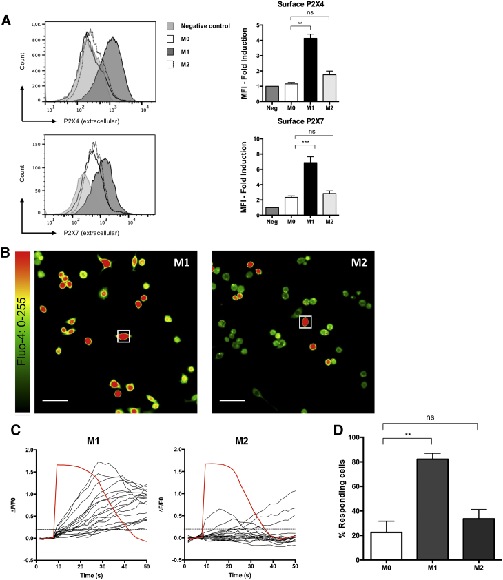
Macrophage Polarization Status Affects Calcium Signal Propagation (A) The surface expression of P2X4R (top) and P2X7R (bottom) was analyzed by flow cytometry in resting, IFNu03b3-treated (10u00a0ng/mL...
Figureu00a04
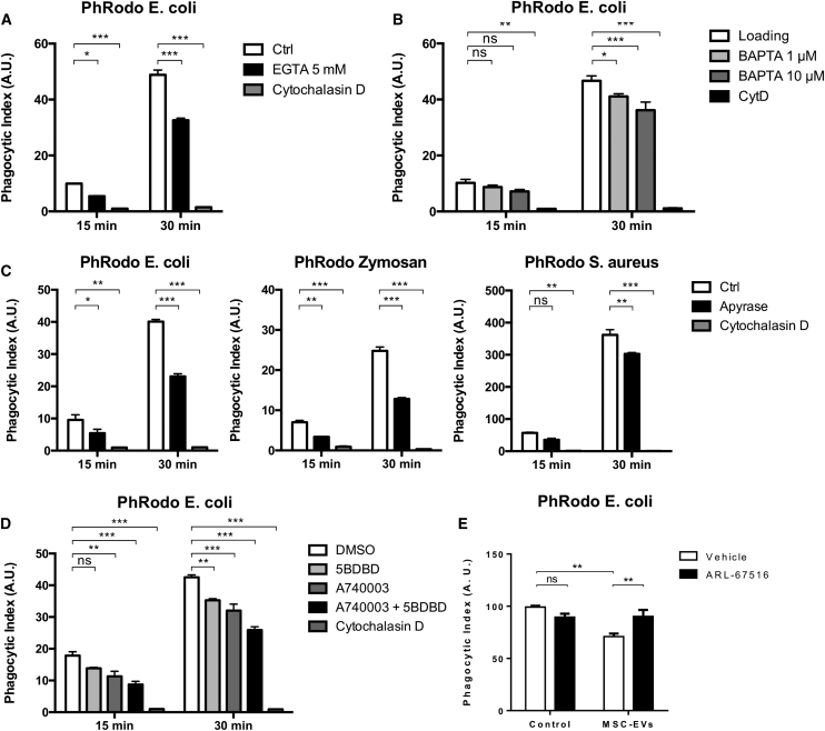
Extracellular ATP Is Required for Efficient Phagocytosis (A) Primary BMDMs were incubated with PhRodo E.u00a0coli fluorescent bioparticles in the presence or absence of 5u00a0mM EGTA to chelate extrac...
Figure images are served from the NIH/NLM PubMed Central Open Access Subset or Europe PMC; copyright remains with the publishers and authors.
💬 Discussion
0 commentsNo comments yet. Be the first to start a discussion!
Leave a Comment