Abstract
Combining the strength of flow cytometry with fluorescence imaging and digital image analysis, imaging flow cytometry is a powerful tool in diverse fields including cancer biology, immunology, drug discovery, microbiology, and metabolic engineering. It enables measurements and statistical analyses of chemical, structural, and morphological phenotypes of numerous living cells to provide systematic insights into biological processes. However, its utility is constrained by its requirement of fluorescent labeling for phenotyping. Here we present label-free chemical imaging flow cytometry to overcome the issue. It builds on a pulse pair-resolved wavelength-switchable Stokes laser for the fastest-to-date multicolor stimulated Raman scattering (SRS) microscopy of fast-flowing cells on a 3D acoustic focusing microfluidic chip, enabling an unprecedented throughput of up to ∼140 cells/s. To show its broad utility, we use the SRS imaging flow cytometry with the aid of deep learning to study the metabolic heterogeneity of microalgal cells and perform marker-free cancer detection in blood.
🔬 Techniques
🧪 Sample Preparation
🔬 Cell Lines
🏭 Microscope Brands
🧪 Reagent Suppliers
📷 Detectors
💾 Data Repositories
🏛️ Research Organizations (ROR)
Affiliated research institutions:
📋 Methods
SRS Imaging Flow Cytometer. A detailed schematic of the SRS imaging flow cytometer is shown in SI Appendix , Fig. S1 . It is composed of a mode-locked Ti:sapphire pulse laser (Coherent, Mira 900D) and a home-built mode-locked Yb fiber pulse laser to generate synchronized pump and Stokes pulse trains at repetition rates of 76 and 38 MHz, respectively. The pump laser has a center wavelength of 790 nm and a spectral width of 0.2 nm, while the Yb fiber laser generates broadband pulses at a wavelength of 1,030 nm with a spectral width of 20 nm. The latter is used to generate 4-color wavelength-switched Stokes pulses as described above. The pump laser and the Yb fiber laser are synchronized by using a feedback loop. Specifically, the intensity cross-correlation between the pump pulses and Yb fiber laser pulses is detected by a GaAsP photodiode (Hamamatsu, G1115) via 2-photon absorption, such that its signal is used to control the intracavity electro-optic modulator and a piezoelectric transducer in the Yb fiber laser. The 2 laser beams are overlapped both spatially and temporally and are focused inside the microchannel via the first objective lens (50×, NA = 0.65). The optical power in the sample plane is
📊 Figures
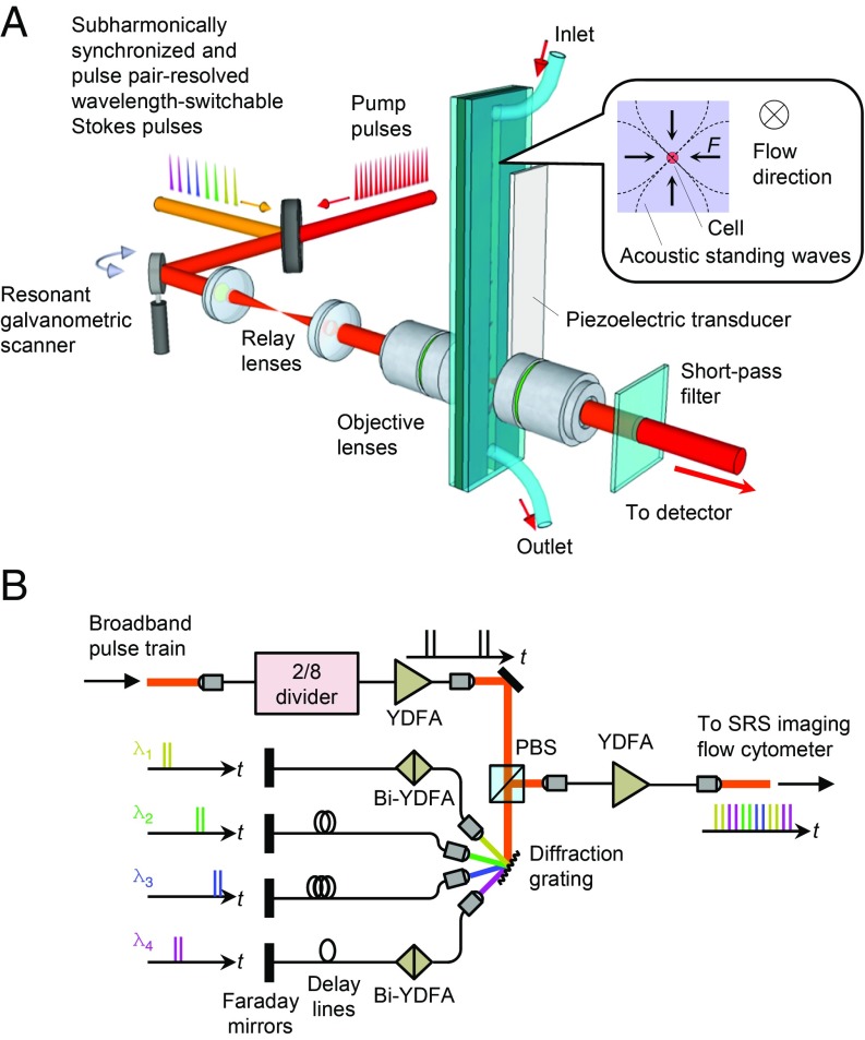
Fig. 1.
Schematic of the SRS imaging flow cytometer. ( A ) Schematic of the SRS imaging flow cytometer. It consists of 1) pulse sources that generate synchronized trains of pump pulses at a repetition rate of...
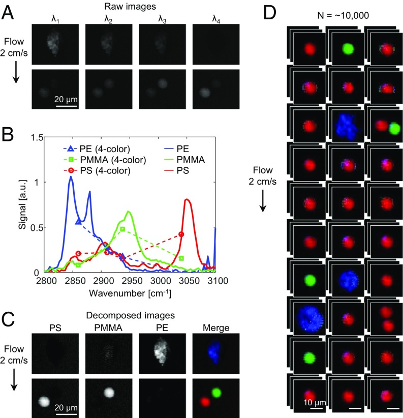
Fig. 2.
Characterization of the SRS imaging flow cytometer with polymer beads. ( A ) Four-color SRS images of PS, PMMA, and PE beads. The Raman shifts used are 2,860 cm u22121 (u03bb 1 ), 2,910 cm u22121 (u03...
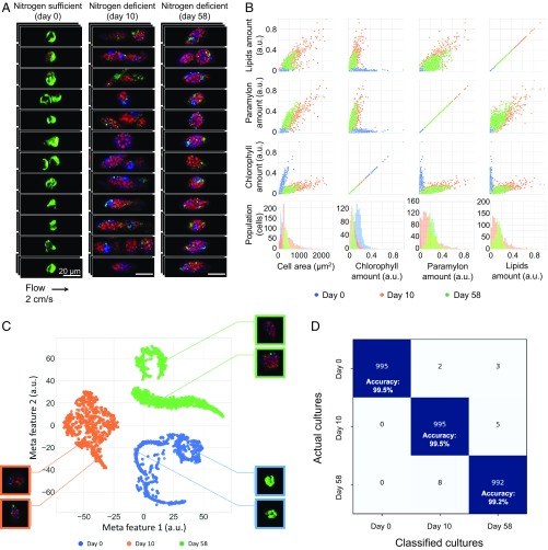
Fig. 3.
SRS imaging flow cytometry and large-scale single-cell analysis of E. gracilis cells. ( A ) SRS images of flowing E. gracilis cells cultured under 3 different conditions: nitrogen-sufficient culture (...
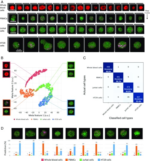
Fig. 4.
SRS imaging flow cytometry and label-free cancer cell detection in liquid biopsy. ( A ) SRS images of whole blood cells, PBMCs, Jurkat cells, and HT29 cells. Green, protein; pink, lipids; red, hemoglo...
Figure images are served from the NIH/NLM PubMed Central Open Access Subset or Europe PMC; copyright remains with the publishers and authors.
💬 Discussion
0 commentsNo comments yet. Be the first to start a discussion!
Leave a Comment