Abstract
Expansion microscopy (ExM) increases the effective resolving power of any microscope by expanding the sample with swellable hydrogel. Since its invention, ExM has been successfully applied to a wide range of cell, tissue, and animal samples. Still, fluorescence signal loss during polymerization and digestion limits molecular-scale imaging using ExM. Here, we report the development of label-retention ExM (LR-ExM) with a set of trifunctional anchors that not only prevent signal loss but also enable high-efficiency labeling using SNAP and CLIP tags. We have demonstrated multicolor LR-ExM for a variety of subcellular structures. Combining LR-ExM with superresolution stochastic optical reconstruction microscopy (STORM), we have achieved molecular resolution in the visualization of polyhedral lattice of clathrin-coated pits in situ.
🔬 Techniques
🔭 Microscopes
✨ Fluorophores
🧪 Sample Preparation
🔬 Cell Lines
🏭 Microscope Brands
🧪 Reagent Suppliers
📷 Detectors
🔎 Objectives
🎨 Filters
💻 Software Details
💾 Data Repositories
🏛️ Research Organizations (ROR)
Affiliated research institutions:
📋 Methods
Trifunctional anchors We synthesized five trifunctional anchors, including HOOC-MA-biotin, HOOC-MA-DIG, BG-MA-biotin, BG-MA-DIG, and BC-MA-DIG ( Fig. 2 ). HOOC-MA-biotin and HOOC-MA-DIG anchors were converted to NHS-MA-biotin and NHS-MA-DIG, respectively, to conjugate antibodies for the immunostaining approach of LR-ExM. The BG-MA-biotin, BG-MA-DIG, and BC-MA-DIG anchors were directly used for the protein tag approach of LR-ExM. The synthetic schemes are shown in Fig. S1 . All reactions were performed in flame- or oven-dried glassware fitted with rubber septa under a positive pressure of nitrogen, unless otherwise noted. All reaction mixtures were stirred throughout the course of each procedure using Teflon-coated magnetic stir bars. Air- and moisture-sensitive liquids were transferred via syringe. Solutions were concentrated by rotary evaporation
📊 Figures
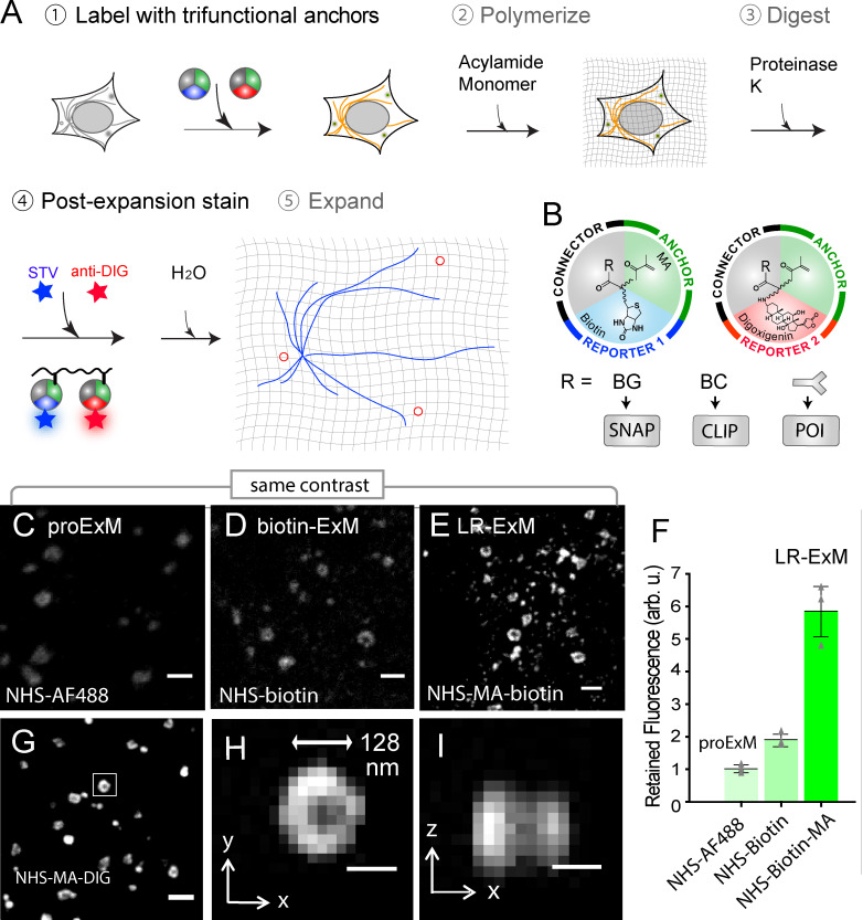
Figure 1.
Workflow and characterization of LR-ExM. (A) Workflow of LR-ExM. (B) Schematic of trifunctional anchors. (Cu2013E) ExM confocal images of CCPs in U2OS cells indirectly immunostained for clathrin heavy...
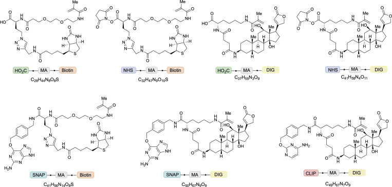
Figure 2.
Structures of trifunctional anchors. HOOC/NHS-MA-biotin, HOOC/NHS-MA-DIG, BG-MA-biotin, BG-MA-DIG, and BC-MA-DIG.
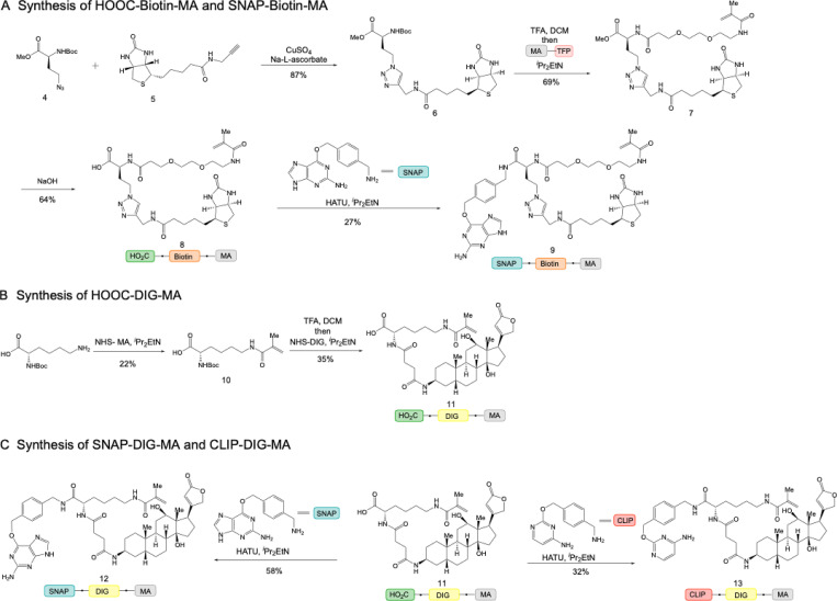
Figure S1.
Synthetic schemes of trifunctional anchors. (A) Synthetic routes of HOOC-biotin-MA and SNAP-biotin-MA. (B) Synthetic route of HOOC-DIG-MA. (C) Synthetic routes of SNAP-DIG-MA and CLIP-DIG-MA.
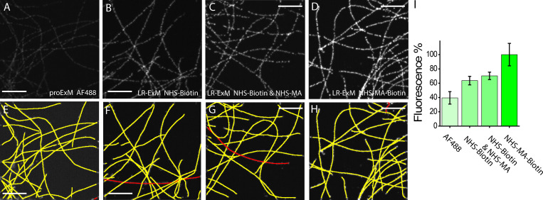
Figure S2.
Comparison of fluorescence intensities resulting from different ExM methods. (Au2013D) Images of microtubules prepared with proExM with AF488-labeled secondary antibody (A), biotin-ExM with the biotin...
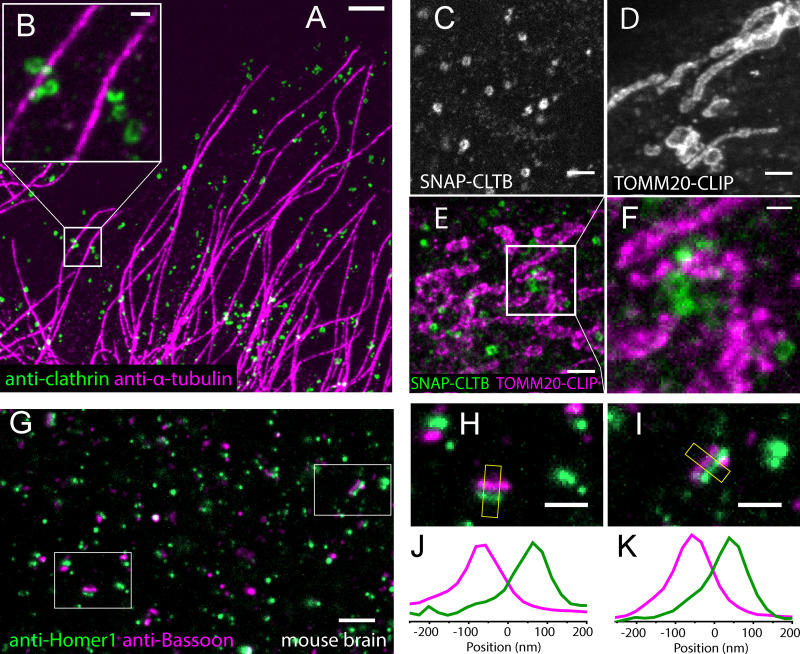
Figure 3.
Two-color LR-ExM images using immunostaining and protein tag approaches . (A) Two-color LR-ExM confocal image of microtubules labeled with NHS-MA-biotinu2013conjugated secondary antibodies (magenta) a...
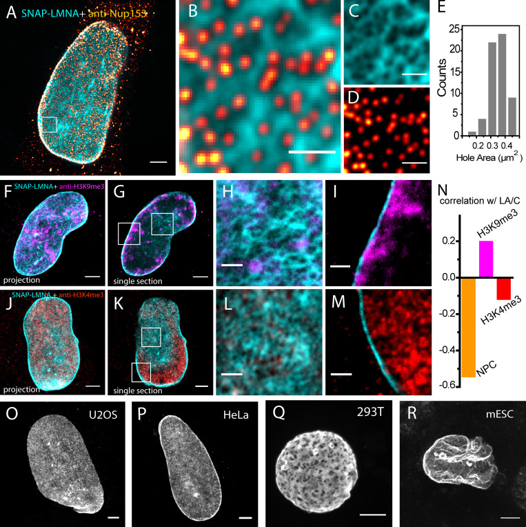
Figure 4.
LR-ExM reveals subcellular protein organizations. (A) Two-color confocal LR-ExM of SNAP-tagged lamin A/C (cyan) and immunostained NPC (red hot) of a HeLa cell. (B) Magnified view. (C and D) Views of i...
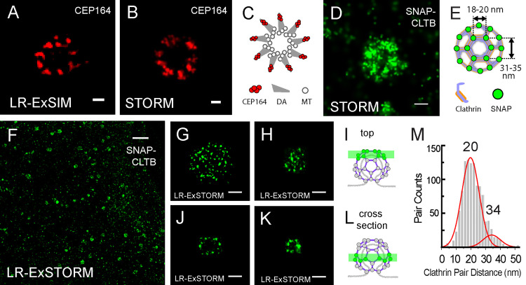
Figure 5.
LR-ExSIM and LR-ExSTORM reveal subcellular protein organization. (A) LR-ExSIM image of Cep164 in distal appendages of a primary cilium indirectly immunostained with NHS-MA-biotin secondary antibodies....
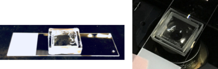
Figure S3.
3D-printed chamber for drift reduction of the hydrogel.
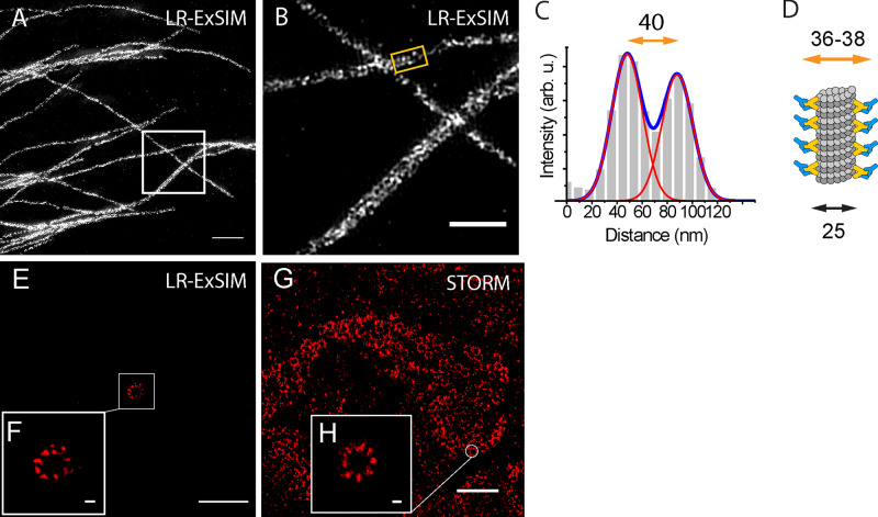
Figure S4.
LR-ExSIM of microtubules. (A) LR-ExSIM image of microtubules in a U2OS cell stained with antibody conjugated with NHS-MA-DIG anchors. (B) Magnification of A. (C) The transverse profile of the microtub...
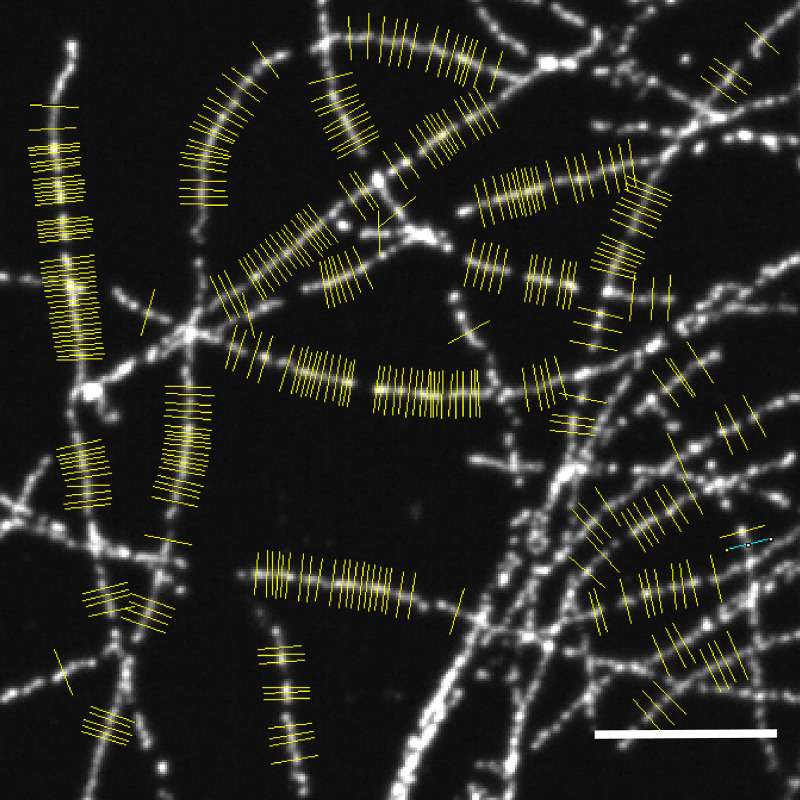
Figure S5.
Resolution measurement for LR-ExM confocal images. The transverse profiles of the microtubule cross sections marked in yellow were used to measure the resolution of LR-ExM using a confocal microscope....
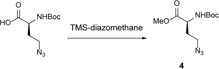
Scheme 1.
Azide 4 ( McLaughlin et al., 2003 ).
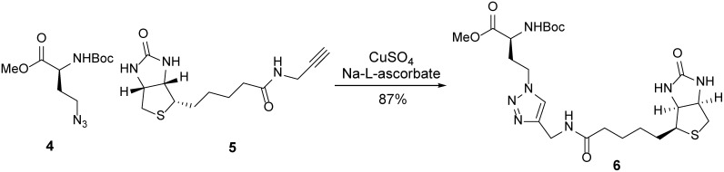
Scheme 2.
Click product 6.
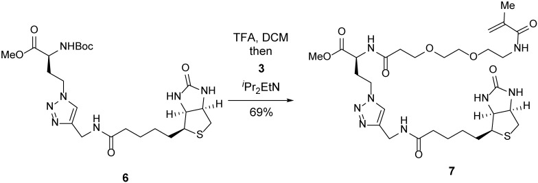
Scheme 3.
Methyl ester 7.
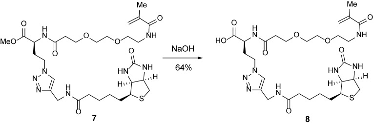
Scheme 4.
Carboxylic acid 8.
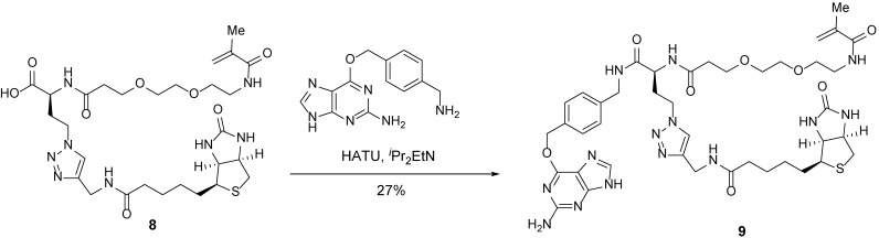
Scheme 5.
Trifunctional anchor 9.

Scheme 6.
MA 10.
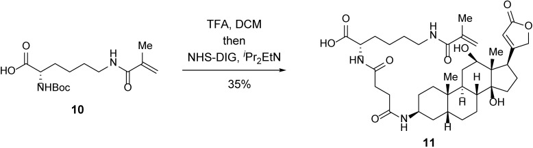
Scheme 7.
Carboxylic acid 11.
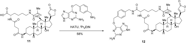
Scheme 8.
Trifunctional anchor 12.
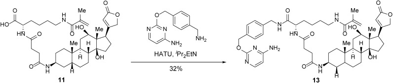
Scheme 9.
Trifunctional anchor 13.
Figure images are served from the NIH/NLM PubMed Central Open Access Subset or Europe PMC; copyright remains with the publishers and authors.
💬 Discussion
0 commentsNo comments yet. Be the first to start a discussion!
Leave a Comment