Abstract
Every day, megakaryocytes produce billions of platelets that circulate for several days and eventually are cleared by the liver. The exact removal mechanism, however, remains unclear. Loss of sialic acid residues is thought to feature in the aging and clearance of platelets. Using state-of-the-art spinning disk intravital microscopy to delineate the different compartments and cells of the mouse liver, we observed rapid accumulation of desialylated platelets predominantly on Kupffer cells, with only a few on endothelial cells and none on hepatocytes. Kupffer cell depletion prevented the removal of aged platelets from circulation. Ashwell-Morell receptor (AMR) deficiency alone had little effect on platelet uptake. Macrophage galactose lectin (MGL) together with AMR mediated clearance of desialylated or cold-stored platelets by Kupffer cells. Effective clearance is critical, as mice with an aged platelet population displayed a bleeding phenotype. Our data provide evidence that the MGL of Kupffer cells plays a significant role in the removal of desialylated platelets through a collaboration with the AMR, thereby maintaining a healthy and functional platelet compartment.
🔬 Techniques
💻 Software
✨ Fluorophores
🧪 Sample Preparation
🔬 Cell Lines
🏭 Microscope Brands
🧪 Reagent Suppliers
💻 Software Details
🏛️ Research Organizations (ROR)
Affiliated research institutions:
📋 Methods
Mice 6- to 8-wk-old
C57BL/6 mice were obtained from The Jackson Laboratory. CD41-YFP ki/+ mice ( Zhang et al., 2007 ) were a generous gift from Dr. Kelly McNagny (University of British Columbia, Vancouver, Canada). All mice were housed in a specific pathogen–free, double-barrier unit at the University of Calgary, with access to water and pelleted food ad libitum. All experimental procedures were approved by the University of Calgary Animal Care Committee and were in compliance with the Canadian Council for Animal Care Guidelines.
Antibodies and reagents
Antibodies against F4/80 (BM8) and CD11b (M1/70) were obtained from eBioscience. Antibodies against Ly6G (1A8), CD45 (30-F11), and Ly6C (HK1.4) were from Biolegend. Antibodies against CD49b (clone HMa2), P-selectin (CD62P, clone RB40.34), and CD41 (MWReg30) were purchased from BD Biosciences PharMingen. The MGL1/2 (CD301a/b) and AMR (ASGR1, clone #352803) antibodies, as well as recombinant human MGL (CD301/CLEC10A), were obtained from R&D Systems and labeled with Alexa647 dye using a Microscale Protein Labeling Kit (Thermo Fisher Scientific). Fluorescein-labeled Ricinus Communis Agglutinin (RCA-FITC) was obtained from Vector Laboratories. JON/A-PE antibody used to detect activated integrin αIIbβ3 and X649, an anti-GPIbβ antibody used to label platelets in vivo , were from Emfret Analytics. ASF, Thiazole Orange, and sialidase (neuraminidase) from Clostridium perfringens were obtained from Sigma-Aldrich. RCA-FITC was obtained from Vector Laboratories. CellTracker Red and Green dyes were from Thermo Fisher Scientific. Clodronate liposomes were from http://www.clodronateliposomes.org . Treatments For Kupffer cell depletion, mice were treated i.v. with clodronate liposomes (0.01 ml/g body weight) 48 h before performing the experiments. For in vivo desialylation, mice were i.v. injected with 50 mU sialidase. ASF (0.2 mg/g body weight) was administered i.v. before sialidase injection, where indicated. For in vivo blockade of the MGL receptors, mice were i.v. injected with 50 µg anti-MGL1/2 antibody. Isolation, ex vivo desialylation, and labeling of platelets Blood was collected in a tube containing 20 U/ml heparin, and platelet-rich plasma (PRP) was obtained by centrifuging twice at 300 g for 6 min. PRP was washed twice at 800 g for 5 min, and the pellet was resuspended in Tyrode’s buffer supplemented with prostacyclin (0.1 µg/ml) and apyrase (0.02 U/ml). Platelets were then resuspended in Tyrode’s buffer and treated with 2.5 mU sialidase for 20 min at 37°C. Desialylation was confirmed by binding of RCA-FITC. Platelets were labeled with 2 µM CellTracker Red or Green dye for 20 min at 37°C and washed twice with Tyrode’s buffer. For experiments investigating the cold-storage phenomenon, platelets were subsequently stored at 4°C for 24 h. Platelet aggregation 400 µl of a washed platelet suspension (3 × 10 5 platelets/µl) in Tyrode’s buffer containing 2 mM CaCl 2 were continuously stirred (1,200 rpm), and light transmission was recorded on a 700 Whole Blood/Optical Lumi-Aggregometer (Chrono-log). Agonists were added at the indicated concentrations to the platelet suspension. Light transmission was expressed as percentages, with buffer representing 100% transmission and washed platelet suspension 0% transmission, respectively. SD-IVM of the liver, spleen, and lung Mice were anesthetized using 10 mg/kg xylazine hydrochloride and 200 mg/kg ketamine hydrochloride, and livers, spleens, and lungs were prepared for IVM as previously described ( Surewaard and Kubes, 2017 ; Deniset et al., 2017 ; Thanabalasuriar et al., 2017 ). A jugular vein catheter was inserted to inject fluorescent antibodies and to maintain anesthesia. Image acquisition (liver and spleen) was performed using an inverted microscope (Olympus IX81) equipped with a focus drive (Olympus) and motorized stage (Applied Scientific Instrumentation) and fitted with a motorized objective turret equipped with 4×/0.16 UPLANSAPO, 10×/0.40 UPLANSAPO, and 20×/0.70 UPLANSAPO objective lenses. Image acquisition (lung) was performed with an upright microscope (BX51; Olympus) using a ×20/0.95W NA water XLUM Plan F1 objective. The microscopes were equipped with a confocal light path (Quorum Technologies WaveFx) based on a modified CSU-10 head (Yokogawa Electric Corporation). Kupffer cells, platelets, and the endothelium were visualized using fluorescently labeled antibodies. For each animal, platelet accumulation was recorded in three randomly selected fields of view. Sialidase was injected i.v. 2 min after image acquisition was started.
Show full methods section
Mice 6- to 8-wk-old
C57BL/6 mice were obtained from The Jackson Laboratory. CD41-YFP ki/+ mice ( Zhang et al., 2007 ) were a generous gift from Dr. Kelly McNagny (University of British Columbia, Vancouver, Canada). All mice were housed in a specific pathogen–free, double-barrier unit at the University of Calgary, with access to water and pelleted food ad libitum. All experimental procedures were approved by the University of Calgary Animal Care Committee and were in compliance with the Canadian Council for Animal Care Guidelines.
Antibodies and reagents
Antibodies against F4/80 (BM8) and CD11b (M1/70) were obtained from eBioscience. Antibodies against Ly6G (1A8), CD45 (30-F11), and Ly6C (HK1.4) were from Biolegend. Antibodies against CD49b (clone HMa2), P-selectin (CD62P, clone RB40.34), and CD41 (MWReg30) were purchased from BD Biosciences PharMingen. The MGL1/2 (CD301a/b) and AMR (ASGR1, clone #352803) antibodies, as well as recombinant human MGL (CD301/CLEC10A), were obtained from R&D Systems and labeled with Alexa647 dye using a Microscale Protein Labeling Kit (Thermo Fisher Scientific). Fluorescein-labeled Ricinus Communis Agglutinin (RCA-FITC) was obtained from Vector Laboratories. JON/A-PE antibody used to detect activated integrin αIIbβ3 and X649, an anti-GPIbβ antibody used to label platelets in vivo , were from Emfret Analytics. ASF, Thiazole Orange, and sialidase (neuraminidase) from Clostridium perfringens were obtained from Sigma-Aldrich. RCA-FITC was obtained from Vector Laboratories. CellTracker Red and Green dyes were from Thermo Fisher Scientific. Clodronate liposomes were from http://www.clodronateliposomes.org . Treatments For Kupffer cell depletion, mice were treated i.v. with clodronate liposomes (0.01 ml/g body weight) 48 h before performing the experiments. For in vivo desialylation, mice were i.v. injected with 50 mU sialidase. ASF (0.2 mg/g body weight) was administered i.v. before sialidase injection, where indicated. For in vivo blockade of the MGL receptors, mice were i.v. injected with 50 µg anti-MGL1/2 antibody. Isolation, ex vivo desialylation, and labeling of platelets Blood was collected in a tube containing 20 U/ml heparin, and platelet-rich plasma (PRP) was obtained by centrifuging twice at 300 g for 6 min. PRP was washed twice at 800 g for 5 min, and the pellet was resuspended in Tyrode’s buffer supplemented with prostacyclin (0.1 µg/ml) and apyrase (0.02 U/ml). Platelets were then resuspended in Tyrode’s buffer and treated with 2.5 mU sialidase for 20 min at 37°C. Desialylation was confirmed by binding of RCA-FITC. Platelets were labeled with 2 µM CellTracker Red or Green dye for 20 min at 37°C and washed twice with Tyrode’s buffer. For experiments investigating the cold-storage phenomenon, platelets were subsequently stored at 4°C for 24 h. Platelet aggregation 400 µl of a washed platelet suspension (3 × 10 5 platelets/µl) in Tyrode’s buffer containing 2 mM CaCl 2 were continuously stirred (1,200 rpm), and light transmission was recorded on a 700 Whole Blood/Optical Lumi-Aggregometer (Chrono-log). Agonists were added at the indicated concentrations to the platelet suspension. Light transmission was expressed as percentages, with buffer representing 100% transmission and washed platelet suspension 0% transmission, respectively. SD-IVM of the liver, spleen, and lung Mice were anesthetized using 10 mg/kg xylazine hydrochloride and 200 mg/kg ketamine hydrochloride, and livers, spleens, and lungs were prepared for IVM as previously described ( Surewaard and Kubes, 2017 ; Deniset et al., 2017 ; Thanabalasuriar et al., 2017 ). A jugular vein catheter was inserted to inject fluorescent antibodies and to maintain anesthesia. Image acquisition (liver and spleen) was performed using an inverted microscope (Olympus IX81) equipped with a focus drive (Olympus) and motorized stage (Applied Scientific Instrumentation) and fitted with a motorized objective turret equipped with 4×/0.16 UPLANSAPO, 10×/0.40 UPLANSAPO, and 20×/0.70 UPLANSAPO objective lenses. Image acquisition (lung) was performed with an upright microscope (BX51; Olympus) using a ×20/0.95W NA water XLUM Plan F1 objective. The microscopes were equipped with a confocal light path (Quorum Technologies WaveFx) based on a modified CSU-10 head (Yokogawa Electric Corporation). Kupffer cells, platelets, and the endothelium were visualized using fluorescently labeled antibodies. For each animal, platelet accumulation was recorded in three randomly selected fields of view. Sialidase was injected i.v. 2 min after image acquisition was started.
Volocity software
(PerkinElmer) was used to quantify platelet accumulation over time.
Quantification of platelet–Kupffer cell interactions under naive conditions
SD-IVM of the mouse liver was performed in naive mice with platelets and Kupffer cells labeled and videos recorded as described above. Videos were then imported into Imaris Image Analysis Software (Bitplane). A surface containing all Kupffer cells was created and converted into a masked channel. Platelets were identified using the “spot” function, and platelet spots that overlapped with the Kupffer cell surface were filtered. Interactions between platelets and Kupffer cells were tracked, and their dwell time was recorded.
Analysis of platelet colocalization after sialidase treatment
SD-IVM of the mouse liver was performed upon sialidase treatment, with endothelium, platelets, and Kupffer cells labeled, and videos were recorded as described above. Videos were then imported into Imaris Image Analysis Software (Bitplane). A surface containing all Kupffer cells was created and converted into a masked channel. Platelets were identified using the “spot” function, and platelet spots that overlapped with the Kupffer cell surface were filtered. To identify platelets bound to the endothelium, platelet spots that colocalized with endothelial signal were filtered. Similarly, platelets that colocalized with the hepatocyte signal were identified. Kupffer cell isolation Mouse Kupffer cells were isolated as previously described ( Lee et al., 2010 ). In brief, the abdominal cavity of anesthetized mice was opened to cannulate the inferior vena cava. The liver was perfused first with Ca 2+ - and Mg 2+ -free HBSS, and then with HBSS containing 0.05% collagenase type IV (Worthington Biochemical Corp.), 0.025% pronase E (US Biological), and 0.02% DNase I (Roche Diagnostics) at a flow rate of 4 ml/min. After perfusion, the liver was removed and further digested in HBSS containing 0.009% collagenase type IV, 0.009% pronase E, and 0.02% DNase I at 37°C with shaking for 30 min. The cell suspension was passed through a 100-micron nylon filter and centrifuged at 25 g for 5 min at room temperature to remove the hepatocytes. The supernatant was transferred to a new tube and centrifuged at 400 g for 10 min at 4°C. The cell pellet was resuspended in 17% iodixanol (Axis-Shield PoC) solution and centrifuged at 400 g for 15 min at 20°C. The band of nonparenchymal liver cells was obtained and washed twice. Red blood cell lysis was performed using ACK lysis buffer (Gibco) for 2 min.
Cell staining and flow cytometry
For determination of platelet count or platelet galactose exposure, 50 µl of heparinized blood was diluted 1:20 in Tyrode’s buffer and incubated for 15 min at room temperature with platelet-specific fluorophore-labeled antibodies or RCA-FITC and analyzed on a FACSCanto flow cytometer (BD Biosciences). For platelet activation studies, samples were washed twice in Tyrode’s buffer, resuspended in Tyrode’s buffer containing 2 mM CaCl 2 , activated with agonists at the indicated concentrations, stained with P-selectin-FITC and JON/A-PE for 6 + 6 min at 37°C and room temperature, respectively, and analyzed on a FACSCanto flow cytometer. For Kupffer cell analysis, single-cell suspensions of nonparenchymal liver cells were resuspended in staining buffer (phosphate buffered saline + 2% BSA) at a concentration of ∼1 × 10 7 cells/ml. Cells were blocked with Fc block reagents (BioXCell) for 10 min. Subsequently, dead cells were labeled using Ghost Dye red 710 (Tonbo Bioscience). After an additional washing step, fluorescent antibodies were added and incubated for 30 min on ice. Cells were subsequently washed and fixed with 1% paraformaldehyde at room temperature for 30 min. Cells were washed twice and resuspended in 200 µl of flow cytometry buffer and analyzed on a FACSCanto flow cytometer. After exclusion of doublets and dead cells, Kupffer cells were identified as CD45 + , Ly6G – , F4/80 + , Ly6C low , CD11b + . Quantification of “young”/reticulated platelets The percentage of young reticulated platelets was determined using Thiazole Orange as described ( Stritt et al., 2017 ; Kienast and Schmitz, 1990 ; Harrison et al., 1997 ). 50 µl of heparinized blood was diluted 1:20 in Tyrode’s buffer and incubated for 15 min at room temperature with platelet-specific antibodies and 1 µg/ml Thiazole Orange and analyzed by flow cytometry. Kupffer cell in vitro phagocytosis assay 50,000 Kupffer cells were incubated with 500,000 desialylated platelets in 96-well plates in DMEM supplemented with 10% FCS. Kupffer cells and platelets were stained using F4/80 antibodies and CellTracker Red dye, respectively. Images were recorded during incubation at 37°C and 5% CO 2 using an IncuCyte ZOOM system (Essen BioScience) equipped for phase contrast and two fluorescent channels (green: excitation, 440–480 nm; emission, 504–544 nm; red: excitation, 565–605 nm; emission, 625–705 nm) and with a 20× air objective (NA 0.40). Image analysis was performed using ImageJ software. Scanning EM Sample preparation for scanning EM was performed as described ( Warren et al., 2006 ). In brief, mice were euthanized, and livers were perfused with fixation solution (1% glutaraldehyde, 4% paraformaldehyde, 2 mM CaCl 2 , 2% (wt/vol) sucrose, and 0.1 M cacodylate buffer pH 7.4). Livers were excised, cut into small pieces, and fixed for an additional 1 h at 4°C. Fixed liver tissue was dehydrated and dried using ethanol and hexamethyl-disilazane series, respectively. Tissue was splutter-coated and examined using a FEI XL30 scanning electron microscope. Bacterial strain, culture, and infection Staphylococcus aureus strain MW2 was obtained from the Network on Antimicrobial Resistance in S. aureus and transformed with pCM29 ( Pang et al., 2010 ) to constitutively express GFP ( Surewaard et al., 2012 ). Bacteria were grown in brain heart infusion medium (+ 10 µg/ml chloramphenicol to maintain the plasmid) overnight with shaking at 37°C. Bacteria were subcultured without antibiotics until exponential growth was achieved (OD 660 nm = 1.0), washed, and resuspended in saline. Mice were infected by i.v. injection of 5 × 10 7 CFUs, and staphylococcal catching by Kupffer cells was observed using SD-IVM as described above. Tail bleeding time Mice were anesthetized, and a 1-mm segment of the tail tip was removed using a scalpel. Tail bleeding was monitored by placing the tail tip into an Eppendorf tube filled with 1 ml of warmed saline. Bleeding was determined to have ceased when no bleeding was observed for >1 min. Experiments were stopped after 20 min, and the amount of blood loss was determined by weighing the Eppendorf tube.
Donor consent and human platelet isolation
Ethical approval for obtaining healthy human volunteer blood was provided by the research ethics committee of the University of Calgary in accordance with the Declaration of Helsinki, and each human subject provided informed consent. Whole blood was drawn from healthy donors who had not taken aspirin products for ≥48 h. Blood was collected via a 21G butterfly needle into a 30-ml propylene syringe. The first 1 ml of blood was discarded. Blood was transferred into 50-ml polypropylene tubes containing 1 ml of acid citrate dextrose for every 5 ml of blood and gently inverted to mix. Samples were centrifuged at 140 g for 10 min at room temperature, and the upper layer (PRP) was carefully transferred to new 50-ml tubes containing 2 ml of acid citrate dextrose, further diluted to 50 ml total volume with room temperature Tyrode’s buffer, and spun at 700 g for 10 min at room temperature. Afterwards, the supernatant was discarded, and the platelet pellet was resuspended in 10 ml of room temperature Tyrode’s buffer. Platelets were treated with sialidase and analyzed using flow cytometry as described above.
Statistical analysis
All experiments were performed with a minimum of three independent replications. Data are presented as means ± SEM. For normally distributed data, unpaired two-tailed t test was used to determine the statistical significance between two groups. One-way ANOVA with post-hoc testing was used to determine statistical significance between multiple groups. All statistical analyses were performed using GraphPad PRISM software (version 8.1.0). Online supplemental material Fig. S1 shows labeling of the vessel lumen with dextran right after injection (related to Fig. 1 ), repopulation of Kupffer cells (related to Fig. 2 ), absolute young and total platelet counts after Kupffer cell depletion (related to Fig. 2 ), galactose exposure on “old” platelets (related to Fig. 2 ), and accumulation of endogenously labeled platelets on Kupffer cells after sialidase injection (related to Fig. 3 ). Fig. S2 shows unaltered Kupffer cell phenotype (size and catching capability) of mice treated with arsenic in drinking water for 5 wk, which display a reduced number of fenestrations in liver sinusoids following this treatment (related to Fig. 3 ). Fig. S3 shows that there is no increase in platelet accumulation in the spleen or lung following sialidase treatment, that sialidase treatment leads to complete platelet removal also in splenectomized mice, and that bone marrow megakaryocytes do not phagocytose platelets. Fig. S4 shows unaltered Kupffer cell phenotype (size, total area, and catching capacity) in ST3Gal-IV Δ/Δ mice (related to Fig. 3 ). Fig. S5 shows that 24 h after sialidase injection, the platelets that clustered on Kupffer cells are gone, that isolated Kupffer cells phagocytose platelets in vitro, that there is no difference in the accumulation of platelets on Kupffer cells following sialidase treatment in Mac1-deficient mice compared with wild-type mice, that AMR and MGL are expressed on mouse Kupffer cells, that Asgr1 −/− mice display unaltered platelet counts, and that human desialylated platelets bind human MGL. Video 1 shows that Kupffer cells are the intravascular, tissue-resident macrophage population of the liver. Video 2 shows that Kupffer cells form both transient and firm interactions with platelets under homeostatic conditions. Video 3 shows that, under naive conditions, platelets interact with and bind to Kupffer cells. Video 4 shows that Kupffer cells phagocytose platelets under naive conditions. Video 5 shows that ex vivo desialylated platelets rapidly adhere to Kupffer cells. Video 6 shows that Kupffer cell depletion prevents accumulation of ex vivo desialylated platelets. Video 7 shows that desialylated platelets rapidly adhere to Kupffer cells. Video 8 shows that platelets do not accumulate on splenic red pulp macrophages after sialidase injection. Video 9 shows that endogenously desialylated platelets from ST3Gal-IV Δ/Δ mice bind to Kupffer cells. Video 10 shows that Kupffer cells phagocytose desialylated platelets. Video 11 shows that desialylated platelets accumulate inside Kupffer cells. Video 12 shows that desialylated platelets are transported to the phagolysosome in Kupffer cells. Video 13 shows that, in control mice, platelets rapidly accumulate on Kupffer cells after sialidase injection. Video 14 shows that, in ASF-pretreated mice, accumulation of platelets on Kupffer cells after sialidase injection is strongly reduced. Video 15 shows that, in AMR-deficient mice, platelets rapidly accumulate on Kupffer cells after sialidase injection. Video 16 shows that, in mice treated with an MGL-blocking antibody, platelets rapidly accumulate on Kupffer cells after sialidase injection. Video 17 shows that combining AMR deficiency with MGL blockade significantly reduced the amount of platelets accumulating on Kupffer cells after sialidase injection.
Online supplemental material Fig. S1 shows labeling of the vessel lumen with dextran right after injection (related to Fig. 1 ), repopulation of Kupffer cells (related to Fig. 2 ), absolute young and total platelet counts after Kupffer cell depletion (related to Fig. 2 ), galactose exposure on “old” platelets (related to Fig. 2 ), and accumulation of endogenously labeled platelets on Kupffer cells after sialidase injection (related to Fig. 3 ). Fig. S2 shows unaltered Kupffer cell phenotype (size and catching capability) of mice treated with arsenic in drinking water for 5 wk, which display a reduced number of fenestrations in liver sinusoids following this treatment (related to Fig. 3 ). Fig. S3 shows that there is no increase in platelet accumulation in the spleen or lung following sialidase treatment, that sialidase treatment leads to complete platelet removal also in splenectomized mice, and that bone marrow megakaryocytes do not phagocytose platelets. Fig. S4 shows unaltered Kupffer cell phenotype (size, total area, and catching capacity) in ST3Gal-IV Δ/Δ mice (related to Fig. 3 ). Fig. S5 shows that 24 h after sialidase injection, the platelets that clustered on Kupffer cells are gone, that isolated Kupffer cells phagocytose platelets in vitro, that there is no difference in the accumulation of platelets on Kupffer cells following sialidase treatment in Mac1-deficient mice compared with wild-type mice, that AMR and MGL are expressed on mouse Kupffer cells, that Asgr1 −/− mice display unaltered platelet counts, and that human desialylated platelets bind human MGL. Video 1 shows that Kupffer cells are the intravascular, tissue-resident macrophage population of the liver. Video 2 shows that Kupffer cells form both transient and firm interactions with platelets under homeostatic conditions. Video 3 shows that, under naive conditions, platelets interact with and bind to Kupffer cells. Video 4 shows that Kupffer cells phagocytose platelets under naive conditions. Video 5 shows that ex vivo desialylated platelets rapidly adhere to Kupffer cells. Video 6 shows that Kupffer cell depletion prevents accumulation of ex vivo desialylated platelets. Video 7 shows that desialylated platelets rapidly adhere to Kupffer cells. Video 8 shows that platelets do not accumulate on splenic red pulp macrophages after sialidase injection. Video 9 shows that endogenously desialylated platelets from ST3Gal-IV Δ/Δ mice bind to Kupffer cells. Video 10 shows that Kupffer cells phagocytose desialylated platelets. Video 11 shows that desialylated platelets accumulate inside Kupffer cells. Video 12 shows that desialylated platelets are transported to the phagolysosome in Kupffer cells. Video 13 shows that, in control mice, platelets rapidly accumulate on Kupffer cells after sialidase injection. Video 14 shows that, in ASF-pretreated mice, accumulation of platelets on Kupffer cells after sialidase injection is strongly reduced. Video 15 shows that, in AMR-deficient mice, platelets rapidly accumulate on Kupffer cells after sialidase injection. Video 16 shows that, in mice treated with an MGL-blocking antibody, platelets rapidly accumulate on Kupffer cells after sialidase injection. Video 17 shows that combining AMR deficiency with MGL blockade significantly reduced the amount of platelets accumulating on Kupffer cells after sialidase injection.
📊 Figures
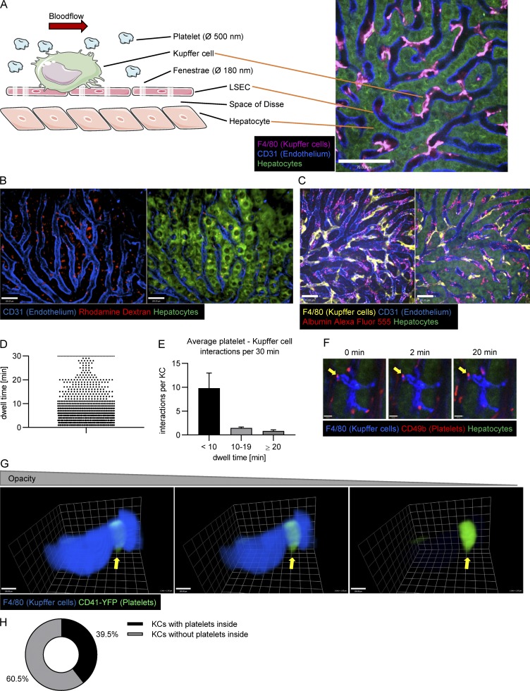
Figure 1.
SD-IVM of the liver allows one to pinpoint the location of platelet clearance and to analyze platelet Kupffer cell interactions in the steady state. (A) Schematic depiction of the liver structure (lef...
Video 1.
Kupffer cells are the intravascular, tissue-resident macrophage population of the liver. 3D reconstruction of a z-stack obtained using SD-IVM of the mouse liver. Kupffer cells (purple), endothelial ce...
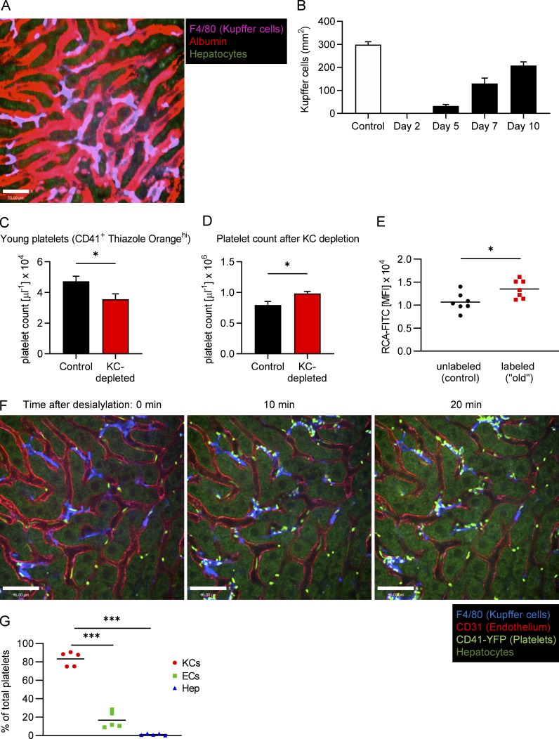
Figure S1.
Following clodronate treatment, Kupffer cells start repopulating the liver from day 7, the number of young platelets decreases while the total platelet count increases and upon desialylation, and CD41...
Video 2.
Kupffer cells form both transient and firm interactions with platelets under homeostatic conditions. Imaris image analysis software was used to quantify interactions between Kupffer cells (turquoise, ...
Video 3.
Under naive conditions, platelets interact with and bind to Kupffer cells. SD-IVM of the mouse liver of CD41-YFP ki/+ mice. Kupffer cells (blue) were stained using an anti-F4/80 antibody. Endogenous p...
Video 4.
Kupffer cells phagocytose platelets under naive conditions. 3D reconstruction of a Kupffer cell from CD41-YFP ki/+ mice. Kupffer cells (blue) were stained using an anti-F4/80 antibody. Endogenous plat...
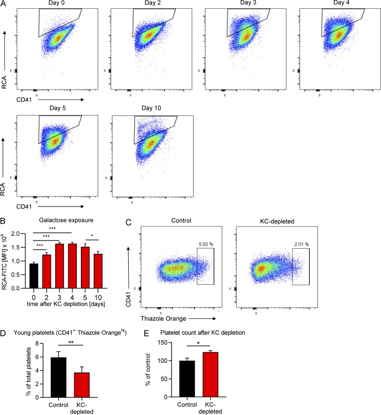
Figure 2.
Kupffer cells are central to the removal of desialylated platelets under homeostatic conditions. Mice were Kupffer cell depleted using clodronate liposomes, and the platelet population was followed ov...
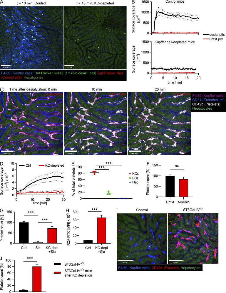
Figure 3.
Desialylated platelets rapidly accumulate on Kupffer cells. (A) Ex vivo desialylated (desial. plts) and untreated control platelets were labeled with CellTracker Green and Red dye, respectively, and t...
Video 5.
Ex vivo desialylated platelets rapidly adhere to Kupffer cells. Time-lapse SD-IVM of the mouse liver after i.v. injection of ex vivo desialylated platelets (CellTracker Green) and control platelets (C...
Video 6.
Kupffer cell depletion prevents accumulation of ex vivo desialylated platelets. Time-lapse SD-IVM video of the liver of mice pretreated with clodronate liposomes to deplete Kupffer cells after i.v. in...
Video 7.
Desialylated platelets rapidly adhere to Kupffer cells. Time-lapse SD-IVM video of the mouse liver after i.v. injection of 50 mU sialidase. Kupffer cells (magenta), endothelial cells (blue), and plate...
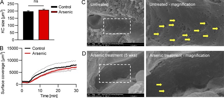
Figure S2.
Unaltered Kupffer cell size and recruitment of desialylated platelets but reduced fenestrations in mice after arsenic treatment. Mice were treated with arsenic in their drinking water for 5 wk. (A) Qu...
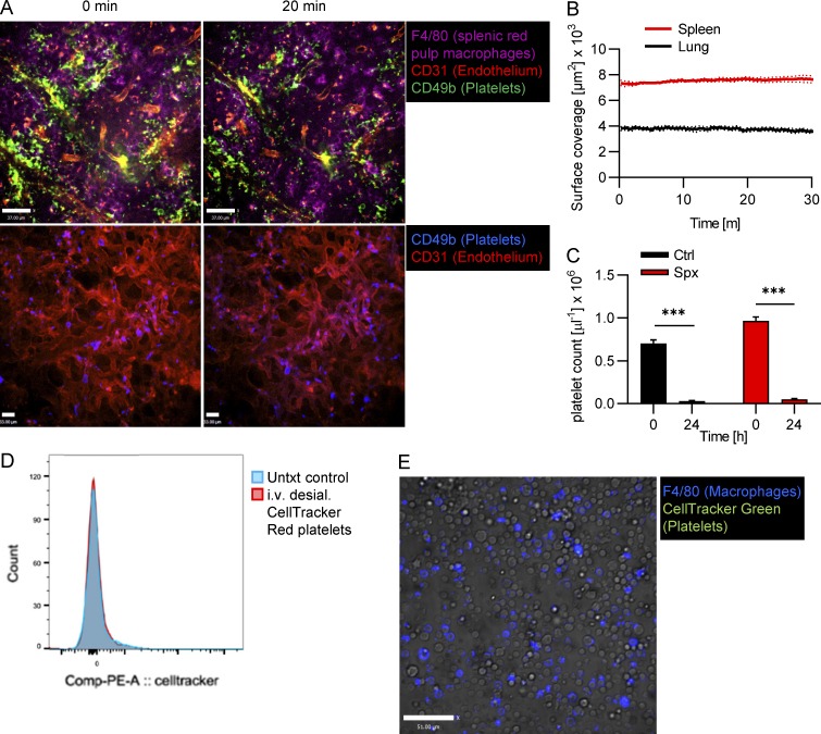
Figure S3.
No accumulation of desialylated platelets in the spleen, lung, or bone marrow. (A) Mice were i.v. injected with 50 mU sialidase, and platelet recruitment in the spleen (upper panel) and lung (lower pa...
Video 8.
Platelets do not accumulate on splenic red pulp macrophages after sialidase injection. Time-lapse SD-IVM video of the spleen of control mice after i.v. injection of 50 mU sialidase. Red pulp macrophag...
Video 9.
Endogenously desialylated platelets from ST3Gal-IV u0394/u0394 mice bind to Kupffer cells. Time-lapse SD-IVM video of the liver of ST3Gal-IV u0394/u0394 mice. Kupffer cells (blue) were stained using a...
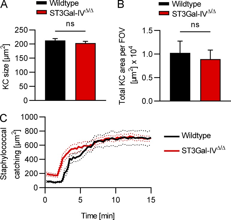
Figure S4.
Unaltered Kupffer cell size, number, and function in ST3Gal-IV u0394/u0394 mice. (A and B) Kupffer cell (KC) size (A) and total Kupffer cell area per field of view (FOV; B) of wild-type and ST3Gal-IV ...
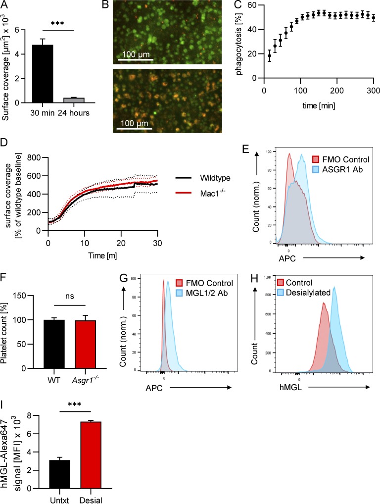
Figure S5.
Desialylated platelets are phagocytosed by Kupffer cells in vitro, Mac1 is dispensable for the clearance of desialylated platelets, Kupffer cells express Asgr1 and MGL1/2, but Asgr1-deficiency alone h...
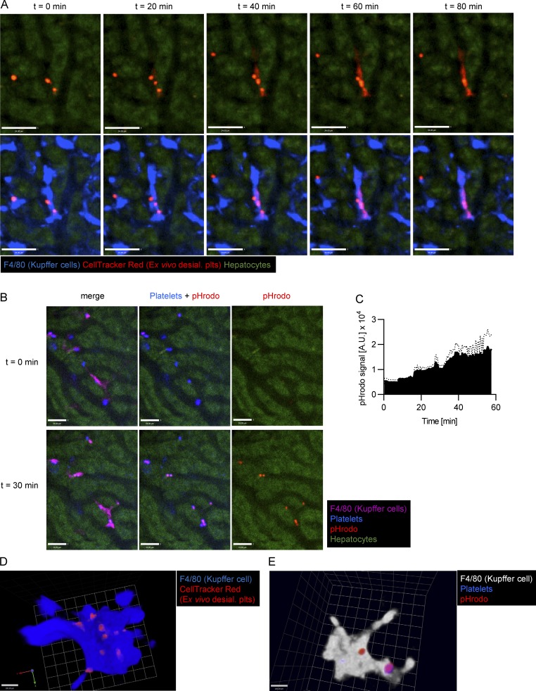
Figure 4.
Kupffer cells phagocytose desialylated platelets. (A) SD-IVM of the liver of wild-type mice i.v. injected with desialylated platelets labeled with CellTracker Red dye. Representative images taken at t...
Video 10.
Kupffer cells phagocytose desialylated platelets. SD-IVM of the mouse liver after i.v. injection of ex vivo desialylated platelets (CellTracker Red). Kupffer cells (blue) were stained using an anti-F4...
Video 11.
Desialylated platelets accumulate inside Kupffer cells. 3D reconstruction of a z-stack obtained using SD-IVM after i.v. injection of ex vivo desialylated platelets. Kupffer cells (blue) were stained u...
Video 12.
Desialylated platelets are transported to the phagolysosome in Kupffer cells. 3D reconstruction of a z-stack obtained using SD-IVM after i.v. injection of ex vivo desialylated platelets loaded with th...
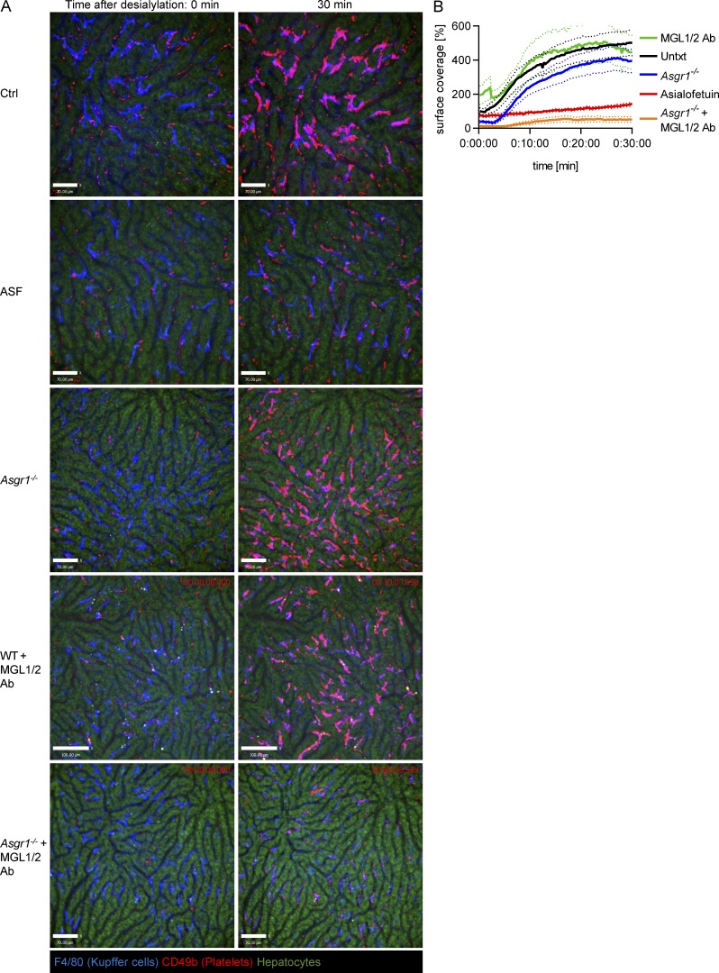
Figure 5.
A collaboration between MGL and AMR facilitates binding of desialylated platelets to Kupffer cells. (A) SD-IVM was performed on the liver of control mice, mice treated with ASF, Asgr1 u2212/u2212 mice...
Video 13.
In control mice, platelets rapidly accumulate on Kupffer cells after sialidase injection. Time-lapse SD-IVM video of the liver of control mice after i.v. injection of 50 mU sialidase. Kupffer cells (b...
Video 14.
In ASF-pretreated mice, accumulation of platelets on Kupffer cells after sialidase injection is strongly reduced. Time-lapse SD-IVM video of the liver of mice pretreated with ASF after i.v. injection ...
Video 15.
In AMR-deficient mice, platelets rapidly accumulate on Kupffer cells after sialidase injection. Time-lapse SD-IVM video of the liver of Asgr1 u2212/u2212 mice after i.v. injection of 50 mU sialidase. ...
Video 16.
In mice treated with an MGL - blocking antibody, platelets rapidly accumulate on Kupffer cells after sialidase injection. Time-lapse SD-IVM video of the liver of wild-type mice treated with MGL1/2 blo...
Video 17.
Combining AMR deficiency with MGL blockade significantly reduced the amount of platelets accumulating on Kupffer cells after sialidase injection. Time-lapse SD-IVM video of the liver of Asgr1 u2212/u2...
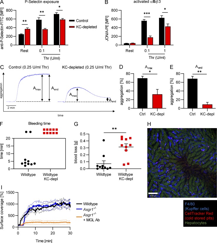
Figure 6.
Old platelets show significantly reduced hemostatic capacity. (A and B) Flow cytometric analysis of platelets from mice 5 d after Kupffer cell depletion shows increased P-selectin exposure in resting ...
Figure images are served from the NIH/NLM PubMed Central Open Access Subset or Europe PMC; copyright remains with the publishers and authors.
💬 Discussion
0 commentsNo comments yet. Be the first to start a discussion!
Leave a Comment