Abstract
AbstractHigh-quality, 25 nm octahedral-shaped Fe3O4 magnetite nanocrystals are epitaxially grown on 9 nm Au seed nanoparticles using a modified wet-chemical synthesis. These Fe3O4-Au Janus nanoparticles exhibit bulk-like magnetic properties. Due to their high magnetization and octahedral shape, the hybrids show superior in vitro and in vivo T2 relaxivity for magnetic resonance imaging as compared to other types of Fe3O4-Au hybrids and commercial contrast agents. The nanoparticles provide two functional surfaces for theranostic applications. For the first time, Fe3O4-Au hybrids are conjugated with two fluorescent dyes or the combination of drug and dye allowing the simultaneous tracking of the nanoparticle vehicle and the drug cargo in vitro and in vivo. The delivery to tumors and payload release are demonstrated in real time by intravital microscopy. Replacing the dyes by cell-specific molecules and drugs makes the Fe3O4-Au hybrids a unique all-in-one platform for theranostics.
🔬 Techniques
🔭 Microscopes
💻 Software
✨ Fluorophores
🧪 Sample Preparation
🔬 Cell Lines
🏭 Microscope Brands
🧪 Reagent Suppliers
🔎 Objectives
🎨 Filters
💻 Software Details
🏛️ Research Organizations (ROR)
Affiliated research institutions:
📋 Methods
Synthesis of Fe 3 O 4 -Au NPs Fe 3 O 4 -Au NPs were synthesized according to a modified protocol 76 , 77 . 0.28 ml of Fe(CO) 5 were injected in the pre-heated (120 °C) mixture of phenyl ether and oleic acid under the argon atmosphere. Afterwards, 0.5 ml of oleylamine and 2 ml of pre-synthesized 9 nm Au NPs (20 mg ml −1 in hexane, synthesized along the procedure established by Liu et al . 75 ) were added. The final solution was boiled at 260°С for 3 h and cooled to room temperature, followed by 1 h of room-temperature oxidation in ambient air. The NPs were isolated via centrifugation, washed with isopropanol and dispersed in toluene or chloroform. Synthesis of NP-PEG Fe 3 O 4 -Au NPs were transferred into water medium with 1,2-distearoyl-sn-glycero-3-phosphoethanolamine-N-[carboxy(polyethylene glycol)−5000] ammonium salt (DSPE-PEG-COOH) 119 . Briefly, equal volumes of Fe 3 O 4 -Au NPs and DSPE-PEG-COOH solutions (both 1 mg ml −1 in chloroform) were mixed by sonication for 5 minutes. Then the obtained mixture was left under weak N 2 flow overnight. After the solvent evaporated, 1 ml of DI H 2 O was added to the precipitate and then it was resuspended in water aided by ultrasound for 5–10 minutes. Unbound polymer was removed by centrifugation. Finally, the sample was passed through a 0.45 μm syringe filter.
Synthesis of NP-Cy5
NP-Cy5 were obtained by conjugation of NP-PEG with Sulfo-Cyanine5 NHS ester derivative (Cy5) fluorescent dye. 0.1 mg of Cy5 in 100 µl of DI H 2 O was added to 5 mg of cystamine dihydrochloride in 900 µl of PBS (pH = 8.3–8.5), shaken on a vortex well and kept on ice overnight. The resulting conjugate of Cy5 was mixed with 1 ml of NP-PEG solution overnight at room temperature. After that, unbound dye was firstly removed by multiple centrifugations, and then the resulting NP-Cy5 solution was dialyzed against water (MWCO = 12 kDa) for 24 h. To confirm that washing steps effectively remove unbound dye, an aliquot of NP-Cy5 was passed through Amicon Ultra-4 Centrifugal Filter Units (100 kDa). The quantity of conjugated dye in NP-Cy5 (33 µg of Cy5 per 1000 µg of Fe 3 O 4 /154 µg of Au) was estimated with a calibration graph.
Show full methods section
Synthesis of Fe 3 O 4 -Au NPs Fe 3 O 4 -Au NPs were synthesized according to a modified protocol 76 , 77 . 0.28 ml of Fe(CO) 5 were injected in the pre-heated (120 °C) mixture of phenyl ether and oleic acid under the argon atmosphere. Afterwards, 0.5 ml of oleylamine and 2 ml of pre-synthesized 9 nm Au NPs (20 mg ml −1 in hexane, synthesized along the procedure established by Liu et al . 75 ) were added. The final solution was boiled at 260°С for 3 h and cooled to room temperature, followed by 1 h of room-temperature oxidation in ambient air. The NPs were isolated via centrifugation, washed with isopropanol and dispersed in toluene or chloroform. Synthesis of NP-PEG Fe 3 O 4 -Au NPs were transferred into water medium with 1,2-distearoyl-sn-glycero-3-phosphoethanolamine-N-[carboxy(polyethylene glycol)−5000] ammonium salt (DSPE-PEG-COOH) 119 . Briefly, equal volumes of Fe 3 O 4 -Au NPs and DSPE-PEG-COOH solutions (both 1 mg ml −1 in chloroform) were mixed by sonication for 5 minutes. Then the obtained mixture was left under weak N 2 flow overnight. After the solvent evaporated, 1 ml of DI H 2 O was added to the precipitate and then it was resuspended in water aided by ultrasound for 5–10 minutes. Unbound polymer was removed by centrifugation. Finally, the sample was passed through a 0.45 μm syringe filter.
Synthesis of NP-Cy5
NP-Cy5 were obtained by conjugation of NP-PEG with Sulfo-Cyanine5 NHS ester derivative (Cy5) fluorescent dye. 0.1 mg of Cy5 in 100 µl of DI H 2 O was added to 5 mg of cystamine dihydrochloride in 900 µl of PBS (pH = 8.3–8.5), shaken on a vortex well and kept on ice overnight. The resulting conjugate of Cy5 was mixed with 1 ml of NP-PEG solution overnight at room temperature. After that, unbound dye was firstly removed by multiple centrifugations, and then the resulting NP-Cy5 solution was dialyzed against water (MWCO = 12 kDa) for 24 h. To confirm that washing steps effectively remove unbound dye, an aliquot of NP-Cy5 was passed through Amicon Ultra-4 Centrifugal Filter Units (100 kDa). The quantity of conjugated dye in NP-Cy5 (33 µg of Cy5 per 1000 µg of Fe 3 O 4 /154 µg of Au) was estimated with a calibration graph.
Synthesis of NRed-NP-Cy5
NRed-NP-Cy5 were synthesized by conjugation of Fe 3 O 4 -Au NPs with two fluorescent dyes, Nile Red (NRed) and Sulfo-Cyanine5 NHS ester derivative (Cy5). 100 μl of NRed (1 mg ml −1 in chloroform) was added to the mixture of 1 ml of Fe 3 O 4 -Au NPs and 1 ml of DSPE-PEG-COOH solutions (both 1 mg ml −1 in chloroform), further procedure was performed by the same protocol as for NP-PEG synthesis, resulting in the stabilization of NRed-NP in water solution (0.5 mg ml −1 ). In parallel with this, 0.1 mg of Cy5 in 100 µl of DI H 2 O was added to 5 mg of cystamine dihydrochloride in 900 µl of PBS (pH = 8.3–8.5), shaken on a vortex well and kept on ice overnight. The resulting conjugate of Cy5 was mixed with 1 ml of NRed-NP solution overnight at room temperature. The process of purification used was the same as for NP-Cy5. The quantity of conjugated dyes in NRed-NP-Cy5 (86 µg of Nile Red and 33 µg of Cy5 per 1000 µg of Fe 3 O 4 /154 µg of Au) was estimated with a calibration graph.
Synthesis of DOX-NP-Cy5
DOX-NP-Cy5 were obtained by doxorubicin loading to NP-Cy5. 100 μl of doxorubicin hydrochloride solution (DOX, 5 mg ml −1 in DI H 2 O) was added to 1 ml of NP-Cy5 (1 mg ml −1 in 1×PBS), and the mixture was shaken overnight at room temperature. Non-bound DOX was removed by centrifugation. The quantity of loaded drug (285 µg of DOX per 1000 µg of Fe 3 O 4 ) was estimated with a calibration graph. X-ray diffraction Patterns were measured from 2 θ = 30° to 120° at a scan rate 0,1° per step and 3 s per point using the X-ray powder diffractometer Rigaku Ultima IV with Co-K α radiation and graphite monochromator on the diffracted beam. Quantitative XRD analysis (including crystal size evaluation by determination of the coherent scattering region, OCD) was performed using PHAN% and SPECTRUM programs developed by Physical Materials Science Department of NUST «MISiS» (modification of Rietveld method), based on the minimization of the difference between the experimental spectrum, taken from the points, and model (calculated) one. For fitting the spectra, the lattice parameters, the amount of each phase and their crystallite diameter are optimized.
Electron Microscopy
Experiments were conducted using a FEI Tecnai F20 and a probe-side Cs-corrected JEOL JEM 2200FS microscopes, both operated at 200 kV acceleration voltage. Overview images were taken in conventional bright-field TEM mode while the high-angle annular dark-field STEM mode was used for the high-resolution micrographs. Samples were prepared by casting and evaporating a droplet of solution onto a carbon-coated copper grid (300 mesh). The average diameter of NPs was calculated from TEM images by analysis of about 1000 NPs for each sample using ImageJ software. EDX elemental mapping was carried out in the scanning mode utilizing an Oxford X-max detector. Magnetometry Dried powder of Fe 3 O 4 -Au NPs or NP-PEG solution (1 mg ml −1 in DI H 2 O/ 1× PBS) was compressed in synthetic capsules and the hysteresis loops and temperature dependent magnetization were measured in a Quantum Design PPMS DynaCool system.
Mössbauer spectroscopy
Mössbauer spectra of 57 Fe nuclei at room temperature were recorded with a MS-1104Em spectrometer in transmission geometry with a 57 Co(Rh) radiation source. Spectra analysis was performed by Univem MS program, the relative intensities (area) of elementary spectra were determined.
Physical-chemical characterization
The hydrodynamic size and ζ–potential of NP-PEG, NP-Cy5, NRed-NP-Cy5, DOX-NP-Cy5 were measured by dynamic light scattering (DLS) using a Nano ZS Zetasizer (Malvern Instruments). The average NPs sizes with error ranges were obtained from three measurements of each sample. Recording of optical spectra of NPs in the visible range (400–800 nm) was performed using Thermo Scientific Multiscan GO instrument. Doxorubicin release 2.5 ml of DOX-NP-Cy5 were suspended in RPMI cell medium (pH = 7.2) or 0.1 M acetate buffer, (pH = 4.7) to a final 1000 μg·ml −1 Fe 3 O 4 , 154 μg·ml −1 Au, 33 μg·ml −1 Cy5, 285 μg·ml −1 DOX concentration and incubated at 37 °C. 30 min, 2 h, 6 h, 24 h or 48 h after the incubation start, 500 µl of solution were centrifuged (10 min, 14100 g) for complete DOX-NP-Cy5 precipitation and supplemented with 3–5 min of magnetic decantation. Afterwards the supernatant absorbance at 480 nm wavelength, corresponding to released DOX, was measured. NP-Cy5 sample of the same concentration after the same centrifugation/decantation procedure was used as a negative control (0%). 100% release was achieved by 30 s treatment of DOX-NP-Cy5 with ultrasonic band (positive control). After background subtraction, the absorbance was normalized to the total absorbance of initially loaded drug.
Antibodies BV421-conjugated rat anti-mouse
Ly6g (clone 1A8) were purchased from BD Biosciences Pharmingen (San Diego, CA). PE-conjugated Armenian hamster anti-mouse CD49b (clone HMα2) were purchased from Biolegend (San Diego, CA). Cell culture 4T1 (mouse breast cancer) and B16-F10 (mouse melanoma) cells were purchased from the American Type Culture Collection (ATCC, Manassas, VA, USA). Cells were cultured in RPMI-1640 (for 4T1) and DMEM (for B16-F10) medium (Gibco) supplemented with 10% fetal bovine serum (FBS) (Gibco) and 2 mM L-glutamine (Gibco) at 37 °C in a humidified incubator supplied with 5% CO 2 . GFP expressing cell line was obtained by 4T1 lentivirus transduction (MOI = 50) using LVT-TagGFP (Eurogen, Russia). All cell lines were routinely tested negative for mycoplasma.
Animals and tumor models
All animal experiments were reviewed at the bioethical commission by the Federal State Budgetary Educational Institution of Higher Education “The Russian National Research Medical University named after N.I. Pirogov” of the Ministry of Health of the Russian Federation and approved for conducting (conclusion of the bioethical commission No. 25/2017 and 26/2017). All methods were performed in accordance with Directive 2010/63/EU of the European Parliament and of the Council of 22 September 2010 on the protection of animals used for scientific purposes (Annex VIII). Six to eight-week-old female BALB/c and C57/bl6 mice were obtained from Andreevka Animal Center (Andreevka, Russia) and maintained in specific-pathogen free facility. At the time of investigations, animals were between 7 and 11 weeks old and weighed 20–25 g. 4T1 or 4T1-GFP tumours were established by injecting 1 × 10 6 cells and B16-F10-6 × 10 6 cells (in 50 µl PBS) subcutaneously into the right/left hind flanks. When tumours reached about 25 mm 2 (after 7–10 days) NPs were injected through a tail vein. Cytotoxicity assay Standard MTS test was performed as described elsewhere 120 . Briefly, 4T1 cells were seeded in 96-well plates (10 4 cells/well). 24 h after, serial dilutions of NP-PEG, NP-Cy5, DOX-NP-Cy5 or free doxorubicin (each point in 50 μl of 1×PBS, pH = 7.4, Gibco) were added to cells. When comparing the cytotoxicity of NP-PEG to NP-Cy5, the equal concentrations of Fe 3 O 4 were used (Supplementary Fig. S8,c ). When comparing the cytotoxicity of DOX-NP-Cy5 to free doxorubicin (DOX), the same concentrations of DOX were used (Fig. 5d ). Cell culture medium with addition of 50 μl 1× PBS was used as a negative control. DMSO (25%) was added in culture medium as a positive control. Afterwards cells were incubated during 48 h at 37 °C and 5% CO 2 , then the medium with NPs was carefully removed, cells were washed with PBS, and 20 μl of 3-(4,5-dimethylthiazol-2-yl)-5-(3-carboxymethoxyphenyl)-2-(4-sulfophenyl)-2H-tetrazolium (MTS reagent, CellTiter 96 AQueous Non-Radioactive Cell Proliferation Assay, Promega, USA) was added to each well with 100 μl of culture medium according to manufacturer’s instructions. After 4 h of incubation at 37 °C in darkness, plates were placed on a permanent magnet for 3–5 min to remove the NPs from solution, and 100 μl of culture medium with MTS from each well were carefully replaced in new plates. The absorbance of the solution was measured at 490 nm wavelength using Thermo Scientific Multiskan GO spectrometer. Survival was calculated as percent compared to cells treated with PBS. MTS assay revealed 100% cell death after incubation with DMSO, data not shown. The absorbance of MTS-reagent in culture medium without cells was taken as zero. ROS detection by 2′,7′-dichlorodihydrofluorescein diacetate (H2DCFDA) Cells were seeded in 24-well plates at a concentration 10 5 cells ml −1 and cultured at 37 °C in a humidified incubator supplied with 5% CO 2 . 24 h after, NP-PEG, dispersed in 1×PBS (pH = 7.4), were added to cells for 4 h and 24 h with final concentrations 16; 49; 91; 193 and 333 µg ml −1 of cell medium in the well. Cells, incubated with addition of 1× PBS and 1 mM H 2 O 2 , were used as negative and positive controls, correspondingly. To detect ROS in cells after incubation with NPs, the culture medium was removed and HBSS (pH = 7.4, Gibco) with 2 mM L-glutamine and 10 mM HEPES was added to cells. Then unfixed cells were stained with 2 µM H2DCFDA (life technologies) for 30 min at 37 °C in a humidified incubator. After this cells were carefully washed with new portions of HBSS 3 × 5 min and investigated in the fourth portion. The obtained preparations were analysed at fluorescence microscope EVOS (life technologies), objective PlanFluor 20×/0.45, GFP channel (470/22 nm Excitation; 510/42 nm Emission).
Determination of the hemolysis
NPs hemolytic activity was assessed ex vivo , as previously described with modifications 121 . In brief, 0.5 ml of mouse blood was obtained by cardiac puncture and centrifuged (10 min, 900 g). Supernatants were ruled out and the red blood cells (RBC) were resuspended in PBS to remove traces of plasma. This washing step was repeated twice and then RBC were dispersed in PBS, at a concentration of 4 x 10 9 cells ml −1 . 0.5 ml of NP-PEG in serial dilutions in 1× PBS (3–330 µg ml −1 Fe 3 O 4 ) were mixed with 25 µl of RBC. The mixtures were incubated at 25 °C, under continuous agitation for the required time (10 min or 24 h) and then centrifuged (5 min, 900 g). The absorbance of the solution was measured at 540 nm wavelength, and the percentage of hemolysis was assessed by comparing with the positive (0.5 ml of distilled water) and negative (0.5 ml of 1×PBS) controls. The results were expressed as the percentage of hemolysis caused. Dynamic of NP-Cy5 and doxorubicin accumulation in cells 4T1 cells were seeded on the coverslip glasses in Petri dishes at concentration 10 5 cells ml −1 . After 24 h, NP-Cy5 (136 μg ml −1 Fe 3 O 4 ; 8 μg ml −1 Cy5) or free Cy5 (8 μg ml −1 Cy5) dye were added to cells and incubated for 15; 30; 45 min; 1; 2; 4; 6 and 24 h. Alternatively, DOX-NP-Cy5 (63 μg·ml −1 Fe 3 O 4 ; 2 μg·ml −1 Cy5; 18 μg·ml −1 (31 μM) DOX) or free DOX (31 μM) were added to cells. At required time points, cells were fixed in 4% formaldehyde (Sigma) for 15 min and imaged using EVOS (life technologies, objective PlanFluor 60×/0,75) in Cy5 channel (628/40 nm Excitation; 692/40 nm Emission). DOX accumulation was quantified by measuring its fluorescence intensity in RFP channel (531/40 nm Excitation; 593/40 nm Emission) in cells nuclei (60–70 cells/time point) in ImageJ software. Fluorescence intensity in untreated cells was taken for zero. The absence of NP-PEG (136 μg ml −1 Fe 3 O 4 ) instrinsic fluorescence in Cy5/RFP/GFP channels and the absence of NP-Cy5 (136 μg ml −1 Fe 3 O 4 ; 8 μg ml −1 Cy5) fluorescence in RFP/GFP channels after the identical incubation with cells were also checked (Supplementary Fig. S13 ). The exposure was adjusted separately for each channel and kept constant during all measurements. Confocal imaging 4T1 cells were seeded 10 5 cells·ml −1 in 30 mm SPL coverglass bottom dish (Biolab, Korea) and 24 h later treated with NP-Cy5 (33 μg·ml −1 Fe 3 O 4 ). Z-stacks (8 steps, 500 nm each) were captured after 30 min of co-incubation using a Nikon A1r MP inverted microscope (Nikon, Japan; oil immersion objective x60/1,49). Maximum projections along X, Y and Z-axis were made using NIS elements AR software.
Atomic emission spectrometry
For in vitro studies 4T1 cells were seeded in 75 cm 2 flasks and cultivated for 24 h. Subsequently, cells were treated with NP-PEG in 1×PBS (final concentration 100 μg·ml −1 Fe 3 O 4 , and 15 μg·ml −1 Au) and incubated for 48 h at 37 °C and 5% CO 2 . After three PBS washing steps, cells were detached with TrypLE (Gibco), resuspended in culture medium and counted. Untreated cells were used as control. Later, cells were dissolved in aqua regia, and the concentrations of Fe and Au were measured by microwave coupled plasma-atomic emission spectrometry (Agilent 4200 MP-AES, USA) using the calibration curve for the standard samples in 0.1–1 mg·ml −1 concentration range. For in vivo studies 24 h after NPs i.v. injection (6.6 mg kg −1 Fe 3 O 4 , 1 mg kg −1 Au) mice (n = 3) were sacrificed after injection by cardiac perfusion with 30 mL PBS under anesthesia, and the liver, spleen, kidney, lung, heart and tumor were collected. The organs were weighed and digested in aqua regia during 24 h. Quantification of the iron and gold concentration was carried out by atomic emission spectrometry as described above. Untreated animals were used as control (n = 3) for measuring endogenous gold and iron levels. Mean gold and iron levels in control organs were subtracted from corresponding gold/iron levels in NPs-treated group to get NPs-associated gold/iron concentration (µg/g tissue). NPs delivery efficiency calculations were based on iron/gold concentration in the tumor tissues, tumor mass and injected dose.
Intravital microscopy
Mice were anesthetized by intraperitoneal injection of 200 mg kg −1 ketamine (Moscow Endocrine Plant, Russia) and 10 mg kg −1 xylazine (Nita-Farm, Russia) and the tail vein was cannulated with polyethylene tubing (0.28 × 0.60 mm, InStech Laboratories, Inc., Plymouth Meeting PA, USA) for delivering fluorescently labeled antibodies (5–10 μg) and maintaining the anaesthetics. Body temperature was maintained using a heated stage. NP-Cy5 or NRed-NP-Cy5 (3 mg kg −1 Fe 3 O 4 ) were injected through a tail vein. Skin and tumor preparations were made as described 122 . Briefly, a midline incision along the spine was made and the skin reflected. The thin connective tissue membrane overlaying the inside surface of the skin was removed and edges of this skin flap were secured using sutures to expose and stabilize the tumor/vessels for imaging. Intravital imaging was performed using a Nikon A1r MP inverted microscope (Nikon, Japan). For kinetic studies fluorescence intensity was measured in intravascular and interstitial ROI using NIS-Elements AR software (Nikon, Japan). In vivo fluorescent imaging Animals with both flanks grafted 4T1-tumors (n = 3) were anesthetized with isoflurane and imaged using IVIS Spectrum CT (Perkin Elmer) on 640/680 nm excitation/emission wavelengths before and 1–24 h after NP i.v. injection (6.6 mg kg −1 Fe 3 O 4 ). For autofluorescense correction spectral unmixing protocol was applied. Average fluorescence intensities were measured in selected ROI in Living Image 4.3 (Perkin Elmer) and tumor/liver ratios were calculated. In vitro and in vivo MRI For in vitro studies the T 2 relaxation rate of water protons in the presence of NP-PEG was measured in 500 μl test tubes at 18 °С in a ClinScan 7 T MRI system (Bruker BioSpin). Image acquisition was performed in the Spin Echo mode with following parameters: MRI system TR = 10 s, TE = 16, 24, …, 256 ms, flip angle = 180°, resolution 640 × 448 pixel, field of view 120 × 82.5 mm 2 . Signal intensities from regions of interest were determined using ImageJ and the T 2 relaxation time was calculated by linear fitting as function of TE. The T 2 relaxivity values were calculated from linear fitting of T 2 −1 relaxation times as function of Fe concentration. The slopes represent the R 2 values for NP-PEG in water and 4T1 cell culture used for MR imaging. Cells were incubated with NP-PEG (100 μg·ml −1 Fe 3 O 4 , 15 μg·ml −1 Au) during 48 h. Cells, cultivated in free medium, were used as control. Non-bound NPs were removed by cell washing with PBS as described above for AES sample preparation. Cells with attached NPs were suspended in 2% agarose gel. For in vivo studies, images were obtained using a 20-cm volumetric coil as a transmitter and a 4-segment surface coil as a receiver of the RF signal. BALB/c mice with both flanks grafted 4T1-tumors (n = 3) and C57/bl6 mice with right flanks grafted B16-F10 tumors (n = 2) were anesthetized with isoflurane and scanned before and 0.5–24 h after NPs i.v. injection (6.6 mg kg −1 Fe 3 O 4 ) using the following settings: 1) fat-suppressed T 2 -weighted turbo spin–echo (TSE) images were made in transversal planes (TR = 3000 ms, TE = 38 ms, FOV = 21 × 30 mm, base resolution (136 × 192); 2) T 2 * weighted gradient echo (GRE) images were made in transversal planes (TR = 400 ms, TE = 10 ms, FOV = 27 × 35 mm, base resolution (200 × 256). Images were processed in RadiAnt DICOM Viewer.
Statistical analysis Plotting and calculation of the standard deviation
(SD) and standard error of mean (SEM) values were made using Origin 8.0 and Prism 6 – GraphPad software. Data were analysed using the Analysis of Variance (ANOVA) test. P values < 0.05 were considered significant.
Data availability
The data supporting the plots and other findings of this study are available from the corresponding authors upon reasonable request.
Electronic supplementary material Video S1 Video S2 Video S3 Video S4 Supplementary Information
📊 Figures
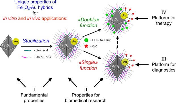
Figure 1
Fe 3 O 4 -Au hybrid NPs serve as a unique theranostics platform. Optimized fundamental properties, i.e. perfect crystallinity, octahedral shape and bulk-like magnetic properties (I), are combined with...
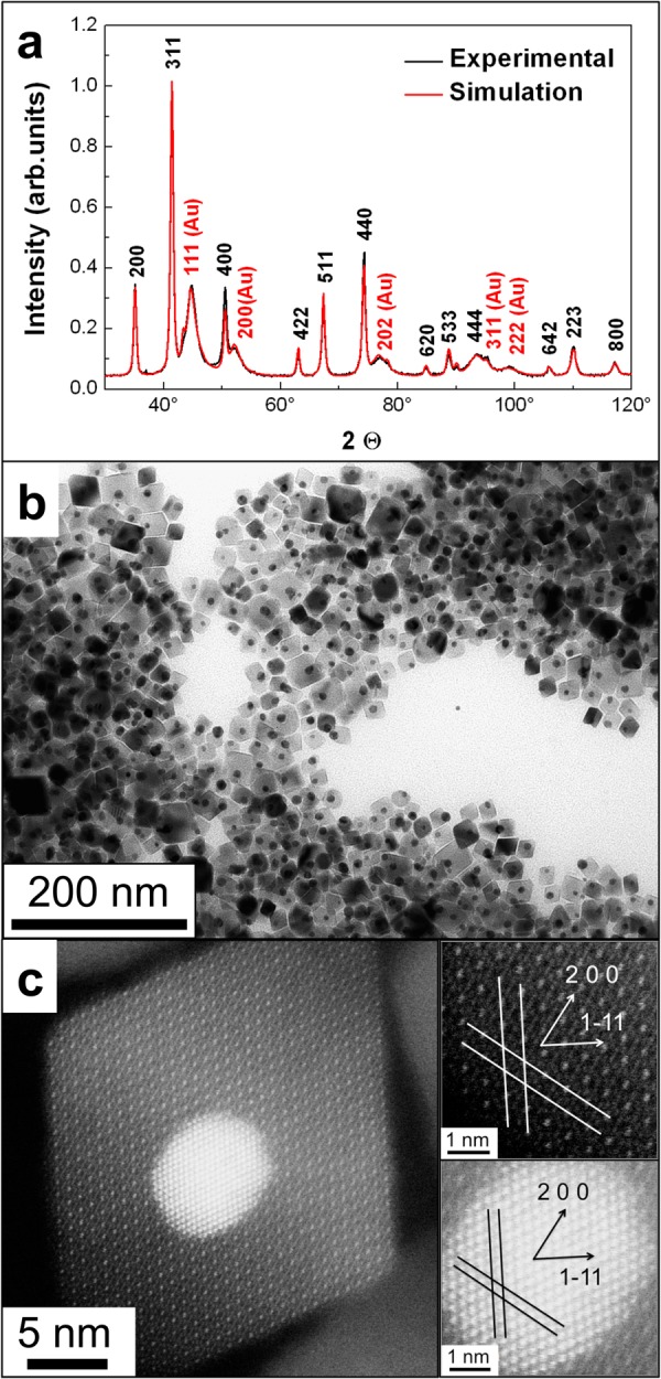
Figure 2
Structural characterization of Fe 3 O 4 -Au hybrid NPs. ( a ) Experimental X-Ray diffraction pattern (black). Black and red Miller indices correspond to Fe 3 O 4 and Au phases, respectively. The red d...
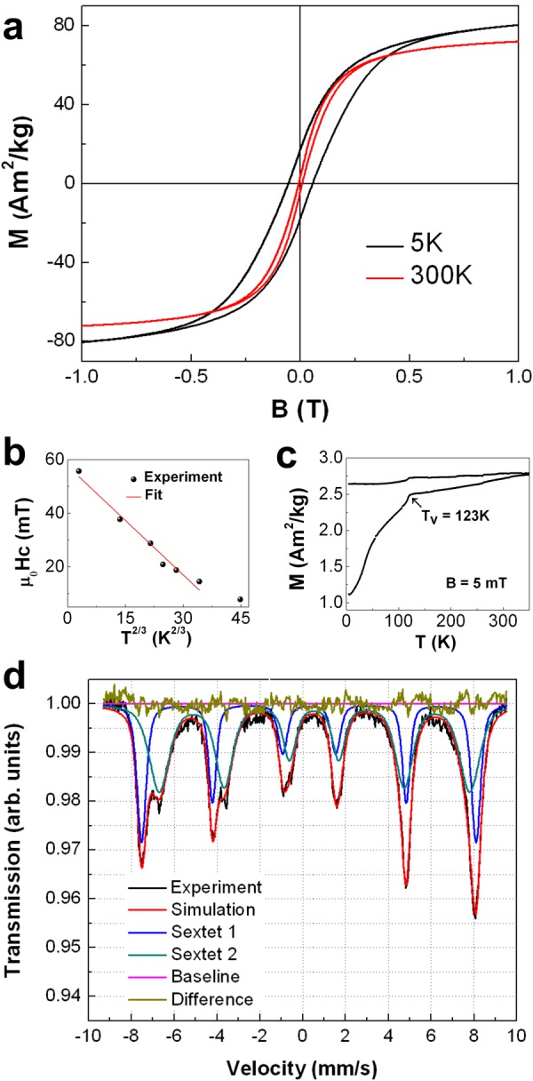
Figure 3
Magnetic characterization of Fe 3 O 4 -Au hybrid NPs. ( a ) Hysteresis loops recorded at 5u2009K and 300u2009K. Both loops are measured in the field range ofu2009u00b1u20099u2009T. ( b ) Coercive fiel...
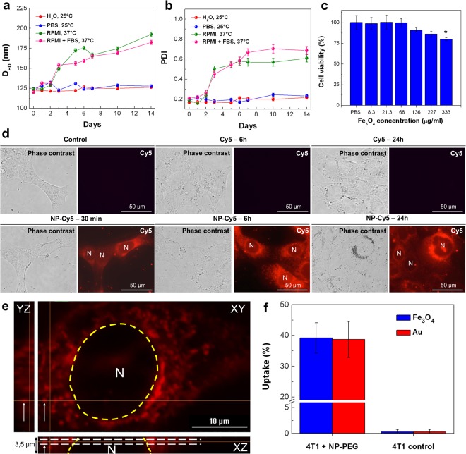
Figure 4
Stability, in vitro toxicity and internalization studies of Fe 3 O 4 -Au hybrid NPs. ( a , b ) Stability of NP-PEG in deionized water, 1u00d7 PBS, RPMI and RPMI with 10% FBS upon the incubation at 25u...
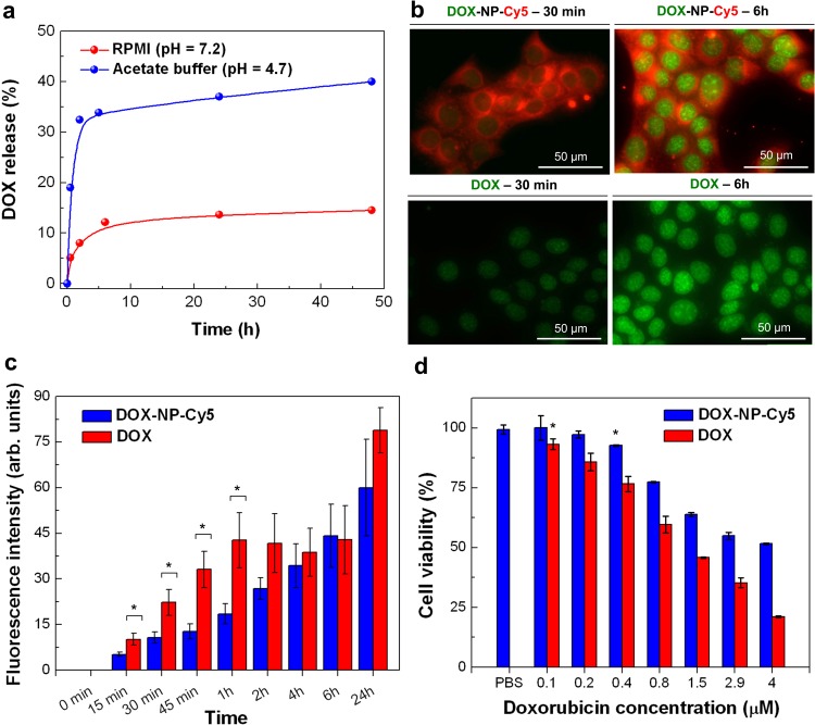
Figure 5
Delivery of doxorubicin to cancer cells by Fe 3 O 4 -Au hybrid NPs. ( a ) pH-dependent kinetics of doxorubicin release from DOX-NP-Cy5 NPs in RPMI (pHu2009=u20097.2) and acetate buffer (pHu2009=u20094...
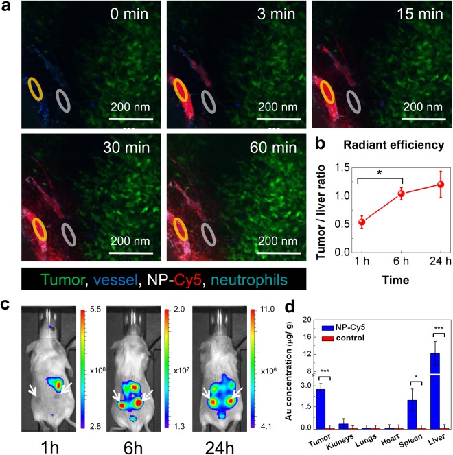
Figure 6
Accumulation of Fe 3 O 4 -Au hybrid NPs in 4T1-tumors. ( a ) Visualization of subcutaneous 4T1-GFP tumor microenvironment by intravital microscopy upon the i.v. injection of NP-Cy5 (6.6u2009mgu00b7kg ...
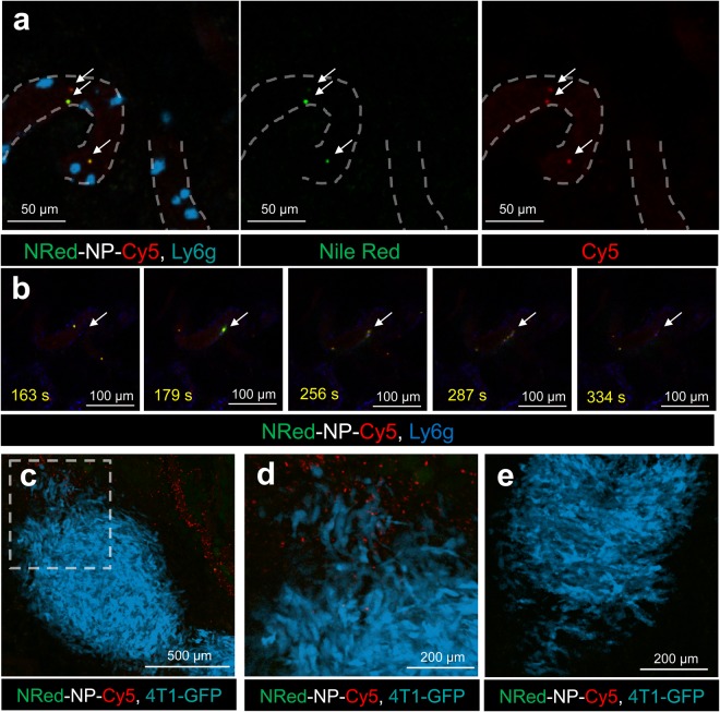
Figure 7
Study of payload delivery by Fe 3 O 4 -Au hybrid NPs. ( a , b ) Confocal intravital microscopy (IVM) of NRed-NP-Cy5 in superficial 4T1 tumor vessels upon i.v. injection to a mouse (6.6u2009mgu00b7kg u...
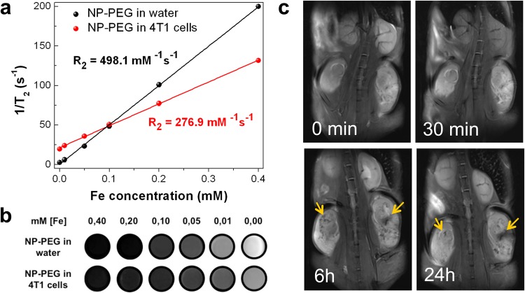
Figure 8
Fe 3 O 4 -Au Janus NPs as MRI contrast agents. ( a ) Proton T 2 relaxation time asu00a0a function of iron concentration for NP-PEG in water and in 4T1 cells. The R 2 value is determined by the slope o...
Figure images are served from the NIH/NLM PubMed Central Open Access Subset or Europe PMC; copyright remains with the publishers and authors.
💬 Discussion
0 commentsNo comments yet. Be the first to start a discussion!
Leave a Comment