Abstract
The drug release and degradation behavior of two double-walled microsphere formulations consisting of a doxorubicin-loaded poly(d,l-lactic-co-glycolic acid) (PLGA) core (∼46 kDa) surrounded by a poly(d,l-lactic acid) (PDLLA) shell layer (∼55 and 116 kDa) were examined. It was postulated that different molecular weights of the shell layer could modulate the erosion of the outer coating and limit the occurrence of water penetration into the inner drug-loaded core on various time scales, and therefore control the drug release from the microspheres. For both microsphere formulations, the drug release profiles were observed to be similar. The degradation of the microspheres was monitored for a period of about nine weeks and analyzed using scanning electron microscopy, laser scanning confocal microscopy, and gel permeation chromatography. Interestingly, both microsphere formulations exhibited occurrence of bulk erosion of PDLLA on a similar time scale despite different PDLLA molecular weights forming the shell layer. The shell layer of the double-walled microspheres served as an effective diffusion barrier during the initial lag phase period and controlled the release rate of the hydrophilic drug independent of the molecular weight of the shell layer.
🔬 Techniques
🔭 Microscopes
💻 Software
🧪 Sample Preparation
🏭 Microscope Brands
🧪 Reagent Suppliers
💻 Software Details
🏛️ Research Organizations (ROR)
Affiliated research institutions:
📋 Methods
2.1. Materials Poly(D,L-lactic-co-glycolic acid) (PLGA) copolymer (50:50 lactic acid:glycolic acid; inherent viscosity (i.v.) = 0.61 dL/g in hexafluoroisopropanol (HFIP)), and poly(lactic acid) (PLA) polymers including poly(D,L-lactic acid) (PDLLA) (i.v. = 0.37 and 0.70 dL/g in chloroform) and poly(L-lactic acid) (PLLA) (i.v. = 1.05 dL/g in chloroform) were purchased from Lactel Absorbable Polymers (Pelham, AL). Poly(vinyl alcohol) (PVA) (M w = 25,000 Da), 88 mol% hydrolyzed, was purchased from Polysciences, Inc. (Warrington, PA). Doxorubicin, in the form of hydrochloride salt with more than 99% purity, was purchased from LC Laboratories (Woburn, MA). Dichloromethane (DCM) and HFIP were acquired from Sigma-Aldrich Corp. (St. Louis, MO) while HPLC-grade tetrahydrofuran (THF) was acquired from Tedia (Fairfield, OH). Phosphate-buffered saline (PBS) with a pH of 7.4 was acquired from Mediatech, Inc. (Manassas, VA). 2.2. Fabrication of double-walled PLA(PLGA) microspheres Double-walled PLA(PLGA) microspheres consisting of a PLGA core surrounded by a PLA shell were produced by using the established precision particle fabrication (PPF) technique ( Fig. 1 ). Solutions containing 20 to 40% (w/v) PLGA and 5% (w/v) PLA in DCM were individually prepared. In this technique, a coaxial nozzle was used to produce a jet of core PLGA surrounded by an annular stream containing PLA. The core-shell polymer jet, protected by a non-solvent 0.5% (w/v) PVA carrier stream, was disrupted into uniform nascent double-walled droplets by an ultrasonic transducer controlled by a frequency generator. In order to control monodispersity of the double-walled microspheres, the fabrication process was monitored to ensure there was a steady disruption of the core-shell polymer jet by adjusting ultrasonic frequency and flow rate of the carrier stream. The droplets were collected in a beaker containing 0.5% (w/v) PVA solution, before they were stirred continuously for ~2 h, filtered and rinsed with an equal volume of distilled water to remove residual PVA from the microspheres. Finally, the microspheres were freeze-dried for 3 days and stored at −20°C under desiccant. To prepare microspheres loaded with doxorubicin in the PLGA core phase, a stock solution of doxorubicin was first prepared in water (50 mg/ml), before an appropriate amount of drug solution was further diluted in water and added to 10 ml of PLGA/DCM solution to obtain the desired drug to polymer loading. The resultant mixture was sonicated using a Model 500 Sonic Dismembrator (Thermo Fisher Scientific, Inc., Pittsburgh, PA) at 30% amplitude in an ice bath for 90 s to form a stable emulsion. 2.3. Particle size distribution The size distributions of the hardened double-walled microspheres were determined using a Multisizer 3 (Beckman Coulter, Inc., Fullerton, CA) with a 120 µm aperture. The microspheres were suspended in Isoton II Diluent (Beckman Coulter, Inc., Fullerton, CA) before measurement. At least 10,000 microspheres were measured for every sample. 2.4.
Show full methods section
2.1. Materials Poly(D,L-lactic-co-glycolic acid) (PLGA) copolymer (50:50 lactic acid:glycolic acid; inherent viscosity (i.v.) = 0.61 dL/g in hexafluoroisopropanol (HFIP)), and poly(lactic acid) (PLA) polymers including poly(D,L-lactic acid) (PDLLA) (i.v. = 0.37 and 0.70 dL/g in chloroform) and poly(L-lactic acid) (PLLA) (i.v. = 1.05 dL/g in chloroform) were purchased from Lactel Absorbable Polymers (Pelham, AL). Poly(vinyl alcohol) (PVA) (M w = 25,000 Da), 88 mol% hydrolyzed, was purchased from Polysciences, Inc. (Warrington, PA). Doxorubicin, in the form of hydrochloride salt with more than 99% purity, was purchased from LC Laboratories (Woburn, MA). Dichloromethane (DCM) and HFIP were acquired from Sigma-Aldrich Corp. (St. Louis, MO) while HPLC-grade tetrahydrofuran (THF) was acquired from Tedia (Fairfield, OH). Phosphate-buffered saline (PBS) with a pH of 7.4 was acquired from Mediatech, Inc. (Manassas, VA). 2.2. Fabrication of double-walled PLA(PLGA) microspheres Double-walled PLA(PLGA) microspheres consisting of a PLGA core surrounded by a PLA shell were produced by using the established precision particle fabrication (PPF) technique ( Fig. 1 ). Solutions containing 20 to 40% (w/v) PLGA and 5% (w/v) PLA in DCM were individually prepared. In this technique, a coaxial nozzle was used to produce a jet of core PLGA surrounded by an annular stream containing PLA. The core-shell polymer jet, protected by a non-solvent 0.5% (w/v) PVA carrier stream, was disrupted into uniform nascent double-walled droplets by an ultrasonic transducer controlled by a frequency generator. In order to control monodispersity of the double-walled microspheres, the fabrication process was monitored to ensure there was a steady disruption of the core-shell polymer jet by adjusting ultrasonic frequency and flow rate of the carrier stream. The droplets were collected in a beaker containing 0.5% (w/v) PVA solution, before they were stirred continuously for ~2 h, filtered and rinsed with an equal volume of distilled water to remove residual PVA from the microspheres. Finally, the microspheres were freeze-dried for 3 days and stored at −20°C under desiccant. To prepare microspheres loaded with doxorubicin in the PLGA core phase, a stock solution of doxorubicin was first prepared in water (50 mg/ml), before an appropriate amount of drug solution was further diluted in water and added to 10 ml of PLGA/DCM solution to obtain the desired drug to polymer loading. The resultant mixture was sonicated using a Model 500 Sonic Dismembrator (Thermo Fisher Scientific, Inc., Pittsburgh, PA) at 30% amplitude in an ice bath for 90 s to form a stable emulsion. 2.3. Particle size distribution The size distributions of the hardened double-walled microspheres were determined using a Multisizer 3 (Beckman Coulter, Inc., Fullerton, CA) with a 120 µm aperture. The microspheres were suspended in Isoton II Diluent (Beckman Coulter, Inc., Fullerton, CA) before measurement. At least 10,000 microspheres were measured for every sample. 2.4.
Optical microscopy
The hardened double-walled microspheres were examined using an Invertoskop inverted microscope (Carl Zeiss Microscopy LLC, Thornwood, NY). A few droplets of the aqueous microsphere suspension were placed directly onto a microscope slide. Images were captured using Digital Microscope Suite software. 2.5. Drug loading The drug loading was determined by dissolving approximately 50 mg of microspheres (2% (w/w) theoretical loading of doxorubicin with respect to PLGA) in 1 ml of HFIP. The samples were allowed to stand until complete dissolution of the polymers. The samples were then centrifuged at 10,000 rpm for 10 min, and the supernatants were carefully extracted. In order to measure doxorubicin concentration, the supernatant was added in triplicate in a 96-well plate, and the absorbance was analyzed using a SpectraMax 340PC spectrophotometer (Molecular Devices LLC, Sunnyvale, CA) at a wavelength of 480 nm. 2.6. In vitro drug release The release profile was determined by suspending approximately 150 mg of microspheres (2% (w/w) theoretical loading of doxorubicin with respect to PLGA) in 5 ml of PBS in centrifuge tubes. The tubes were placed in an incubator maintained at 37°C and shaken at 240 rpm. At selected time points, the tubes were centrifuged at 10,000 rpm for 10 min before 1 ml of supernatant was collected and 1 ml of fresh PBS was replaced. This is done to ensure sink conditions. In order to measure doxorubicin concentration, the supernatant was sufficiently diluted in PBS before adding in triplicate in a 96-well plate, and the fluorescence was analyzed using the fluorescence spectrophotometer at excitation and emission wavelengths of 480 and 590 nm, respectively. 2.7. In vitro degradation The in vitro degradation study was performed by suspending approximately 20 mg of microspheres (0.06% (w/w) loading of doxorubicin with respect to PLGA) in 660 µl of PBS in centrifuge tubes. Similar to the in vitro drug release study, the tubes were placed in an incubator maintained at 37°C and shaken at 240 rpm. Based on the in vitro drug release time points, the tubes were centrifuged at 10,000 rpm for 10 min before 132 µl of supernatant was removed, and 132 µl of fresh PBS was replaced. This is done to ensure sink conditions. At pre-determined time points, the tubes were centrifuged, and PBS was removed. The microspheres were then rinsed twice with distilled water, and imaged using scanning electron microscopy (SEM) and laser scanning confocal microscopy. Samples were also collected for gel permeation chromatography (GPC) analysis. In this case, the samples were further freeze-dried for 3 days before dissolving them in HPLC-grade THF. 2.8.
Scanning electron microscopy
The surface morphology of the degrading double-walled microspheres was examined using a JEOL JSM-5600LV scanning electron microscope (SEM) (JEOL Ltd., Tokyo, Japan). A few droplets of the aqueous microsphere suspension were placed directly onto a SEM sample holder coated with conductive carbon tape and air-dried overnight. The samples were sputter-coated with gold palladium prior to imaging at 10 kV. 2.9.
Laser scanning confocal microscopy
The distribution of doxorubicin in the degrading double-walled microspheres was examined using a Fluoview FV1000 laser scanning confocal microscope (Olympus Corp., Tokyo, Japan) equipped with argon ion laser tuned to 488 nm. A few droplets of the aqueous microsphere suspension were placed directly onto a microscope slide and sealed with a cover glass. The samples were then visualized using an oil immersion objective lens under ×60 magnification and ×1 zoom with the following calibrations: 4.0 µs/pixel sampling speed, laser at 720 V and transmissivity of 10%. The fluorescence emission was collected using a 505 nm long pass interference filter. These settings were used for all the samples to ensure consistency. Optical cross-sections were taken at various depths for each sample in order to determine drug distribution at the centerline of the microspheres. Images were captured using Olympus Fluoview software. The fluorescence intensity profiles were obtained using ImageJ software. 2.10.
Molecular weight analysis
The molecular weights of pure polymers and double-walled microspheres were determined using a gel permeation chromatography (GPC) system consisting of a Waters 1515 Isocratic HPLC Pump, Waters 717plus Autosampler and Waters 2414 Refractive Index Detector (Waters Corp., Milford, MA). The samples were eluted in HPLC-grade THF through 5 µm Jordi Gel DVB 1,000, 10,000 and 100,000 Ǻ columns (Jordi Labs LLC, Bellingham, MA) connected in series at a flow rate of 1 ml/min and a temperature of 35°C. The samples were filtered before injecting into the column to remove insoluble particulates when present. The calibration curve was generated using polystyrene standards (Polymer Laboratories Ltd., Church Stretton, Shropshire, UK) prepared at concentrations of 1 mg/ml. The semi-logarithmic calibration curve of molecular weight versus elution time is linear ( R 2 = 0.998). The weight-averaged and peak molecular weights of the samples were obtained using Waters Breeze software. The molecular weights were reported based on the average of two measurements.
2.1. Materials Poly(D,L-lactic-co-glycolic acid) (PLGA) copolymer (50:50 lactic acid:glycolic acid; inherent viscosity (i.v.) = 0.61 dL/g in hexafluoroisopropanol (HFIP)), and poly(lactic acid) (PLA) polymers including poly(D,L-lactic acid) (PDLLA) (i.v. = 0.37 and 0.70 dL/g in chloroform) and poly(L-lactic acid) (PLLA) (i.v. = 1.05 dL/g in chloroform) were purchased from Lactel Absorbable Polymers (Pelham, AL). Poly(vinyl alcohol) (PVA) (M w = 25,000 Da), 88 mol% hydrolyzed, was purchased from Polysciences, Inc. (Warrington, PA). Doxorubicin, in the form of hydrochloride salt with more than 99% purity, was purchased from LC Laboratories (Woburn, MA). Dichloromethane (DCM) and HFIP were acquired from Sigma-Aldrich Corp. (St. Louis, MO) while HPLC-grade tetrahydrofuran (THF) was acquired from Tedia (Fairfield, OH). Phosphate-buffered saline (PBS) with a pH of 7.4 was acquired from Mediatech, Inc. (Manassas, VA).
📊 Figures
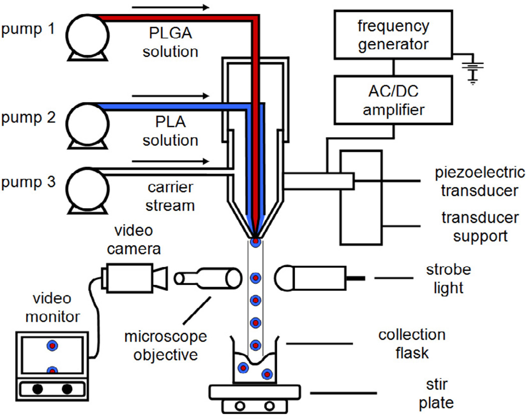
Figure 1
Schematic diagram of precision particle fabrication apparatus for the production of uniform double-walled microspheres of controlled shell thickness.
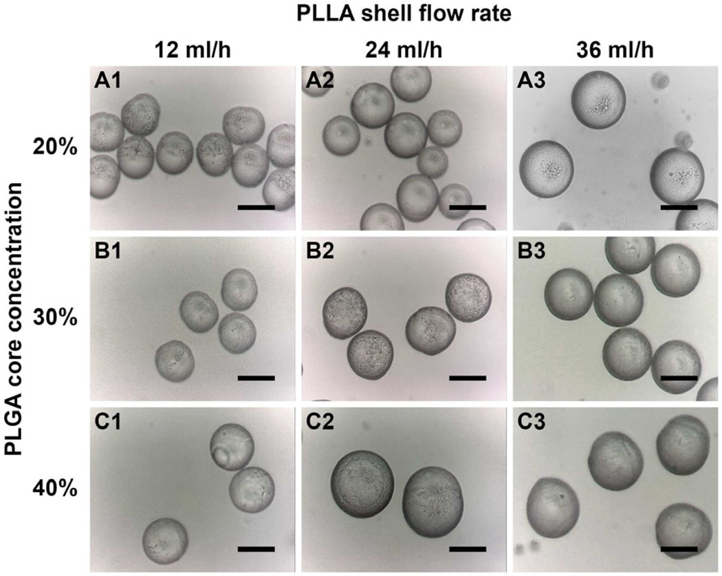
Figure 2
Optical images depicting the surface morphology of double-walled PLLA(PLGA) microspheres for various microsphere samples listed in Table 1 . Partial encapsulation was observed for samples A1, A2, B1, ...
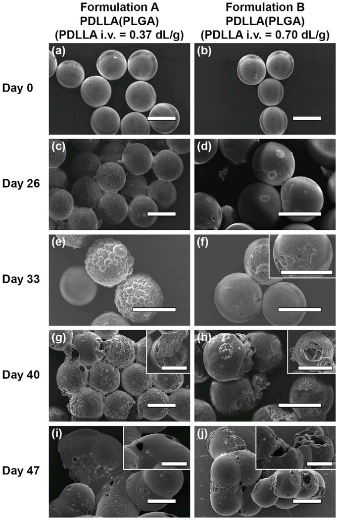
Figure 3
SEM images depicting the surface morphology of double-walled PDLLA(PLGA) microspheres with a low PDLLA molecular weight shell layer (formulation A) and a high PDLLA molecular weight shell layer (formu...
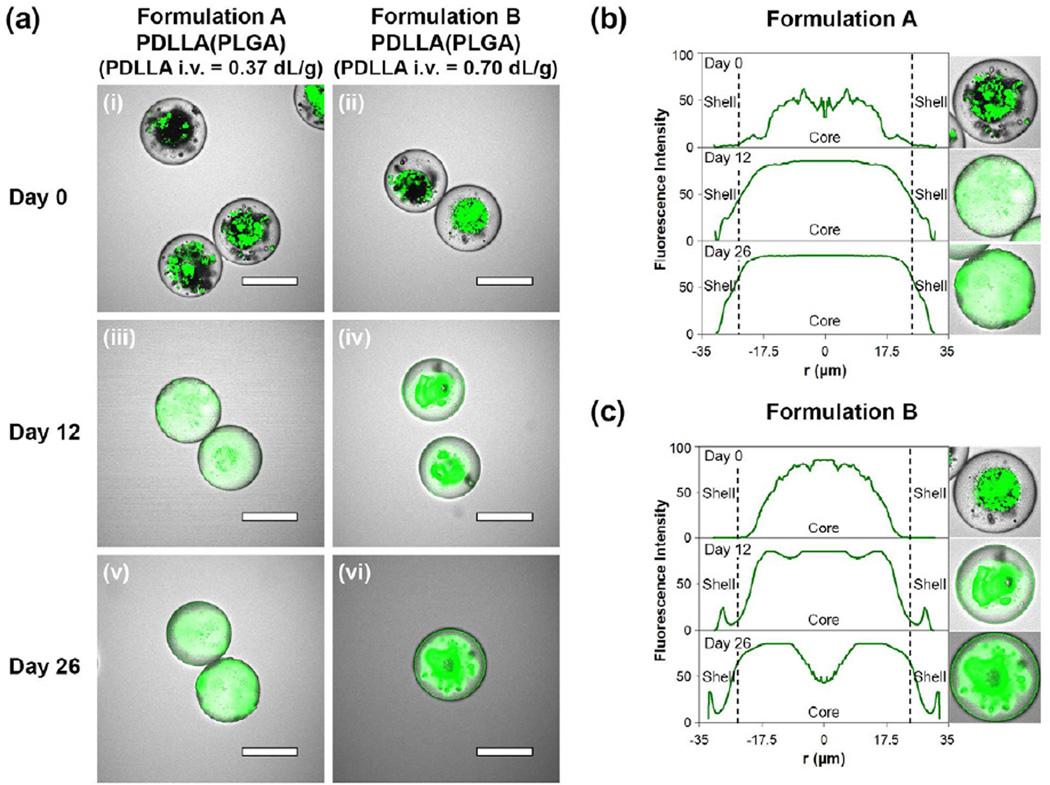
Figure 4
Laser scanning confocal images and fluorescence intensity profiles depicting the distribution of doxorubicin in the double-walled PDLLA(PLGA) microspheres during the initial stage of the degradation p...
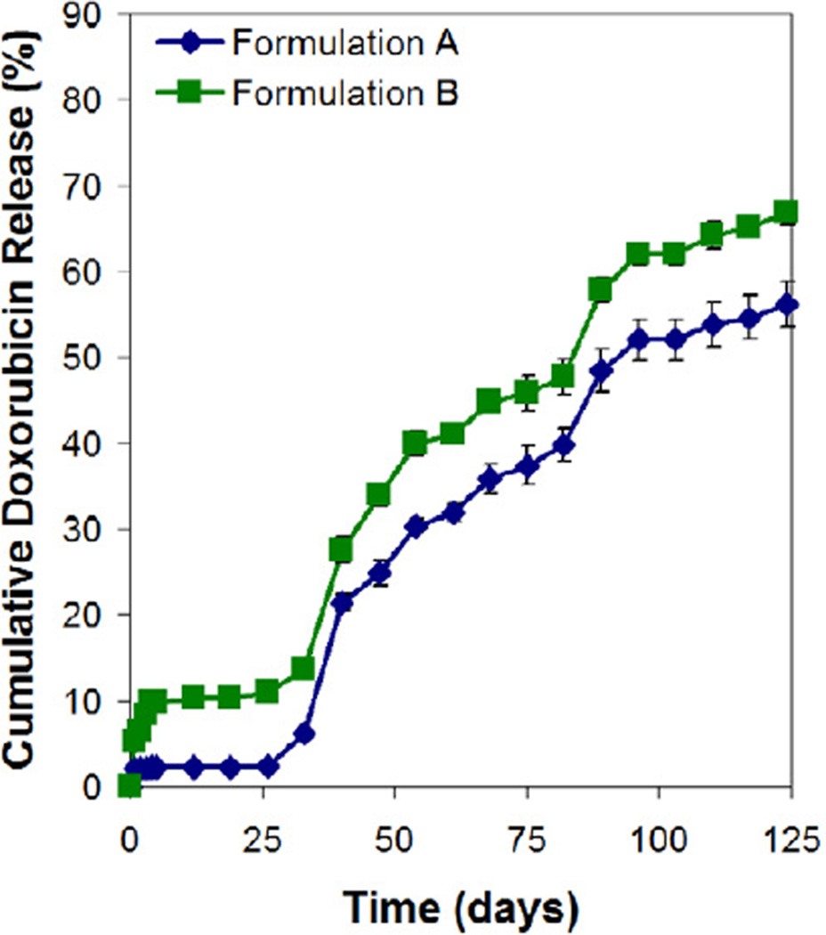
Figure 5
In vitro release of doxorubicin from double-walled PDLLA(PLGA) microspheres.
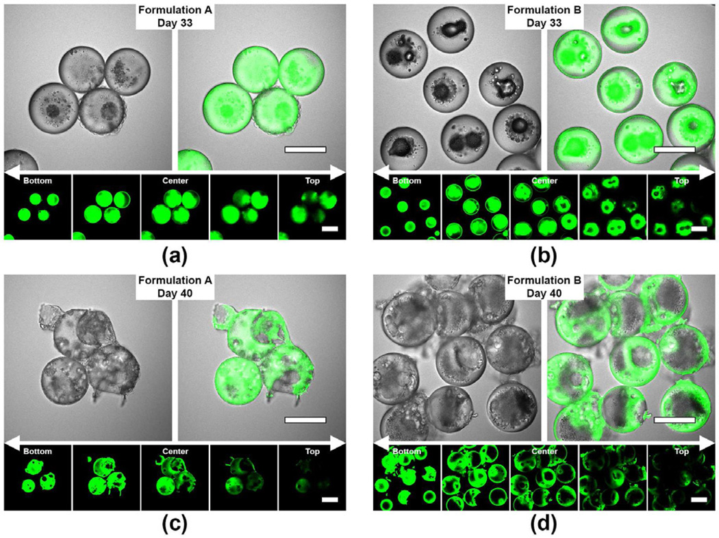
Figure 6
Laser scanning confocal images depicting the development of multiple pores and/or cavities in the double-walled PDLLA(PLGA) microspheres during the later stage of the degradation process (33 to 40 day...
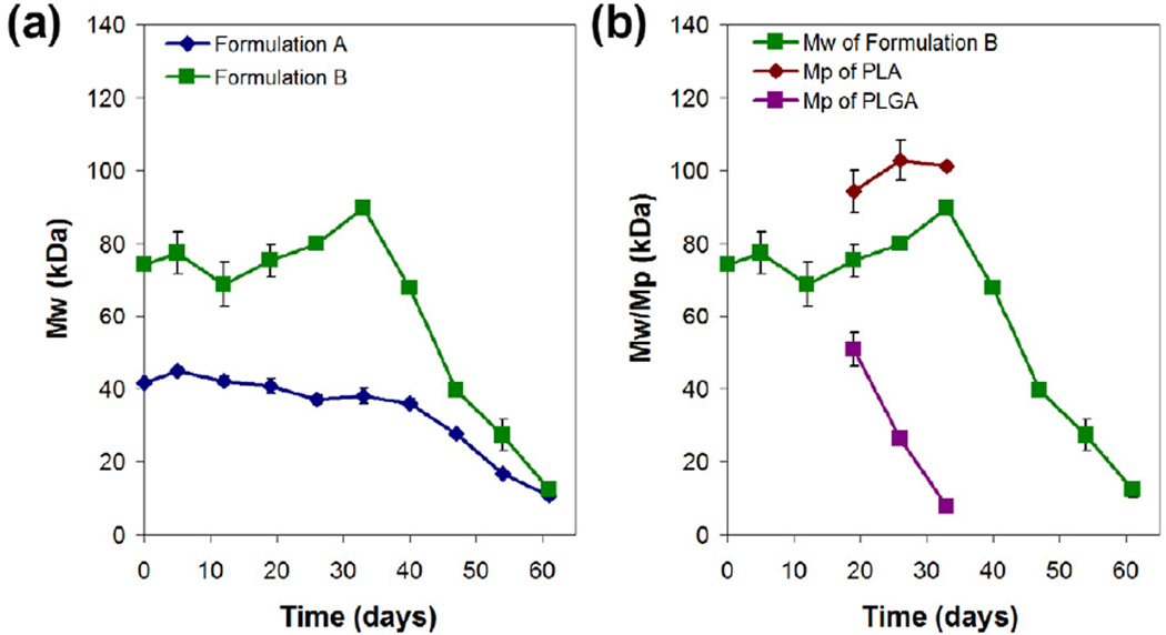
Figure 7
Molecular weight profiles as a function of incubation time for double-walled PDLLA(PLGA) microspheres during degradation. (a) Weight-averaged molecular weight (M w ) profiles for formulations A and B ...
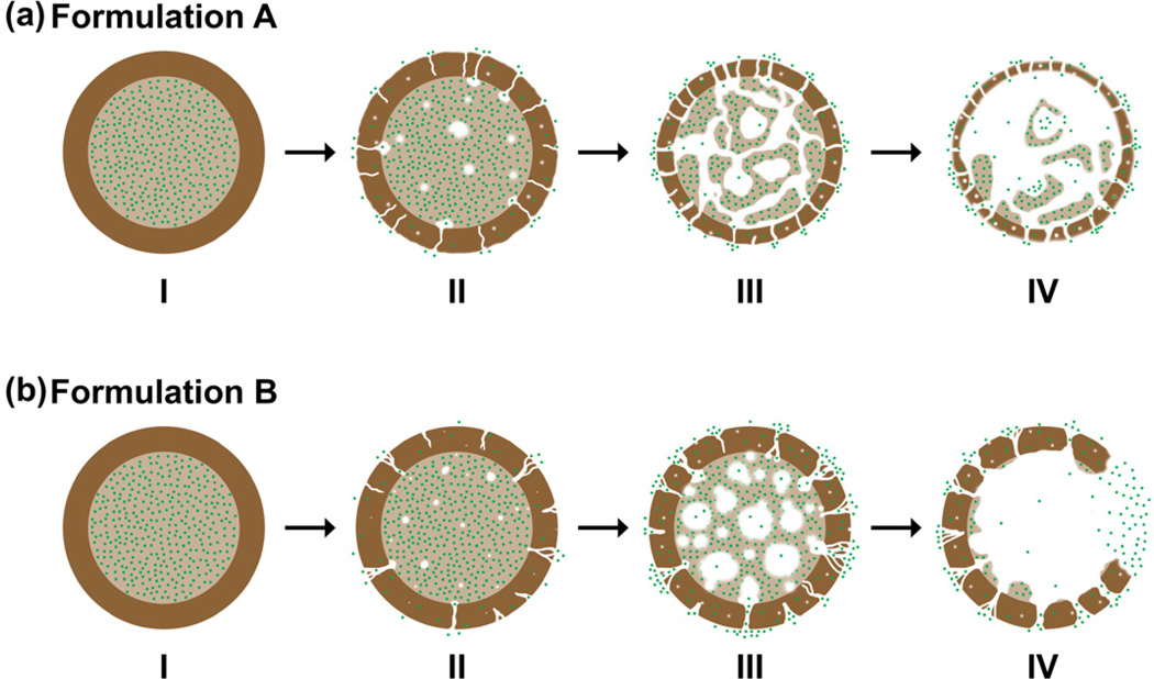
Figure 8
Schematic illustration of the proposed mechanism for the release of doxorubicin from double-walled PDLLA(PLGA) microspheres. PLGA core and PDLLA shell layer are represented by light and dark brown res...
Figure images are served from the NIH/NLM PubMed Central Open Access Subset or Europe PMC; copyright remains with the publishers and authors.
💬 Discussion
0 commentsNo comments yet. Be the first to start a discussion!
Leave a Comment