Abstract
Mitotic chromosome assembly remains a big mystery in biology. Condensin complexes are pivotal for chromosome architecture yet how they shape mitotic chromatin remains unknown. Using acute inactivation approaches and live-cell imaging in Drosophila embryos, we dissect the role of condensin I in the maintenance of mitotic chromosome structure with unprecedented temporal resolution. Removal of condensin I from pre-established chromosomes results in rapid disassembly of centromeric regions while most chromatin mass undergoes hyper-compaction. This is accompanied by drastic changes in the degree of sister chromatid intertwines. While wild-type metaphase chromosomes display residual levels of catenations, upon timely removal of condensin I, chromosomes present high levels of de novo Topoisomerase II (TopoII)-dependent re-entanglements, and complete failure in chromosome segregation. TopoII is thus capable of re-intertwining previously separated DNA molecules and condensin I continuously required to counteract this erroneous activity. We propose that maintenance of chromosome resolution is a highly dynamic bidirectional process.
🔬 Techniques
🧬 Organisms
✨ Fluorophores
🧪 Sample Preparation
🏭 Microscope Brands
🧪 Reagent Suppliers
📷 Detectors
💻 Software Details
📋 Protocols
💾 Data Repositories
🏷️ Research Resource Identifiers (RRIDs)
Verified research resources used in this paper:
🏛️ Research Organizations (ROR)
Affiliated research institutions:
📋 Methods
Fly strains
To destroy condensin by TEV protease-mediated cleavage, strains carrying solely TEV-sensitive Barren versions were produced. A construct carrying a ~4.7 kb Barren genomic region was used as a starting point (kindly provided by Beat Suter, Institute of Cell Biology, University of Bern). This region contains the regulatory sequences and was previously shown to restore Barren function ( Masrouha et al., 2003 ). This construct was engineered to add a 10xMyc sequence at the C-terminus of Barren. Three consecutive TEV recognition sites were placed at different positions (corresponding to a.a. 175, a.a. 389, a.a. 437 and a.a 600). Cloning details are available upon request. Each variant of genomic Barren with different TEV sites was cloned into pCaSpeR4 vector used for fly transformation. Transgenic flies were produced by P-element integration (BestGene Inc, Chino Hills, CA). Transgenes were placed in a Barr L305 background, a Barren null allele ( Bhat et al., 1996 ), over a deficiency for the corresponding genomic region ( Df(2L)Exel7077 , stock #7850 from Bloomington stock center). To destroy cohesin by TEV-protease we used strains carrying Rad21 TEV , previously described ( Oliveira et al., 2010 ; Pauli et al., 2008 ). Fly strains also expressed His2AvD–mRFP1 or polyubiquitin His2B–RFP, to monitor DNA and EGFP–Cid to monitor centromeres ( Schuh et al., 2007 ). A list with detailed genotypes can be found in Table 1 . 10.7554/eLife.26120.028 Table 1. List of fly strains used in this study DOI: http://dx.doi.org/10.7554/eLife.26120.028 CHR# * Genotype Reference 1418 Barr L305 /CyO Bhat et al. (1996) (RRID: BDSC_4402 ) 1421 Df(2L)Exel7077/CyO Blommington #7850 (RRID: BDSC_7850 ) 1513 w;; Barr(175 - 3TEV)-myc10 III.5 This study 1509 w; Barr(175 - 3TEV)-myc10 II.1; This study 1522 w;; Barr(389 - 3TEV)-myc10 III.2 This study 1514 w;; Barr(437 - 3TEV)-myc10 III.1 This study 1520 w;; Barr(600 - 3TEV)-myc10 III.3 This study 1525 w;; Barr(wt)-myc10 III.1 This study 1560 w; Barr L305 / Df(2L)Exel7077; Barr(175 - 3TEV)-myc10 III.5 This study 820 w;; HisH2AvD-mRFP1 III.1, CGC (CID-EGFP) III.1 Schuh et al. (2007) 1564 Df(2L)Exel7077 / CyO; HisH2AvD-mRFP1 III.1, CGC (CID-EGFP) III.1 This study w; Barr L305 / Df(2L)Exel7077; Barr(175 - 3TEV)-myc10 III.5/ HisH2AvD-mRFP1 III.1, CGC (CID-EGFP) III.1 629 w;; Rad21 ex15 , polyubiq-H2B-RFP, tubpr-Rad21(550-3TEV) -myc10 Oliveira et al. (2010) 1646 w; Barr L305 , Barr(175 - 3TEV)-myc10 II.1; +/+ This study 1648 w; Barr L305 , Barr(175 - 3TEV)-myc10 II.1; Rad21 ex15 , polyubiq-H2B-RFP, tubpr-Rad21(550-3TEV) -myc10 This study * Reference number in our internal lab fly database. Microinjections Microinjection experiments were performed as previously described ( Oliveira et al., 2010 ). 1–1.5 hr old embryos (or 0–30 min for mRNA injections) were collected and processed according to standard protocols, and embryos were injected at the posterior pole (up to three sequential injections) using a Burleigh Thorlabs Micromanipulator, a Femtojet microinjection system (Eppendorf, Germany), and pre-pulled Femtotip I needles (Eppendorf). Embryos were injected with buffer, drugs or proteins purified from E. coli at the following concentrations: Buffer (20 mM Tris-HCl at pH 8.0, 1 mM EDTA, 50 mM NaCl and 2 mM DTT), 13 mg/ml TEV protease in TEV buffer, 12 mg/ml UbcH10 C114S , 14 mg/ml UbcH10 wt and/or 280 μM ICRF-193 (Sigma-Aldrich, St Louis, MO). Protein purification Purified TEV protease was described previously ( Haering et al., 2008 ).
Show full methods section
Fly strains
To destroy condensin by TEV protease-mediated cleavage, strains carrying solely TEV-sensitive Barren versions were produced. A construct carrying a ~4.7 kb Barren genomic region was used as a starting point (kindly provided by Beat Suter, Institute of Cell Biology, University of Bern). This region contains the regulatory sequences and was previously shown to restore Barren function ( Masrouha et al., 2003 ). This construct was engineered to add a 10xMyc sequence at the C-terminus of Barren. Three consecutive TEV recognition sites were placed at different positions (corresponding to a.a. 175, a.a. 389, a.a. 437 and a.a 600). Cloning details are available upon request. Each variant of genomic Barren with different TEV sites was cloned into pCaSpeR4 vector used for fly transformation. Transgenic flies were produced by P-element integration (BestGene Inc, Chino Hills, CA). Transgenes were placed in a Barr L305 background, a Barren null allele ( Bhat et al., 1996 ), over a deficiency for the corresponding genomic region ( Df(2L)Exel7077 , stock #7850 from Bloomington stock center). To destroy cohesin by TEV-protease we used strains carrying Rad21 TEV , previously described ( Oliveira et al., 2010 ; Pauli et al., 2008 ). Fly strains also expressed His2AvD–mRFP1 or polyubiquitin His2B–RFP, to monitor DNA and EGFP–Cid to monitor centromeres ( Schuh et al., 2007 ). A list with detailed genotypes can be found in Table 1 . 10.7554/eLife.26120.028 Table 1. List of fly strains used in this study DOI: http://dx.doi.org/10.7554/eLife.26120.028 CHR# * Genotype Reference 1418 Barr L305 /CyO Bhat et al. (1996) (RRID: BDSC_4402 ) 1421 Df(2L)Exel7077/CyO Blommington #7850 (RRID: BDSC_7850 ) 1513 w;; Barr(175 - 3TEV)-myc10 III.5 This study 1509 w; Barr(175 - 3TEV)-myc10 II.1; This study 1522 w;; Barr(389 - 3TEV)-myc10 III.2 This study 1514 w;; Barr(437 - 3TEV)-myc10 III.1 This study 1520 w;; Barr(600 - 3TEV)-myc10 III.3 This study 1525 w;; Barr(wt)-myc10 III.1 This study 1560 w; Barr L305 / Df(2L)Exel7077; Barr(175 - 3TEV)-myc10 III.5 This study 820 w;; HisH2AvD-mRFP1 III.1, CGC (CID-EGFP) III.1 Schuh et al. (2007) 1564 Df(2L)Exel7077 / CyO; HisH2AvD-mRFP1 III.1, CGC (CID-EGFP) III.1 This study w; Barr L305 / Df(2L)Exel7077; Barr(175 - 3TEV)-myc10 III.5/ HisH2AvD-mRFP1 III.1, CGC (CID-EGFP) III.1 629 w;; Rad21 ex15 , polyubiq-H2B-RFP, tubpr-Rad21(550-3TEV) -myc10 Oliveira et al. (2010) 1646 w; Barr L305 , Barr(175 - 3TEV)-myc10 II.1; +/+ This study 1648 w; Barr L305 , Barr(175 - 3TEV)-myc10 II.1; Rad21 ex15 , polyubiq-H2B-RFP, tubpr-Rad21(550-3TEV) -myc10 This study * Reference number in our internal lab fly database. Microinjections Microinjection experiments were performed as previously described ( Oliveira et al., 2010 ). 1–1.5 hr old embryos (or 0–30 min for mRNA injections) were collected and processed according to standard protocols, and embryos were injected at the posterior pole (up to three sequential injections) using a Burleigh Thorlabs Micromanipulator, a Femtojet microinjection system (Eppendorf, Germany), and pre-pulled Femtotip I needles (Eppendorf). Embryos were injected with buffer, drugs or proteins purified from E. coli at the following concentrations: Buffer (20 mM Tris-HCl at pH 8.0, 1 mM EDTA, 50 mM NaCl and 2 mM DTT), 13 mg/ml TEV protease in TEV buffer, 12 mg/ml UbcH10 C114S , 14 mg/ml UbcH10 wt and/or 280 μM ICRF-193 (Sigma-Aldrich, St Louis, MO). Protein purification Purified TEV protease was described previously ( Haering et al., 2008 ).
Purification of UbcH10 wt and UbcH10
C114S was performed from BL21 cells as previously described ( Oliveira et al., 2010 ), with minor modifications, as follows. Bacterial cells were grown for 16 hr at 37°C, 225 rpm. This pre-culture was used to inoculate fresh LB media and cells were allowed to grow until 0.8/1 ODs. Cultures were then induced with 1 mM IPTG and after 4 hr of induction at 37°C, 225 rpm, cells were harvested. Pellets were ressuspended in Lysis Buffer (20 mM Tris-HCL pH7.5, 0.5M NaCl, 5 mM Imidazole with protease inhibitors) and sonicated 5x on ice in 30 s cycles (power 5- Sonicator XL2020, Misonix, Farmingdale, NY). The soluble fraction of the extracts was then incubated in TALON Metal Affinity Resin (Takara Bio Inc. , Japan), according to manufacturer’s instructions. After several washes with Lysis Buffer, the resin coated with the protein was packed into a Poly-Prep Chromatography Column (Biorad, Hercules, CA). Proteins were eluted in the same buffer with 300 mM imidazole. For buffer exchange, purified UbcH10 wt and UbcH10 C114S proteins were dialyzed overnight, at 4°C, in a Slide-a-Lyzer 7 KDa Dialysis cassettes (Thermo Scientific, Waltham, MA). Final storage buffer was 20 mM Tris-HCL pH7.5, 0.3M NaCl). The purified proteins were concentrated in a Vivaspin 6 Centrifugal Concentrator MWCO 10.000 KDa (GE Healthcare, Issaquah, WA). mRNA synthesis Barren 175TEV- EGFP was cloned into a pRNA plasmid and mRNA was synthesized by in vitro transcription with the mMessage mMachine T3 kit (Ambion, Austin, TX), followed by purification with RNeasy kit (Qiagen, Germany), and elution in RNase-free water. To probe for the efficiency of Barren TEV removal ( Figure 1C ), 0–30 min old embryos surviving only on Barren TEV -Myc were injected with Barren TEV -EGFP mRNA in pure water at ~2.2 μg/μl. Embryos were left to develop at 22°C for 1,5–2 hr, to allow for protein translation, before the subsequent injections. In vitro cleavage experiments Ovaries were dissected from females and homogenized in PBS. Extracts were sonicated for 2 min in a water-bath (power 5- Sonicator XL2020, Misonix). After centrifugation for 10 min at 15.000 rpm at 4°C, the supernatant was removed and adjusted to a final concentration of 2 μg/μl. For cleavage experiments, 80 μl of extract were incubated with 2 μg of TEV protease. At the indicated time points, 10 μl of the reaction were diluted with sample buffer, boiled and stored at −20°C. Western-blot Samples were loaded on a 10% SDS-gel for electrophoresis and transferred onto a membrane (Immun-Blot PVDF, Biorad). Western-blot analysis was performed according to standard protocols using the following antibodies: anti myc-tag (1:200, Santa Cruz Biotechnology, Dallas, TX, Cat# sc-47694 RRID: AB_627266 ), anti-α-tubulin (1:50.000, DM1A, Sigma-Aldrich Cat# T9026 RRID: AB_477593 ) and anti-Barren (1:3000, kindly provided by Hugo Bellen, ( Bhat et al., 1996 ), RRID: AB_2567044 ).
Microscopy
Aligned embryos on coverslips were covered with Series 700 halocarbon oil (Sigma-Aldrich). Imaging of embryos after mRNA injection ( Figure 1c ) was performed with a spinning disc Revolution XD microscope (Andor, UK) at 22°C. Stacks of around 20 frames 1 μm were taken at indicated times using a 100 × 1.4 oil immersion objective (Nikon, Japan) and iXon +512 EMCCD camera (Andor). Time-lapse microscopy was performed with an inverted wide-field DeltaVision microscope (Applied Precision Inc., Issaquah, WA) at 18–20°C in a temperature-controlled room. One stack of ~20 frames (0.8 μm apart) was acquired every 1 or 2 min using a 100 × 1.4 oil immersion objective (Olympus, Japan) and an EMCCD camera (Roper Cascade 1024, Roper Technologies, Inc., Sarasota, FL). Widefield images were restored by deconvolution with the Huygens v15.10/16.10 deconvolution software using a calculated point-spread function (RRID: SCR_014237 , Scientific Volume Imaging, The Netherlands). Movies were assembled using FIJI software (RRID: SCR_002285 ) ( Schindelin et al., 2012 ) and selected stills were processed with Photoshop CS6 (Adobe Systems Incorporated, San Jose, CA).
Quantitative imaging analysis
For the quantification of chromosome condensation presented in Figure 3g and Figure 3—figure supplement 1 , deconvolved images were analyzed using Imaris v6.1 software (RRID: SCR_007370 , Bitplane, Switzerland). The same metaphase was tracked over time and average values for mean voxel intensity, volume and surface area were normalized to the first frame after injection. For the fluorescence profiles presented in Figures 3f and 6b–e,a wide 15 μm-long line was placed manually along the segregation plane and measured using the ‘Plot Profile’ function on FIJI software. For each data set, values were normalized to the maximum. Measurements of single chromatids width and length were performed on projected images (maximum intensity projection), using FIJI software and single chromatids mean voxel intensity measurements were performed using Imaris software. Quantification of chromosome movement ( Figure 4 ) was performed as previously described ( Mirkovic et al., 2015 ). Briefly, HisH2B-RFP was imaged at 1 min intervals. Images were segmented to select the chromosomal regions, based on an automatic threshold (set in the first frame after TEV injection), to create binary images. For each movie, a walking average of 3 frames was produced (using kymograph plug-in, written by J. Rietdorf and A. Seitz, EMBL, Heidelberg, Germany) creating a merged image in which the intensity is proportional to the overlap between consecutive frames. Intensity profiles were used to estimate the percentage of non-overlapping, 2- frame overlap and 3-frame overlap pixels. Graphic representation was performed using Prism seven software (RRID: SCR_002798 , GraphPad, La Jolla, CA).
Statistical analysis
To compare the average of the centromere distances between each experimental condition ( Figure 6f ), at least 10 independent embryos were analyzed. Statistical analysis was performed using Prism seven software (RRID: SCR_002798 ). Given that some datasets did not pass the normality test (D'Agostino and Pearson normality test), multiple comparisons were performed using the non-parametric Krustal-Wallis test.
📊 Figures
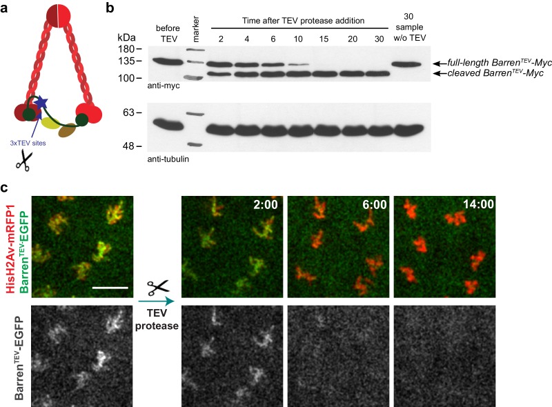
Figure 1.
TEV-mediated cleavage of Barren disrupts condensin I function within a few minutes.
( a ) Schematic representation of condensin complex indicating the position of the 3xTEV cleavage sites in the kleisin subunit Barren (aa175). ( b ) In vitro cleavage of Barren TEV -myc. Extracts were...
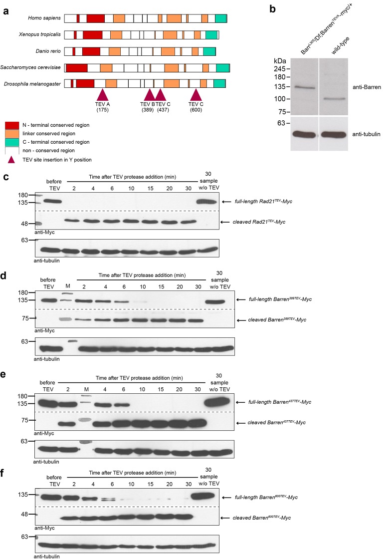
Figure 1u2014figure supplement 1.
A TEV-cleavable system to destroy condensin I.
( a ) graphic representation of the four positions used to introduce three consecutive TEV-protease consensus sites within the linker region of Drosophila Barren. Conservation with other species is sh...
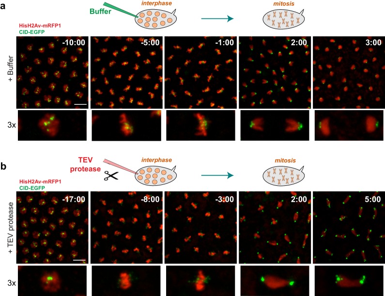
Figure 2.
Condensin I inactivation prior to mitotic entry.
Embryos surviving solely on Barren TEV were injected with buffer ( a ) or 13 mg/ml TEV protease ( b )u00a0~10u201315 min before mitosis; Embryos also express His2Au2013mRFP1 (red) and Cid-EGFP (green)...
Video 1.
Mitosis in Drosophila embryos.
Embryos were injected with buffer in early interphase and monitored throughout the subsequent mitosis. Embryos express HisH2Av-mRFP1 (red) and Cid-EGFP (green). Times are relative to injection time. S...
Video 2.
Mitosis upon condensin I inactivation in Drosophila embryos.
Embryos surviving solely on Barren TEV were injected with TEV protease in early interphase and monitored in the subsequent mitosis. Embryos express HisH2Av-mRFP1 (red) and Cid-EGFP (green). Times are ...
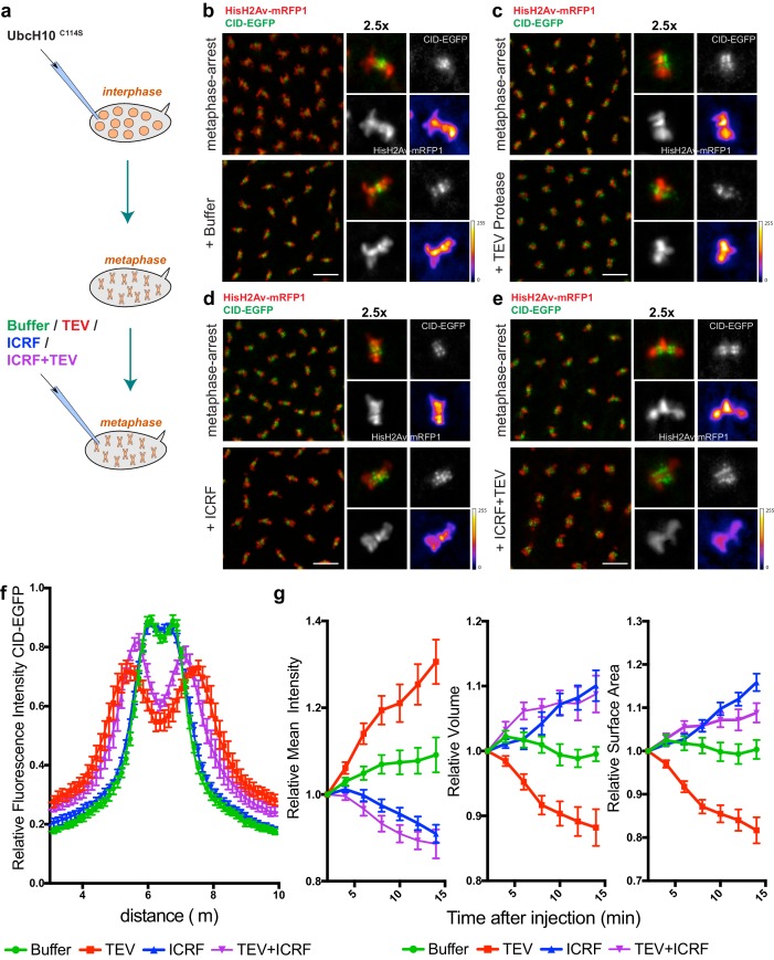
Figure 3.
Condensin I inactivation in pre-assembled chromosomes leads to disruption of centromere structure and hyper-compaction of mitotic chromosomes.
( a ) Schematic representation of the experimental layout. Embryos expressing solely Barren TEV were injected with 12 mg/ml of a dominant-negative form of the human E2 ubiquitin-conjugating enzyme (Ub...
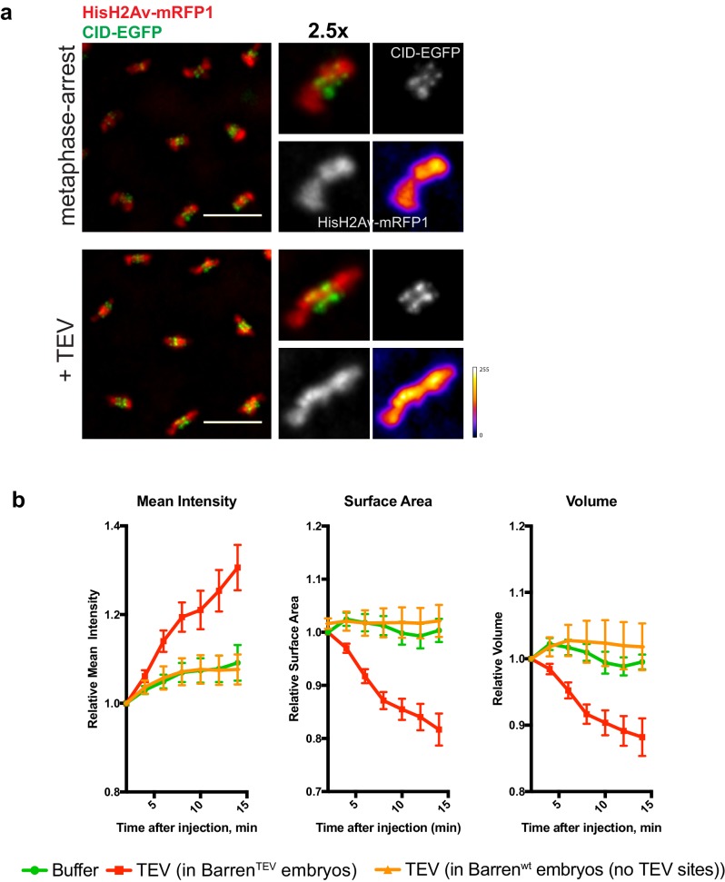
Figure 3u2014figure supplement 1.
- Chromosome condensation induced by TEV-protease depends on TEV cleavage sites present in Barren TEV .
( a ) Representative images from embryos that do not contain TEV-cleavage sites in Barren. Embryos were injected with UbcH10 C114S to induce a metaphase arrest and subsequently injected with 13 mg/ml ...
Video 3.
Buffer injection in metaphase-arrested embryos.
Embryos expressing solely Barren TEV were injected with 12 mg/ml of a dominant-negative form of the human E2 ubiquitin-conjugating enzyme (UbcH10 C114S ), to induce a metaphase arrest, and subsequentl...
Video 4.
Condensin I inactivation in metaphase-arrested embryos.
Embryos expressing solely Barren TEV were injected with 12 mg/ml of a dominant-negative form of the human E2 ubiquitin-conjugating enzyme (UbcH10 C114S ), to induce a metaphase arrest, and subsequentl...
Video 5.
Topoisomerase II inhibition in metaphase-arrested embryos.
Embryos expressing solely Barren TEV were injected with 12 mg/ml of a dominant-negative form of the human E2 ubiquitin-conjugating enzyme (UbcH10 C114S ), to induce a metaphase arrest, and subsequentl...
Video 6.
Concomitant inactivation of Topoisomerase II and Condenin I in metaphase-arrested embryos.
Embryos expressing solely Barren TEV were injected with 12 mg/ml of a dominant-negative form of the human E2 ubiquitin-conjugating enzyme (UbcH10 C114S ), to induce a metaphase arrest, and subsequentl...
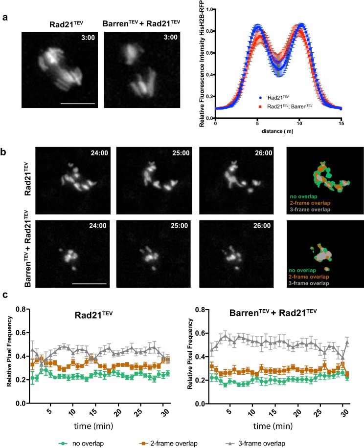
Figure 4.
Condensin I inactivation in separated sister chromatids reduces their movement.
( a ) Representative images of the initial separation after TEV-mediated cleavage of Rad21 TEV and Rad21 TEV + Barren TEV . Graph plots the relative distribution of HisH2B-RFP at the maximal state of ...
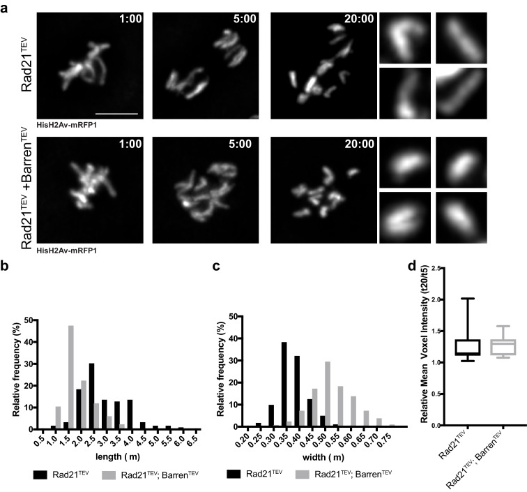
Figure 5.
Chromosome over-compaction depends on sister-chromatid proximity.
( a ) Stills from metaphase-arrested embryos after injection of TEV protease in strains surviving solely on Rad21 TEV (cohesin cleavage) or Rad21 TEV +Barren TEV (cohesin and condensin cleavage); embr...
Video 7.
Artificial induction of sister chromatid separation in metaphase-arrested embryos.
Embryos expressing solely Rad21 TEV and wild-type Barren were injected with 12 mg/ml of a dominant-negative form of the human E2 ubiquitin-conjugating enzyme (UbcH10 C114S ), to induce a metaphase arr...
Video 8.
Effect of condensin I inactivation on isolated sister chromatids.
Embryos expressing uniquely TEV-sensitive Rad21 and Barren were injected with 12 mg/ml of a dominant-negative form of the human E2 ubiquitin-conjugating enzyme (UbcH10 C114S ), to induce a metaphase a...
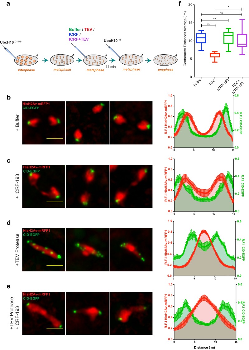
Figure 6.
Condensin I inactivation results in TopoII-dependent sister chromatids intertwines and segregation failure.
( a ) Schematic representation of the experimental set-up. Embryos were arrested with 12 mg/ml UbcH10 C114S and injected with buffer ( b ), 280 u03bcM ICRF-193 ( c ), 13 mg/ml TEV protease ( d ) or TE...
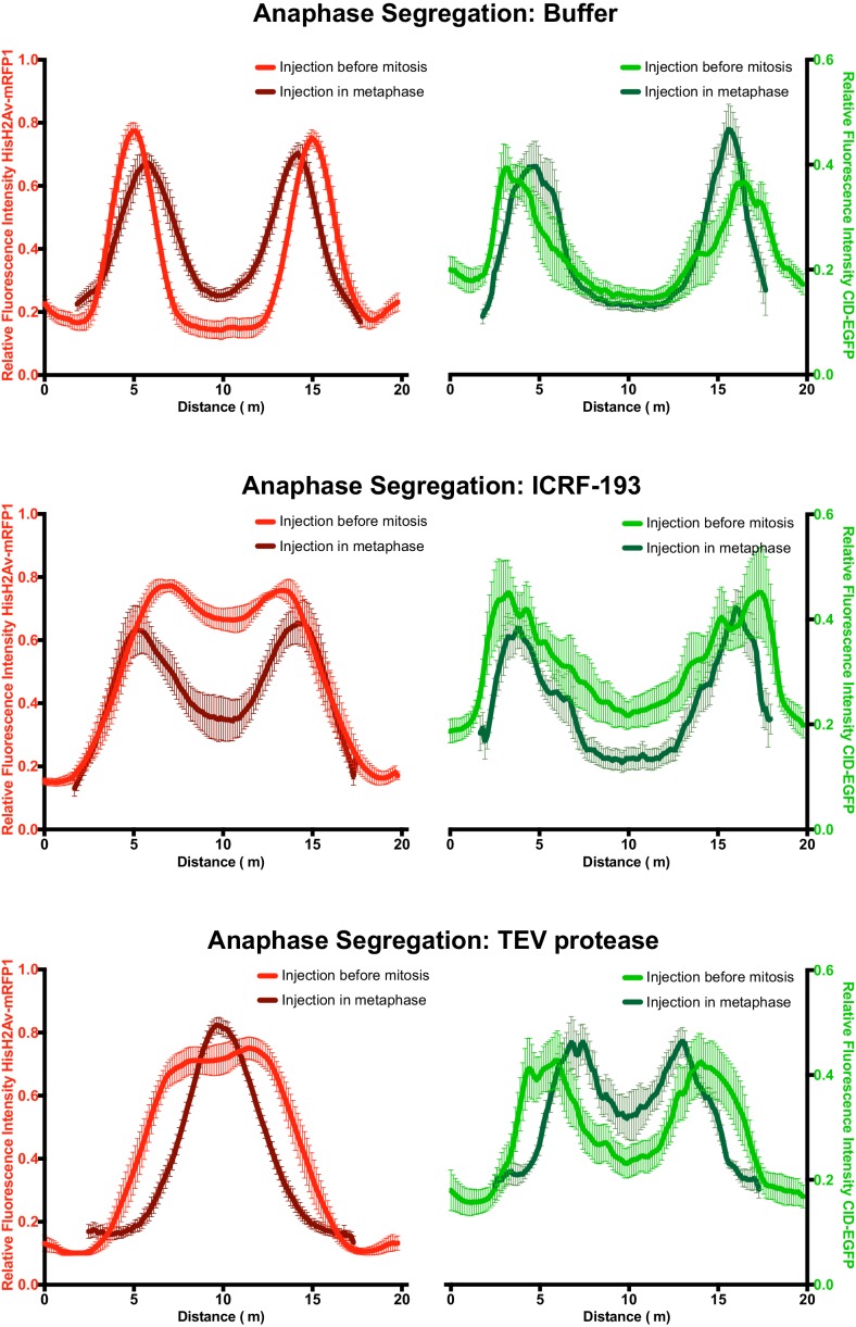
Figure 6u2014figure supplement 1.
u2013 Comparative analysis of segregation efficiency for condensin and/TopoII inhibition before mitosis (light colour) and during metaphase arrest/release (dark colour); Graphs plot the relative distribution of HisH2Av-mRFP1 (red) and Cid-EGFP (green) across a 20 u03bcm segregation plane, measured 4u20136 min after anaphase onset.
Graphs plot the average +_SEM of individual embryos (nu00a0u2265u00a08 embryos for each experimental condition). For each embryo, at least eight anaphases were analysed. DOI: http://dx.doi.org/10.7554...
Video 9.
Induced anaphase in control embryos.
Embryos expressing solely Barren TEV were injected with 12 mg/ml of a dominant-negative form of the human E2 ubiquitin-conjugating enzyme (UbcH10 C114S ), to induce a metaphase arrest, and subsequentl...
Video 10.
Induced anaphase after timely inhibition of topoisomerase II.
Embryos expressing solely Barren TEV were injected with 12 mg/ml of a dominant-negative form of the human E2 ubiquitin-conjugating enzyme (UbcH10 C114S ), to induce a metaphase arrest, and subsequentl...
Video 11.
Induced anaphase after timely inhibition of Condensin I.
Embryos expressing solely Barren TEV were injected with 12 mg/ml of a dominant-negative form of the human E2 ubiquitin-conjugating enzyme (UbcH10 C114S ), to induce a metaphase arrest, and subsequentl...
Video 12.
Induced anaphase after timely inhibition of Condensin I and topoisomerase II.
Embryos expressing solely Barren TEV were injected with 12 mg/ml of a dominant-negative form of the human E2 ubiquitin-conjugating enzyme (UbcH10 C114S ), to induce a metaphase arrest, and subsequentl...
Figure images are served from the NIH/NLM PubMed Central Open Access Subset or Europe PMC; copyright remains with the publishers and authors.
💬 Discussion
0 commentsNo comments yet. Be the first to start a discussion!
Leave a Comment