Abstract
The mitotic spindle is a complex and dynamic structure. Although much has been learned about how spindles assemble and mediate chromosome segregation, how spindles rapidly and irreversibly disassemble during telophase is less clear. We used synthetic lethal screens in budding yeast to identify mutants defective in spindle disassembly. Real-time, live cell imaging analysis of spindle disassembly was performed on nine mutants defective in this process. Results of this analysis suggest that spindle disassembly is achieved by mechanistically distinct but functionally overlapping subprocesses: disengagement of the spindle halves, arrest of spindle elongation, and initiation of interpolar microtubule depolymerization. These subprocesses are largely governed by the anaphase-promoting complex, Aurora B kinase, and kinesin-8. Combinatorial inhibition of these subprocesses yielded cells with hyperstable spindle remnants and dramatic defects in cell cycle progression, establishing that rapid spindle disassembly is crucial for cell proliferation.
🔬 Techniques
🧬 Organisms
💻 Software
✨ Fluorophores
🧪 Sample Preparation
🏭 Microscope Brands
🧪 Reagent Suppliers
📷 Detectors
💻 Software Details
🏛️ Research Organizations (ROR)
Affiliated research institutions:
📋 Methods
Yeast strains
The yeast strains used in this study are derivatives of S288C and are listed in Table S1 . doc1Δ , cdh1Δ , dbf2Δ , dcc1Δ , ctf8Δ , mcm21Δ , and kip3Δ null mutants originated from the Research Genetics Collection. The Ase1-4GFP and Cin8-4GFP strains were provided by E. Schiebel (ZMBH, Heidelberg, Germany). The phosphomutant alleles kip3-4A (S74A, T172A, S402A, and S792A) and she1-5A (T14A, S165A, S269A, T280A, and S325A) were generated using QuikChange site-directed mutagenesis. Ipl1 phosphorylation sites selected for mutation matched the (R/K)X(T/S)(I/L/V/T) consensus motif ( Cheeseman et. al, 2002 ). Plasmids containing full-length kip3-4A and she1-5A genes were integrated at each gene’s endogenous locus in kip3Δ and she1Δ strains, respectively. The bim1-6A and bim1-6D strains were provided by S. Westermann (Research Institute of Molecular Pathology, Vienna, Austria), and the Kip3-GFP strain was provided by K. Bloom (University of North Carolina at Chapel Hill, Chapel Hill, NC).
Fluorescence microscopy
Live cell microscopy at room temperature was performed using a microscope (IX-71; Olympus), a 100× NA 1.4 objective, and a camera (Orca-ER; Hamamatsu Photonics). All microscopy at 37°C was performed using a microscope (IX-81; Olympus) equipped with a temperature-controlled enclosure (Precision Control Weather Station), a 100× NA 1.4 objective, and a camera (Orca-ER). Two-color images were obtained by sequential switching between RFP and GFP filter sets. For time-lapse microscopy of shrinking half-spindles, actinomyosin ring contraction, and Bim1-3GFP localization, images were collected at 10-s intervals with 300-ms exposures. Each image represents a maximum intensity projection from a z stack containing six planes 0.2 µm apart. All image processing was performed using MetaMorph software (MDS Analytical Technologies).
Show full methods section
Yeast strains
The yeast strains used in this study are derivatives of S288C and are listed in Table S1 . doc1Δ , cdh1Δ , dbf2Δ , dcc1Δ , ctf8Δ , mcm21Δ , and kip3Δ null mutants originated from the Research Genetics Collection. The Ase1-4GFP and Cin8-4GFP strains were provided by E. Schiebel (ZMBH, Heidelberg, Germany). The phosphomutant alleles kip3-4A (S74A, T172A, S402A, and S792A) and she1-5A (T14A, S165A, S269A, T280A, and S325A) were generated using QuikChange site-directed mutagenesis. Ipl1 phosphorylation sites selected for mutation matched the (R/K)X(T/S)(I/L/V/T) consensus motif ( Cheeseman et. al, 2002 ). Plasmids containing full-length kip3-4A and she1-5A genes were integrated at each gene’s endogenous locus in kip3Δ and she1Δ strains, respectively. The bim1-6A and bim1-6D strains were provided by S. Westermann (Research Institute of Molecular Pathology, Vienna, Austria), and the Kip3-GFP strain was provided by K. Bloom (University of North Carolina at Chapel Hill, Chapel Hill, NC).
Fluorescence microscopy
Live cell microscopy at room temperature was performed using a microscope (IX-71; Olympus), a 100× NA 1.4 objective, and a camera (Orca-ER; Hamamatsu Photonics). All microscopy at 37°C was performed using a microscope (IX-81; Olympus) equipped with a temperature-controlled enclosure (Precision Control Weather Station), a 100× NA 1.4 objective, and a camera (Orca-ER). Two-color images were obtained by sequential switching between RFP and GFP filter sets. For time-lapse microscopy of shrinking half-spindles, actinomyosin ring contraction, and Bim1-3GFP localization, images were collected at 10-s intervals with 300-ms exposures. Each image represents a maximum intensity projection from a z stack containing six planes 0.2 µm apart. All image processing was performed using MetaMorph software (MDS Analytical Technologies).
Synthetic lethal profile comparison
To examine the synthetic interactions for SHE1 , we used the synthetic lethal and synthetic sick dataset described previously by Costanzo et al. (2010) and Tong et al. (2004) . Although synthetic interactions between SHE1 and CDH1 and between SHE1 and DOC1 did not appear in these lists, we had previously identified such interactions through tetrad analysis. When comparing the synthetic interaction profiles of KIP3 , CDH1 , and SHE1 , we used only the raw dataset with an intermediate cutoff applied (|ε| > 0.08, P < 0.05) Costanzo et al., 2010) , plus several interactions we had identified in our laboratory.
Detection of protein phosphorylation
To detect phosphorylation-dependent mobility shifts of She1-13Myc, Kip3-13Myc, and Bim1-HA, whole cell extracts were loaded onto 6–8% SDS polyacrylamide gels containing either 20 µM or 30 µM Phos-tag acrylamide (Hiroshima University, Hiroshima, Japan) and 40 µM or 60 µM MnCl 2 , respectively. Membranes were then probed with 1:500 mouse anti-Myc (9E10; our laboratory) or 1:1,000 mouse anti-HA (12CA5; Roche).
Immunoprecipitation
Immunoprecipitation was performed essentially as described previously ( Woodruff et al., 2009 ) with a few minor modifications. Lysates were incubated with mouse anti-Myc antibody for 1 h and then passed over protein G–Sepharose beads (GE Healthcare) to purify She1-13Myc. The column was washed with lysis buffer and eluted with SDS loading buffer.
Chromatin immunoprecipitation
Chromatin immunoprecipitation was conducted essentially as described previously ( Kang et al., 2001 ) with a few minor changes. Cells were fixed for 1 h in 1% formaldehyde. Whole cell extracts were passed over IgG Sepharose (GE Healthcare) to pull down She1-TAP and Mif2-TAP. Chromosome spreads Chromosome spreads were prepared as described previously ( Cheeseman et al., 2001 ). Anti-Duo1 antibody ( Hofmann et al., 1998 ) was used at a dilution of 1:2,000 to detect Duo1, and anti-GFP (rabbit; Torrey Pines Institute) was used at a dilution of 1:2,000 to detect She1-3GFP. Online supplemental material Fig. S1 describes spindle morphology in cells compromised for Kip3 and APC Cdh1 activity. Fig. S2 depicts the pairwise comparison of previously described synthetic genetic interactions shared between CDH1 , KIP3 , and SHE1 . Fig. S3 shows She1 phosphorylation and localization to the kinetochore and localization of Cdc14 and Kip3 in an she1Δ mutant. Fig. S4 shows Ipl1-3GFP localization in dbf2Δ cells. Videos 1–3 show spindle disassembly relative to cytokinetic ring contraction in wild-type, cdh1Δ , and kip3Δ cells, respectively.
Table
S1 lists the strains used in this study. Online supplemental material is available at http://www.jcb.org/cgi/content/full/jcb.201006028/DC1 .
Online supplemental material Fig. S1 describes spindle morphology in cells compromised for Kip3 and APC Cdh1 activity. Fig. S2 depicts the pairwise comparison of previously described synthetic genetic interactions shared between CDH1 , KIP3 , and SHE1 . Fig. S3 shows She1 phosphorylation and localization to the kinetochore and localization of Cdc14 and Kip3 in an she1Δ mutant. Fig. S4 shows Ipl1-3GFP localization in dbf2Δ cells. Videos 1–3 show spindle disassembly relative to cytokinetic ring contraction in wild-type, cdh1Δ , and kip3Δ cells, respectively.
Table
S1 lists the strains used in this study. Online supplemental material is available at http://www.jcb.org/cgi/content/full/jcb.201006028/DC1 .
📊 Figures
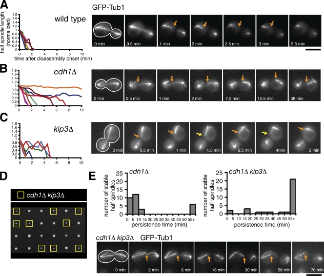
Figure 1.
The combined activities of APC Cdh1 and Kip3 are required to depolymerize ipMTs completely. (Au2013C) Time-lapse fluorescence images of wild-type, cdh1u0394 , and kip3u0394 cells expressing GFP-Tub1 d...
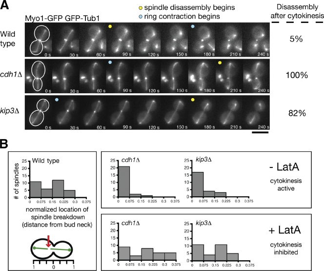
Figure 2.
Cytokinetic ring contraction breaks the mitotic spindle when normal disassembly mechanisms are impaired. (A) Time-lapse images of cells expressing GFP-Tub1 and the cytokinetic ring marker Myo1-GFP dur...
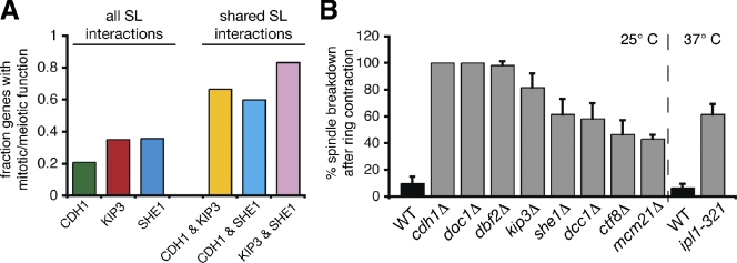
Figure 3.
Synthetic lethal screens identify additional genes important for spindle disassembly. (A) Fraction of the genes found in the CDH1 , KIP3 , and SHE1 synthetic interaction profiles that have previously ...
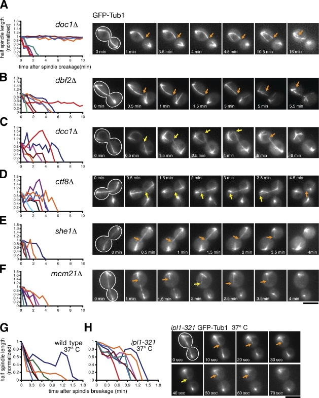
Figure 4.
Dynamics of ipMT depolymerization in spindle disassembly mutants. (Au2013F) Cells expressing the spindle marker GFP-Tub1 were analyzed by time-lapse microscopy during mitotic exit similar to the exper...
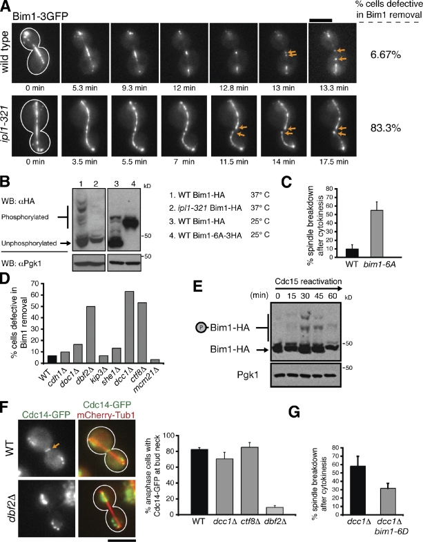
Figure 5.
The MEN and the A-RFC complex regulate Ipl1-mediated removal of Bim1 from the spindle midzone. (A) Time-lapse fluorescence images of Bim1-3GFP localization in wild-type and ipl1-321 cells incubated at...
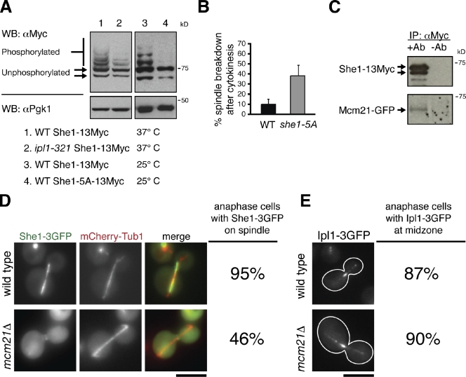
Figure 6.
Ipl1 and Mcm21 mediate spindle disassembly by regulating She1 activity. (A) Protein extracts isolated from asynchronous populations of the indicated strains were separated by SDS-PAGE supplemented wit...
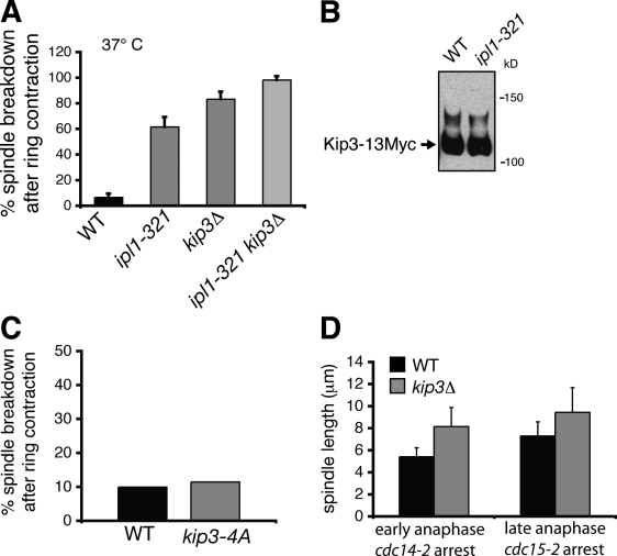
Figure 7.
Kip3 and Ipl1 operate in separate pathways during spindle disassembly. (A) Frequency in which spindles disassembled after initiation of cytokinetic ring contraction in wild-type (WT) cells and selecte...
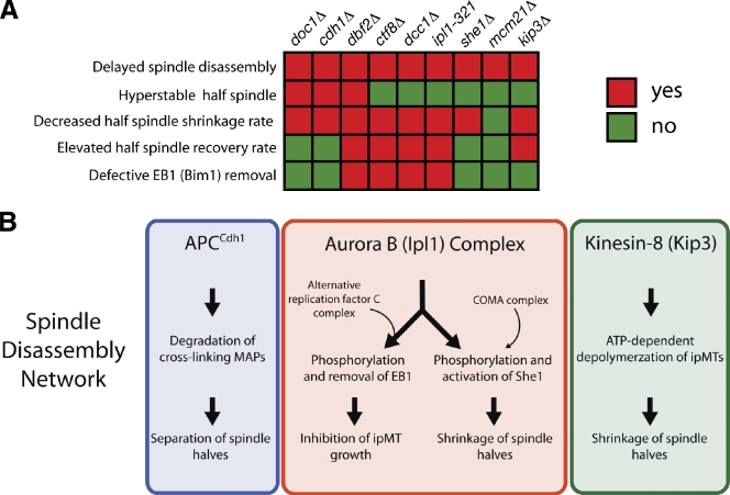
Figure 8.
Multiple pathways regulating distinct subprocesses drive spindle disassembly. (A) Summary of phenotypic analysis for nine spindle disassembly mutants. Decreased half-spindle shrinkage rate indicates t...
Figure images are served from the NIH/NLM PubMed Central Open Access Subset or Europe PMC; copyright remains with the publishers and authors.
💬 Discussion
0 commentsNo comments yet. Be the first to start a discussion!
Leave a Comment