Abstract
In the eukaryotic replisome, DNA unwinding by the Cdc45-MCM-Go-Ichi-Ni-San (GINS) (CMG) helicase requires a hexameric ring-shaped ATPase named minichromosome maintenance (MCM), which spools single-stranded DNA through its central channel. Not all six ATPase sites are required for unwinding; however, the helicase mechanism is unknown. We imaged ATP-hydrolysis-driven translocation of the CMG using cryo-electron microscopy (cryo-EM) and found that the six MCM subunits engage DNA using four neighboring protomers at a time, with ATP binding promoting DNA engagement. Morphing between different helicase states leads us to suggest a non-symmetric hand-over-hand rotary mechanism, explaining the asymmetric requirements of ATPase function around the MCM ring of the CMG. By imaging of a higher-order replisome assembly, we find that the Mrc1-Csm3-Tof1 fork-stabilization complex strengthens the interaction between parental duplex DNA and the CMG at the fork, which might support the coupling between DNA translocation and fork unwinding.
🔬 Techniques
🔭 Microscopes
🧬 Organisms
💻 Software
✨ Fluorophores
🧪 Sample Preparation
🏭 Microscope Brands
🧪 Reagent Suppliers
📷 Detectors
💻 Software Details
💾 Data Repositories
🏷️ Research Resource Identifiers (RRIDs)
Verified research resources used in this paper:
🏛️ Research Organizations (ROR)
Affiliated research institutions:
📋 Methods
Key Resources Table REAGENT or RESOURCE SOURCE IDENTIFIER Antibodies
Anti-CBP antibody Sigma-Aldrich RRID: AB_10743822 Anti-FLAG antibody Sigma-Aldrich RRID: AB_2811010 Chemicals, Peptides, and Recombinant Proteins 3X FLAG peptide Sigma F4799 Anti-FLAG M2 affinity gel Sigma A2220 Calmodulin-Sepharose 4B GE Healthcare 17-0529-01 cOmplete, EDTA-free Roche 5056489001 yeast CMG Zhou et al., 2017 N/A Drosophila CMG Ilves et al., 2010 N/A Mrc1 Yeeles et al., 2017 N/A Csm3/Tof1 Yeeles et al., 2017 N/A Ctf4 Gambus et al., 2009 N/A Pol epsilon(exo-) Goswami et al., 2018 N/A M.HpaII NEB M0214S Deposited Data Drosophila CMG-DNA state 1A cryo-EM map This paper EMD-4785 Drosophila CMG-DNA state 1A PDB coordinates This paper 6RAW Drosophila CMG-DNA state 1B cryo-EM map This paper EMD-4786 Drosophila CMG-DNA state 1B PDB coordinates This paper 6RAX Drosophila CMG-DNA state 2A cryo-EM map This paper EMD-4787 Drosophila CMG-DNA state 2A PDB coordinates This paper 6RAY Drosophila CMG-DNA state 2B cryo-EM map This paper EMD-4788 Drosophila CMG-DNA state 2B PDB coordinates This paper 6RAZ Experimental Models: Cell Lines High Five cells Thermofisher B855-0202 Sf9 cells Thermofisher A38841 Experimental Models: Organisms/Strains yJCZ3 (yeast CMG purification) Zhou et al., 2017 N/A yAE99 (Pol epsilon exo-) Goswami et al., 2018 N/A yAE48 (Csm3/Tof1 purification) Yeeles et al., 2017 N/A yJY32 (Mrc1 purification) Yeeles et al., 2017 N/A Oligonucleotides A list of oligonucleotides is provided in Table S3 . N/A Recombinant DNA pFastBac1 Mcm2 Ilves et al., 2010 N/A pFastBac1 Mcm3 Ilves et al., 2010 N/A pFastBac1 Mcm4 Ilves et al., 2010 N/A pFastBac1 Mcm5 Ilves et al., 2010 N/A pFastBac1 Mcm6 Ilves et al., 2010 N/A pFastBac1 Mcm7 Ilves et al., 2010 N/A pFastBac1 Cdc45 Ilves et al., 2010 N/A pFastBac1 Psf1 Ilves et al., 2010 N/A pFastBac1 Psf2 Ilves et al., 2010 N/A pFastBac1 Psf3 Ilves et al., 2010 N/A pFastBac1 Sld5 Ilves et al., 2010 N/A pFastBac1 RA Mcm2 This study; Ilves et al., 2010 N/A pFastBac1 RA Mcm3 This study; Ilves et al., 2010 N/A pFastBac1 RA Mcm4 This study; Ilves et al., 2010 N/A pFastBac1 RA Mcm5 This study; Ilves et al., 2010 N/A pFastBac1 RA Mcm6 This study; Ilves et al., 2010 N/A pFastBac1 RA Mcm7 This study; Ilves et al., 2010 N/A Software and Algorithms RELION v2.1 and v3 Scheres, 2012 https://www2.mrc-lmb.cam.ac.uk/relion/index.php?title=Main_Page MotionCor2 Zheng et al., 2017 https://msg.ucsf.edu/em/software/motioncor2.html cryoSPARC v2 Punjani et al., 2017 https://www.nature.com/articles/nmeth.4169 Coot v0.8.8 Emsley et al., 2010 http://scripts.iucr.org/cgi-bin/paper?S0907444910007493 PHENIX v1.13 Adams et al., 2010 http://www.phenix-online.org/ UCSF Chimera UCSF Resource for Biocomputing, Visualization, and Informatics https://www.cgl.ucsf.edu/chimera/ Lead Contact and Materials Availability Further information and requests for resources and reagents should be directed to and will be fulfilled by the Lead Contact, Alessandro Costa ( alessandro.costa@crick.ac.uk ). Material will be made available upon reasonable request. This study did not generate new unique reagents.
Show full methods section
Key Resources Table REAGENT or RESOURCE SOURCE IDENTIFIER Antibodies
Anti-CBP antibody Sigma-Aldrich RRID: AB_10743822 Anti-FLAG antibody Sigma-Aldrich RRID: AB_2811010 Chemicals, Peptides, and Recombinant Proteins 3X FLAG peptide Sigma F4799 Anti-FLAG M2 affinity gel Sigma A2220 Calmodulin-Sepharose 4B GE Healthcare 17-0529-01 cOmplete, EDTA-free Roche 5056489001 yeast CMG Zhou et al., 2017 N/A Drosophila CMG Ilves et al., 2010 N/A Mrc1 Yeeles et al., 2017 N/A Csm3/Tof1 Yeeles et al., 2017 N/A Ctf4 Gambus et al., 2009 N/A Pol epsilon(exo-) Goswami et al., 2018 N/A M.HpaII NEB M0214S Deposited Data Drosophila CMG-DNA state 1A cryo-EM map This paper EMD-4785 Drosophila CMG-DNA state 1A PDB coordinates This paper 6RAW Drosophila CMG-DNA state 1B cryo-EM map This paper EMD-4786 Drosophila CMG-DNA state 1B PDB coordinates This paper 6RAX Drosophila CMG-DNA state 2A cryo-EM map This paper EMD-4787 Drosophila CMG-DNA state 2A PDB coordinates This paper 6RAY Drosophila CMG-DNA state 2B cryo-EM map This paper EMD-4788 Drosophila CMG-DNA state 2B PDB coordinates This paper 6RAZ Experimental Models: Cell Lines High Five cells Thermofisher B855-0202 Sf9 cells Thermofisher A38841 Experimental Models: Organisms/Strains yJCZ3 (yeast CMG purification) Zhou et al., 2017 N/A yAE99 (Pol epsilon exo-) Goswami et al., 2018 N/A yAE48 (Csm3/Tof1 purification) Yeeles et al., 2017 N/A yJY32 (Mrc1 purification) Yeeles et al., 2017 N/A Oligonucleotides A list of oligonucleotides is provided in Table S3 . N/A Recombinant DNA pFastBac1 Mcm2 Ilves et al., 2010 N/A pFastBac1 Mcm3 Ilves et al., 2010 N/A pFastBac1 Mcm4 Ilves et al., 2010 N/A pFastBac1 Mcm5 Ilves et al., 2010 N/A pFastBac1 Mcm6 Ilves et al., 2010 N/A pFastBac1 Mcm7 Ilves et al., 2010 N/A pFastBac1 Cdc45 Ilves et al., 2010 N/A pFastBac1 Psf1 Ilves et al., 2010 N/A pFastBac1 Psf2 Ilves et al., 2010 N/A pFastBac1 Psf3 Ilves et al., 2010 N/A pFastBac1 Sld5 Ilves et al., 2010 N/A pFastBac1 RA Mcm2 This study; Ilves et al., 2010 N/A pFastBac1 RA Mcm3 This study; Ilves et al., 2010 N/A pFastBac1 RA Mcm4 This study; Ilves et al., 2010 N/A pFastBac1 RA Mcm5 This study; Ilves et al., 2010 N/A pFastBac1 RA Mcm6 This study; Ilves et al., 2010 N/A pFastBac1 RA Mcm7 This study; Ilves et al., 2010 N/A Software and Algorithms RELION v2.1 and v3 Scheres, 2012 https://www2.mrc-lmb.cam.ac.uk/relion/index.php?title=Main_Page MotionCor2 Zheng et al., 2017 https://msg.ucsf.edu/em/software/motioncor2.html cryoSPARC v2 Punjani et al., 2017 https://www.nature.com/articles/nmeth.4169 Coot v0.8.8 Emsley et al., 2010 http://scripts.iucr.org/cgi-bin/paper?S0907444910007493 PHENIX v1.13 Adams et al., 2010 http://www.phenix-online.org/ UCSF Chimera UCSF Resource for Biocomputing, Visualization, and Informatics https://www.cgl.ucsf.edu/chimera/ Lead Contact and Materials Availability Further information and requests for resources and reagents should be directed to and will be fulfilled by the Lead Contact, Alessandro Costa ( alessandro.costa@crick.ac.uk ). Material will be made available upon reasonable request. This study did not generate new unique reagents.
Experimental Model and Subject Details Yeast Expression
Yeast proteins were purified from Saccharomyces cerevisiae strains (genotypes: MATa ade2-1 ura3-1 his3-11,15 trp1-1 leu2-3,112 can1- 100 bar1::Hyg pep4::KanMX or MATa pep4::KanMx4 bar1::Hph-NT1 yJCZ1: MATα pep4::KanMx4 bar1::Hph-NT1 ade2-1::pJCZ3 (ADE2)) containing integrated expression constructs and grown at 30°C in YEP media supplemented with 2% raffinose.
Baculovirus Expression
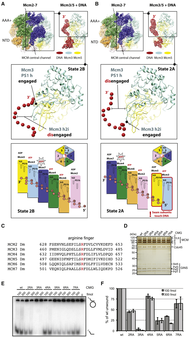
Drosophila melanogaster proteins were purified from baculovirus-infected female High-five cells incubated at 27°C, as previously described ( Abid Ali et al., 2016 , Ilves et al., 2010 ). Method Details Cloning and Construction of Baculoviruses Viruses used in the studies were constructed following the manufacturer’s manual for the Bac-to-Bac expression system from Invitrogen. The pFastBac1 recombination vectors containing the cDNAs for all 11 wild-type CMG subunits were described in detail before ( Ilves et al., 2010 ). These vector templates were used for generation of CMG mutants using PCR-based mutagenesis. Arginine finger residues were targeted and alanine substitutions were introduced to generate the RA point mutant. These include R641 in MCM2, R473 in MCM3, R645 in MCM4, R510 in MCM5, R521 in MCM6, and R514 in MCM7.
Protein Expression and Purification
Expression of S. Cerevisiae Proteins All yeast proteins (except Ctf4) were expressed in S. cerevisiae cells and harvested following the same procedure. Cells were grown at 30°C in YEP media supplemented with 2% raffinose. At a cell density of ∼2-3x10 7 cells/ml, expression was induced for 3 hours by the addition of 2% galactose. Cells were harvested by centrifugation at 5,020 x g for 30 min at 4°C. After washing pellets in lysis buffer (see individual protein purifications for buffer details) cells were re-centrifuged at 4,000 x g for 20 min at 4°C. Cells were subsequently resuspended in lysis buffer at half pellet volumes, flash frozen in liquid nitrogen and crushed at −80°C using a 6875D Freezer/Mill® Dual Chamber Cryogenic Grinderfreezer mill (SPEX SamplePrep) at intensity 15 (6 cycles of 2 min milling with 1 min rest).
Purification of Yeast CMG Sc
CMG was expressed and purified as previously described using the yeast strain yJCZ3 ( Zhou et al., 2017 ). Following harvesting in CMG lysis buffer (25 mM HEPES pH 7.6, 15 mM KCl, 2 mM MgCl 2 , 0.02% Tween-20, 1 mM EDTA, 1 mM EGTA, 10% glycerol, 2 mM β-Mercaptoethanol, complete protease inhibitor tablets (Roche)) the cell powder was resuspended in Buffer C-100 (25 mM HEPES pH 7.6, 100 mM KCl, 0.02% Tween-20, 1 mM EDTA, 1 mM EGTA, 10% glycerol, 1 mM DTT) supplemented with 10 mM Mg(OAc) 2 , 25 units/ml benzonase (Sigma Aldrich) and complete protease inhibitor tablets (Roche). The lysate was incubated at 4°C for 45 minutes and cleared by ultracentrifugation at 235,000 x g for 60 minutes at 4°C. Clear supernatants were incubated for 3 hours at 4°C with 4 mL anti-FLAG M2 agarose beads (Sigma Aldrich) pre-equilibrated in Buffer C-100. Beads were subsequently washed with 150 mL Buffer C-100 after which bound proteins were eluted by incubation at room temperature for 30 minutes with the same buffer supplemented with 500 μg/ml FLAG peptide (DYKDDDDK) and complete protease inhibitor tablets (Roche). The eluate was collected and further proteins were eluted by repeating the FLAG peptide incubation for an additional 20 minutes. Combined eluates were passed through a 1 mL HiTrap SPFF column (GE Healthcare) and injected onto a MonoQ 5/50 GL column (GE Healthcare), both equilibrated in Buffer C-100. Proteins were washed with 10 CV of the same buffer and eluted with a 100-550 mM KCl gradient over 20 CV in Buffer C (25 mM HEPES pH 7.6, 0.02% Tween-20, 1 mM EDTA, 1 mM EGTA, 10% glycerol, 1 mM DTT). CMG peak fractions were diluted in Buffer C to 150 mM KCl and injected onto a MonoQ 1.6/5 PC column (GE Healthcare) equilibrated in Buffer C-150 (25 mM HEPES pH 7.6, 150 mM KCl, 0.02% Tween-20, 1 mM EDTA, 1 mM EGTA, 10% glycerol, 1 mM DTT). Proteins were washed with 10 CV of the same buffer and eluted with a 150-550 mM KCl gradient over 15 CV in Buffer C. CMG peak fractions were dialysed against Protein Binding Buffer (25 mM HEPES pH 7.6, 100 mM KOAc, 2 mM Mg(OAc) 2 , 5% glycerol, 0.02% NP-40, 1 mM DTT) for 3 hours at 4°C.
Purification of DNA Polymerase ε Sc
Polε was expressed and purified as previously described using the yeast strain yAE99 (Polε exo- mutant)( Goswami et al., 2018 ). Following harvesting in Buffer E-500 (25 mM HEPES pH 7.6, 400 mM KOAc, 10% glycerol, 1 mM DTT) supplemented with complete protease inhibitor tablets (Roche), the cell powder was resuspended in Buffer E-400 (25 mM HEPES pH 7.6, 400 mM KOAc, 10% glycerol, 1 mM DTT) supplemented with complete protease inhibitor tablets (Roche). The lysate was incubated at 4°C for 45 minutes and cleared by ultracentrifugation at 235,000 x g for 60 minutes at 4°C. Clear supernatants were supplemented with 2 mM CaCl 2 and incubated for 2 hours at 4°C with 3 mL Calmodulin Affinity Resin (Agilent) pre-equilibrated in Buffer E-400. Beads were subsequently washed with 300 mL Buffer E-400 supplemented with 2 mM CaCl 2 after which bound proteins were eluted by incubation at 4°C with Buffer E-400 supplemented with 2 mM EDTA and 2 mM EGTA. Pooled elutions were injected onto an SP Sepharose Fast Flow 1 mL column (GE Healthcare) attached to a MonoQ 5/50 GL column (GE Healthcare) and washed with 20 CV Buffer E-400. Following removal of the SP Sepharose Fast Flow column, proteins were eluted with a 400-1,000 mM KOAc gradient over 15 CV in Buffer E (25 mM HEPES pH 7.6, 10% glycerol, 1 mM DTT). Polε fractions were pooled, dialysed against Buffer E-400 and concentrated using a 30,000 MWCO cut-off spin column. 50 μl concentrated sample was subsequently passed over a Superose 6 3.2/300 gel filtration column in Buffer E-400.
Purification of Mrc1 Sc
Mrc1 was expressed and purified as previously described using the yeast strain yJY32( Yeeles et al., 2017 ). Cells were harvested, lysed and resuspended in Buffer T-400 (25 mM Tris-HCl pH 7.5, 400 mM NaCl, 10% glycerol, 0.01% Tween-20, 1 mM DTT) supplemented with complete protease inhibitor tablets (Roche). The lysate was incubated at 4°C for 45 minutes and cleared by ultracentrifugation at 235,000 x g for 60 minutes at 4°C. Clear supernatants were incubated for 2 hours at 4°C with 2 mL anti-FLAG M2 agarose beads (Sigma Aldrich) pre-equilibrated in Buffer T-400. Beads were subsequently washed with 50 CV Buffer T-400 and 25 CV Buffer T-200 (25 mM Tris-HCl pH 7.5, 200 mM NaCl, 10% glycerol, 0.01% Tween-20, 1 mM DTT) followed by incubation for 10 min in Buffer T-200 supplemented with 1 mM ATP and 10 mM Mg(OAc) 2 . After washing beads in 10 CV Buffer T-200, bound proteins were eluted by incubation at room temperature for 45 minutes with the same buffer supplemented with 500 μg/ml FLAG peptide (DYKDDDDK) and complete protease inhibitor tablets (Roche). The eluate was collected and further proteins were eluted by repeating the FLAG peptide incubation for an additional 30 minutes. Combined eluates were subsequently injected onto a MonoQ 1.6/5 PC column (GE Healthcare) equilibrated in Buffer T-200. Proteins were washed with 10 CV of the same buffer and eluted with a 200-600 mM NaCl gradient over 15 CV in Buffer T (25 mM Tris-HCl pH 7.5, 10% glycerol, 0.01% Tween-20, 1 mM DTT). Mrc1 peak fractions were dialysed against Buffer T-200.
Purification of Csm3-Tof1 Sc
Csm3/Tof1 was co-expressed and co-purified as previously described using the yeast strain yAE48( Yeeles et al., 2017 ). Cells were harvested, lysed and resuspended in CBP lysis buffer (25 mM Tris-HCl pH 7.5, 200 mM NaCl, 10% glycerol, 0.01% NP-40, 1 mM DTT) supplemented with complete protease inhibitor tablets (Roche). The lysate was incubated at 4°C for 45 minutes and cleared by ultracentrifugation at 235,000 x g for 60 minutes at 4°C. Clear supernatants were supplemented with 2 mM CaCl 2 and incubated for 2 hours at 4°C with 2 mL Calmodulin Affinity Resin (Agilent) pre-equilibrated in CBP lysis buffer. Beads were subsequently washed with 75 CV CBP lysis buffer supplemented with 2 mM CaCl 2 after which bound proteins were eluted by incubation at 4°C with CBP lysis buffer supplemented with 2 mM EDTA and 2 mM EGTA. Pooled elutions were concentrated to 500 μl using a 30,000 MWCO cut-off spin column and passed over a Superdex 200 10/300 gel filtration column equilibrated in CBP Gel Filtration Buffer (25 mM Tris-Hcl pH 7.5, 150 mM NaCl, 1 mM DTT). Csm3-Tof1 peak fractions were pooled and concentrated to 100 μl using a 30,000 MWCO cut-off spin column.
Expression and Purification of Ctf4 Trimer Sc
Ctf4 expression plasmids( Gambus et al., 2009 ) were transformed into BL21 (DE3) E. coli cells. Cells were grown in LB media at 37°C to an optical density (OD = 600) of 0.5 before expression was induced with 1 mM IPTG for 3 hours. Cells were harvested by centrifugation at 5,020 x g for 20 min at room temperature. Pelleted cells were subsequently resuspended in Ctf4 lysis buffer (50 mM Tris-HCl pH 7.5, 500 mM NaCl, 10 mM MgCl 2 , 10% glycerol, 1 mM DTT) supplemented with complete protease inhibitor tablets (Roche) and lysed by sonication. Lysate was cleared by centrifugation at 27,216 xg for 30 min at 4°C and incubated for 90 min at 4°C with 1 mL Ni-NTA Agarose Resin (QIAGEN) pre-equilibrated in Buffer A-20 (50 mM Tris-HCl pH 7.5, 300 mM NaCl, 20 mM imidazole). After washing resin with 20 CV Buffer A-20, proteins were eluted five times with 1 mL Buffer A-250 (50 mM Tris-HCl pH 7.5, 300 mM NaCl, 250 mM imidazole). Elutions were pooled and dialysed against Buffer B-100 (20 mM Tris-HCl pH 8.0, 100 mM NaCl, 1 mM DTT) before injection onto a MonoQ 5/50 GL column (GE Healthcare) equilibrated in Buffer B-100. Proteins were washed with 10 CV of the same buffer and eluted with a 100-1,000 mM NaCl gradient over 30 CV in Buffer B (20 mM Tris-HCl pH 8.0, 1 mM DTT). Ctf4 peak fractions were pooled and concentrated to 450 μl using a 30,000 MWCO cut-off spin column before being passed over a Superdex 200 16/600 gel filtration column equilibrated in Buffer B-150 (20 mM Tris-HCl pH 8.0, 150 mM NaCl, 1 mM DTT). Ctf4 trimer peak fractions were pooled and concentrated to 4 mg/ml using a 30,000 MWCO cut-off spin column.
Expression and Purification of Drosophila Melanogaster CMG Drosophila melanogaster
CMG was expressed and purified as previously described ( Abid Ali et al., 2016 , Ilves et al., 2010 ). Following bacmid generation for each subunit of Dm CMG, Sf21 cells were used for transfection and virus amplification stages to generate P2 stocks using serum-free Sf-900TM III SFM insect cell medium (Invitrogen/GIBCO). In the P3 virus amplification stage, 100 mL Sf9 cell (0.5x10 5 /ml) cultures were infected with 0.5 mL of P2 stocks with an approximate MOI of 0.1 for each virus and incubated in 500 mL Erlenmeyer sterile flasks (Corning) for 4 days at 27°C, shaking at 100 rpm. After 4 days, 4 L of Hi-Five cells (10 6 /ml) supplemented with 10% FCS were infected using fresh P3 stocks with MOI of 5. Cells were incubated at 27°C and harvested after 60 hours. Cell pellets were washed with PBS supplemented with 5 mM MgCl 2, resuspended in lysis buffer and frozen in 10 mL aliquots on dry ice. Protein purification was performed at 4°C. Cell pellets were thawed and lysed by applying at least 50 strokes per 30 mL of cell pellets using tissue grinders (Wheaton, 40 mL Dounce Tissue Grinder) after which the lysate was cleared by centrifugation at 24,000 x g for 10 min. Supernatants were incubated for 2.5 hours with 2 mL ANTI-FLAG M2 agarose beads (Sigma Aldrich) pre-equilibrated with Buffer C. Non-bound proteins were removed by centrifugation at 200 x g for 5 minutes followed by bead washing with 30 mL of Buffer C-100. Bound proteins were subsequently eluted by incubation at room temperature for 15 min with Buffer C-100 supplemented with 200 μg/ml FLAG peptide (DYKDDDDK). The eluate was passed through a 1 mL HiTrap SPFF column (GE Healthcare) and injected onto a MonoQ 5/50 GL column (GE Healthcare), both equilibrated in buffer C-100. Proteins were washed with 10 CV of the same buffer and eluted with a 100-550 mM KCl gradient over 20 CV in buffer C. CMG peak fractions were diluted in buffer C to 150 mM KCl and injected onto a MonoQ 1.6/5 PC column (GE Healthcare) equilibrated in buffer C-150. Proteins were washed with 10 CV of the same buffer and eluted with a 150-550 mM KCl gradient over 15 CV in Buffer C. CMG peak fractions were dialysed into Protein Binding Buffer (25 mM HEPES pH 7.6, 100 mM KOAc, 2 mM Mg(OAc) 2 , 5% glycerol, 0.02% NP-40, 1 mM DTT) for 2 hours.
Forked DNA Unwinding Assay
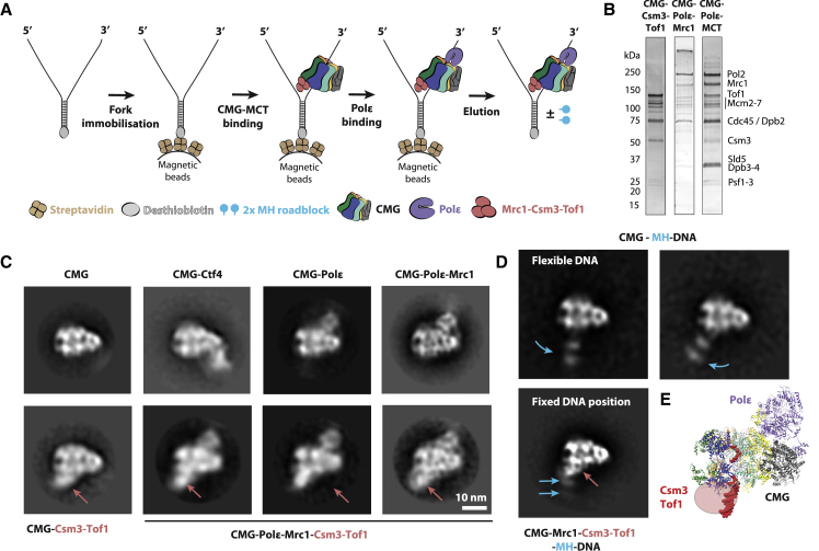
A list of oligonucleotides is provided in Table S3 . Roadblock experiments. To prepare Cy5-labeled fork DNA substrate containing a single MH roadblock on the leading-strand template, oligonucleotides A, B and C were annealed at equimolar concentrations, and the resulting nick was sealed with T4 DNA ligase. DNA was purified via electroelution after separating on 8% PAGE. M.HpaII (NEB) was crosslinked in methyltransferase buffer (50 mM Tris-HCl pH 7.5, 0.5 mM 2-mercaptoethanol, 10 mM EDTA, NEB) supplemented with 100 μM S-adenosylmethionine (NEB), and incubated at 37°C for 3 hours. M.HpaII crosslinked substrate was separated on 8% PAGE and purified via electroelution. Cy5-labeled fork DNA with a lagging-strand MH roadblock was prepared by annealing oligonucleotides D and E. The substrate was gel purified, crosslinked to MH, and re-purified as described above. For unwinding assays, Drosophila CMG was first bound to fork DNA by incubating 3-5 nM DNA substrate with 30 nM Drosophila CMG in CMG-binding buffer (25 mM HEPES pH 7.5, 5 mM NaCl, 10 mM magnesium acetate, 5 mM DTT, 0.1 mg/ml BSA) supplemented with 0.1 mM ATPγS in 5 μL volume at 37°C for 2 hours. To initiate unwinding, 15 μL ATP mix (CMG-binding buffer with 3.3 mM ATP) was added into the reaction. The ATP mix contained 1.5 μM 40 nt polyT oligonucleotide to capture free CMG and 150 nM oligonucleotide with the sequence 5′-GGATGCTGAGGCAATGGGAATTCGCCAACC-3′ to prevent re-annealing of DNA. After further 30 min incubation at 37°C, reactions were stopped with SDS-containing buffer, separated on 8% PAGE, and imaged on Fujifilm SLA-5000 scanner using 635-nm laser and LPR/R665 filter. M13-Based DNA Unwinding Assay A 70-mer oligonucleotide (“T”) was designed such that 40 nucleotides anneal to a M13mp18ssDNA plasmid (New England Biolabs), leaving a 30-mer polyT extension at the 5′ end. The 5′ end was previously radioactively labeled with γ- 32 P ATP (MP Biomedicals or Perkin Elmer) and T4 polynucleotide kinase (New England Biolabs), subsequently purified through a llustra MicroSpin G-50 column (GE Healthcare) and mixed with the M13mp18 ssDNA plasmid. The reactions were heat-denatured for 1 minute and annealed through gradual cooling to room temperature. Free oligonucleotide was separated by purification through MicroSpin S-400 HR columns (GE Healthcare). The helicase assays were carried out in 25mM HEPES pH 7.6, 10% glycerol, 50mM sodium acetate, 10mM magnesium acetate, 0.2mM PMSF, 1mM DTT, with addition of 250 μg/ml insulin. Desired protein concentrations were mixed with 1-2fmol of a circular M13 based DNA substrate and unwinding initiated in the presence of 0.3mM ATP in a total reaction volume of 10 μL at 30°C. Reactions were stopped after 30 minutes by addition of 0.1% SDS and 20mM EDTA, and the reaction products were immediately electrophoretically separated on a TBE-acrylamide gel (8% TBE with 0.1%SDS). Fork Affinity Purification of CMG To prepare desthiobiotin-tagged, M.HpaII-labeled DNA fork substrates containing two M.HpaII on the leading-strand template, oligonucleotides F, G, H and I were annealed at a 1:1:2:1 molar ratio, and the resulting nicks were sealed with T4 DNA ligase. DNA was purified via electroelution after separating on 8% PAGE. M.HpaII (NEB) was crosslinked in methyltransferase buffer (50 mM Tris-HCl pH 7.5, 0.5 mM 2-mercaptoethanol, 10 mM EDTA, NEB) supplemented with 100 μM S-adenosylmethionine (NEB), and incubated at 37°C for 5 hours. M.HpaII crosslinked substrate was separated on 8% PAGE and purified via electroelution. To prepare desthiobiotin-tagged MH-labeled DNA fork substrates containing one MH on the leading-strand template and one M.HpaII on the lagging-strand template, oligonucleotides J + K and L + M were annealed separately at equimolar concentrations. The annealed oligonucleotide samples were then mixed and nicks were sealed with T4 DNA ligase. The substrate was gel purified, crosslinked to M.HpaII, and purified as described above. To isolate DNA-bound CMG complexes, a fork affinity purification approach was adapted from a previously published method( Goswami et al., 2018 ). Here, desthiobiotin-tagged DNA forks were immobilised onto streptavidin-coated magnetic beads. 6 μl M-280 Streptavidin Dynabeads® (Thermo Fisher) slurry was added to each reaction tube and washed twice in 20 μl DNA Binding Buffer (25 mM HEPES 7.6, 1M NaCl, 10% glycerol, 0.01% NP-40, 1 mM EDTA). Washed beads were resuspended in 20 μl 250 nM MH-conjugated DNA forks and incubated for 30 minutes at 30°C shaking at 1,250 rpm in a thermomixer. All subsequent incubations were performed at the same conditions. Following fork immobilisation, supernatants were discarded and beads were washed once in DNA Binding Buffer and once in Protein Binding Buffer. Fork-bound beads were subsequently resuspended in 250 nM CMG supplemented with 2 mM ATPγS and incubated for 30 minutes. Supernatants were collected to eliminate non-bound CMG and beads were washed twice in Protein Binding Buffer with 2 mM ATPγS (the second wash was performed without glycerol). CMG-bound DNA-forks were eluted from beads by resuspension in 10 μl Elution Buffer (25 mM HEPES pH 7.6, 100 mM KOAc, 2 mM Mg(OAc) 2 , 5% glycerol, 0.02% NP-40, 1 mM DTT, 400 nM biotin) supplemented with 2 mM ATPγS or 5 mM ATP followed by incubation for 30 minutes. The ATP elution with forks harboring both leading and lagging strand roadblocks was also supplemented by 1 μM of oligonucleotide Q. Supernatants were pooled and used for negative stain or cryo-EM grid preparation. Fork Affinity Purification of CMG-Polε-Mrc1-Csm3-Tof1 To prepare desthiobiotin-tagged fork DNA substrates, oligonucleotides N and O were annealed at a 1:1.2 molar ratio and subsequently immobilised onto streptavidin-coated magnetic beads as described above. 250 nM CMG was mixed with 350 nM MCT (Mrc1 + Csm3-Tof1) in the presence of 2 mM ATPγS and incubated on ice for 5 min. Following washing in Protein Binding Buffer, fork-bound beads were resuspended in the CMG-MCT sample and incubated for 30 min before addition of 80 nM Polε and co-incubation for another 15 min. Protein-bound DNA-forks were washed twice in Protein Binding Buffer (the second wash was performed without glycerol) with 1 mM ATPγS and eluted from beads by resuspension in 12 μl Elution Buffer supplemented with 1 mM ATPγS or ATP. Supernatants were pooled and used for negative stain EM grid preparation. In a parallel experiment, the same affinity purification was performed in the absence of Polε using a DNA fork labeled with two leading strand M.HpaII-conjugates (same constructs used for “Fork affinity purification of CMG”).
DNA Affinity Purification of Mrc1-Csm3-Tof1
To prepare desthiobiotin-tagged duplex DNA substrates, oligonucleotide P was PCR-amplified using primers R and S. Desthiobiotin-tagged fork DNA substrates were prepared as in above CMG-Polε-Mrc1-Csm3-Tof1 affinity purification. 250 nM DNA constructs were immobilised onto streptavidin-coated magnetic beads as described above. Following washing in Protein Binding Buffer, fork-bound beads were resuspended in 20 μl 350 nM Mrc1, 350 nM Csm3-Tof1 or 350 nM MCT pre-incubated on ice for 5 min. Protein-DNA samples were incubated for 30 min at 30°C shaking at 1,250 rpm. Beads were washed twice in Protein Binding Buffer (the second wash was performed without glycerol) after which DNA-bound proteins were eluted by resuspension in 12 μl Elution Buffer. Reconstitution of CMG-Ctf4 100 μl 250 nM CMG supplemented with 2 mM ATPγS was added to 100 μl Calmodulin Affinity Resin (Agilent) equilibrated in Protein Binding Buffer and incubated for 2 hours at 4°C. Beads were subsequently washed in 100 μl PBB with 1 mM ATPγS and resuspended in 100 μl 500 nM Ctf4 supplemented by 1 mM ATPγS. Following incubation for 30 min at 30°C shaking at 1,250 rpm the beads were washed twice in PBB with 1 mM ATPγS and resuspended in 50 μl CBP Elution Buffer (25 mM HEPES pH 7.5, 100 mM KOAc, 2 mM MgOAc, 5 mM EDTA, 5 mM EGTA, 0.01% Tween-20, 1 mM DTT) supplemented by 1 mM ATPγS. After incubation for an additional 30 minutes the supernatant was separated and incubated with 0.01% glutaraldehyde for 5 min. Cross-linked samples were immediately applied to EM grids for negative staining. Reconstitution of CMG-Csm3-Tof1 100 μl 200 nM CMG supplemented with 2 mM ATPγS was added to 100 μl anti-FLAG M2 agarose beads (Sigma Aldrich) equilibrated in Protein Binding Buffer with 5 mM MgOAc and incubated for 2.5 hours at 4°C. Beads were subsequently washed in 450 μl PBB with 5 mM MgOAc and 1 mM ATPγS and resuspended in 100 μl 500 nM Csm3-Tof1 supplemented by 1 mM ATPγS. Following incubation for 30 min at 4°C shaking at 1,250 rpm the beads were washed twice in PBB with 5 mM MgOAc and 1 mM ATPγS and resuspended in 100 μl FLAG Elution Buffer (25 mM HEPES pH 7.5, 100 mM KOAc, 5 mM MgOAc, 0.01% Tween-20, 1 mM DTT, 2.5 mg/ml 3x FLAG peptide) supplemented by 1 mM ATPγS. After incubation for an additional 30 minutes at room temperature the supernatant was separated and applied to EM grids for negative staining. Electrophoretic Mobility Shift Assay Purified Mrc1 and/or Csm3-Tof1 was serially diluted in Protein Binding Buffer (200, 400, 800, 1600 and 3200 nM) and preincubated on ice for 5 min. Diluted samples were subsequently mixed with 300 nM duplex DNA or DNA forks (same DNA constructs used for DNA affinity purification of Mrc1-Csm3-Tof1 ) in 10 μl reactions and incubated for 30 min on ice. Protein-DNA complexes were resolved by polyacrylamide gel electrophoresis (PAGE) using an 4% polyacrylamide gel ran at 100 V for 90 min (4°C) in 0.5x TAE buffer after which nucleic acids were visualized by staining with SYBR Safe. To verify that the DNA binding function is contained in Csm3-Tof1 and Mrc1 and not in uncharacterized contaminant proteins, we performed native PAGE super-shift assays using antibodies specific for the CBP-Csm3 or the FLAG-Mrc1. DNA-Csm3-Tof1 complexes were pre-assembled by mixing Csm3-Tof1 and DNA-fork substate at a concentration of 200nM and 300nm respectively. DNA-Mrc1 complexes were pre-assembled by mixing Mrc1 and fork at a concentration of 1600nM and 300nM respectively. The pre-assembled complexes were mixed with anti-CBP antibody (SIGMA 07-4820) or anti-FLAG antibody (SIGMA F3165-.2MG, prepared according to manufacturer’s instruction) to respectively assay DNA-Csm3-Tof1 or DNA-Mrc1 complex formation. The DNA-Csm3-Tof1 anti-CBP super-shift was resolved using a 4% PAGE run in 0.5x TAE. The DNA-Mrc1 anti-FLAG super-shift was resolved using a 1.2% agarose gel, run in 0.2x TB. Both gels were stained with SYBR Safe. EM Grid Preparation Negative Stain EM Grids 300-mesh copper grids with a continuous carbon film (EM Resolutions, C300Cu100) were glow-discharged for 30 s at 45 mA with a 100x glow discharger (EMS). 4-μl samples were applied to glow-discharged grids and incubated for 1 minute. Following blotting of excess sample, grids were stained by stirring in four 75-μl drops of 2% uranyl acetate for 5, 10, 15 and 20 s respectively. Excess stain was subsequently blotted dry. Cryo-EM Grids 400-mesh lacey grids with a layer of ultra-thin carbon (Agar Scientific) were glow-discharged for 1 min at 45 mA with a 100x glow discharger (EMS). 4-μl fork-bound Dm CMG eluted with ATP was applied to glow-discharged grids and incubated for 2 minutes. Excess sample was subsequently blotted away for 0.5 s using a Vitrobot Mark IV (FEI ThermoFisher) at 4°C and ∼90% humidity. To increase particle concentration, a second 4-μl sample was applied to blotted grids and incubated for 2 minutes. Following blotting for 3 s the sample was plunge-frozen into liquid ethane. EM Data Collection Negative Stain EM Data were acquired on a FEI Tecnai LaB6 G2 Spirit electron microscope operated at 120kV and equipped with a 2K x 2K GATAN UltraScan 1000 CCD camera. Micrographs were collected at x30,000 nominal magnification (3.45 Å pixel size) with a defocus range of −0.5 to −2.5 μm. Cryo-EM High-resolution cryo-EM data were acquired on a Titan Krios operated at 300kV and equipped with a K2 Summit detector operated in counting mode with 30 frames per movie. Micrographs were collected at x130,000 nominal magnification (1.08 Å pixel size) using a total electron dose of 50 e/Å 2 and a defocus range of −2.0 to −4.1 μm (see Table S1 for further details). Image Processing Negative Stain EM Image Processing All particles were picked semi-automatically using e2boxer in EMAN2 v2.07 ( Tang et al., 2007 ) and contrast transfer function parameters were estimated by Gctf v1.18 ( Zhang, 2016 ). All further image processing was performed in RELION v2.1 ( Fernandez-Leiro and Scheres, 2017 , Kimanius et al., 2016 ). Particles were extracted with a box size of 128 pixels for initial reference-free 2D classification and CTF was corrected using the additional argument–only_flip_phases. To allow visualization of roadblocks in fork affinity purified CMG samples, helicase side views were selected for further rounds of 2D classification following particle re-extraction using a larger (192-pixel) box size. Sc CMG samples (double leading strand M.HpaII) eluted with ATPγS or ATP showed 5,558 and 3,933 side-view particles respectively, out of which 587 (10.6%) and 468 (11.9%) displayed roadblock densities. Similarly, Dm CMG samples (double leading strand M.HpaII) eluted with ATPγS or ATP showed 4,875 and 31,485 side-view particles respectively, out of which 1,712 (35.1%) and 8,227 (26.1%) displayed additional roadblock densities. Dm CMG samples with leading and lagging strand M.HpaII roadblocks eluted with ATPγS or ATP showed 16,420 and 25,397 side-view particles respectively, out of which 820 (5.0%) and 4,162 (16.4%) displayed additional roadblock densities. Cryo-EM Image Processing The 30-frame movies collected were corrected for beam-induced motion using 5 × 5 patch alignment in MotionCor2 ( Zheng et al., 2017 ) whereby all frames were integrated. CTF parameters were estimated on non dose-weighted micrographs by Gctf v.1.18 ( Zhang, 2016 ). Particles were picked using crYOLO of the SPHIRE software package ( Moriya et al., 2017 ). All subsequent image processing was performed in RELION-3 ( Zivanov et al., 2018 ) and cryoSPARC ( Punjani et al., 2017 ). An initial dataset of 3,296,333 binned-by-3 particles were extracted from 19,097 dose-weighted micrographs with a box size of 128 pixels (3.24 Å/pixel). After two rounds of 2D classification 1,151,231 high-resolution CMG averages were selected and re-extracted as unbinned particles with a box size of 384 pixels (1.08 Å/pixel). An initial 3D structure was generated by homogeneous refinement in cryoSPARC using a previous structure of DNA-bound CMG low-pass filtered to 30 Å as a starting model. The resulting CMG structure was subjected to three-dimensional classification with alignment in RELION that yielded 2 high-resolution classes with different DNA-binding modes in the central MCM channel. The remaining structures appeared severely anisotropic and were discarded. The largest of the two structures after initial 3D classification (370,005 particles) was 3D refined in RELION followed by Bayesian particle polishing and one round of CTF refinement to solve a structure at 3.46 Å resolution (State 1). To better resolve DNA densities in the MCM central channel, ATPase domains were subtracted and the resulting particles were analyzed by 3D classification in RELION. In parallel efforts, focused 3D classification was performed on the ATPase domain of State 1 unsubtracted particles. These endeavors resulted in the identification of two states with a one-subunit register shift. 3D refinement, followed by Bayesian particle polishing and one round of CTF refinement of these structures allowed us to solve two structures at 3.70 Å (State 1A: 170,329 particles) and 3.99 Å (State 1B: 92,754 particles) resolution respectively. The smaller of the two structures (State 2) after initial 3D classification (241,490 particles) was 3D refined in RELION and subjected to one additional round of 3D classification with alignment that eliminated some residual anisotropy and led to the determination of a structure from 117,560 particles. Following 3D refinement, Bayesian particle polishing and two rounds of CTF refinement, this structure was solved to 4.23 Å resolution (State 2). Further 3D classification of this particle subset, focused on the AAA+ domain, resulted in the identification of two structures with DNA-binding register shifted by one subunit. 3D refinement, Bayesian particle polishing and one round of CTF refinement of these structures allowed us to refine two structures at 4.28 Å (State 2A: 52,214 particles) and 4.46 Å (State 2B: 61,082 particles) resolution respectively. An alternative initial 3D structure was generated by homogeneous refinement in cryoSPARC following less stringent 2D classification (2,251,730 particles). Further processing of this particle subset, including two rounds of heterogeneous refinement in cryoSPARC, allowed us to determine an alternative structure at 3.88 Å resolution (State 1 ∗ : 152,519 particles) with ATPase DNA-binding similar to that of State 1, but with lagging strand density projecting from the N-terminal side of the helicase.
Model Building and Refinement
Homology models for Drosophila CMG were obtained using Swiss-Model ( Waterhouse et al., 2018 ). The cryo-EM maps generated with RELION ( Zivanov et al., 2018 ) were sharpened with phenix.auto_sharpen using resolution rage between 3.3 and 6 Å. To handle residual anisotropy in the structures three flags were employed, local_sharpening; local_aniso_in_local_sharpening and remove_aniso. While homology models for GINS and Cdc45 were initially docked as rigid bodies, MCM subunits were first split in three rigid bodies (A domain, B-C domains and AAA+ domain and simultaneously fitted into the cryo-EM density for each distinct state. The atomic models were subsequently refined using phenix.real_space_refine ( Adams et al., 2010 ) with restrains for secondary structure elements and for planarity in the base pairing. The atomic models were corrected with Coot ( Emsley et al., 2010 ) according to map density, geometries and chemistry. ATP and ADP molecules were manually fitted into densities. Single-stranded DNA was built by hand following the phosphate backbone and bases densities in Coot. The final atomic models were refined using phenix.real_space_refine with restrains for secondary structure elements and for base pair planarity. The quality of the atomic models was evaluated with the comprehensive cryo-EM validation tool in Phenix using the atomic models corrected with Coot and the maps generated by Relion Refine3D, as recommended in Afonine et al. (2018) . Inter-protomer buried area was measured using the PDBe-PISA webserver ( http://www.ebi.ac.uk/pdbe/pisa/ ), between each pair of neighboring MCM AAA+ domains or between each nucleotide and the opposed, Arg-finger providing ATPase module.
Quantification and Statistical Analysis
Quantification, statistical analysis and validation pertaining to processing of negative stain and cryo-EM images are implemented in the software described in the image processing section of the methods details. Global resolution stimation of refined cryo-EM maps are based on the 0.143 cutoffs of the Fourier Shell Correlation between two half maps refined independently.
Data and Code Availability
CMG-DNA maps and atomic models have been deposited with the Electron Microscopy Data Bank (EMDB) and the Protein Data Bank (PDB) under the following accession codes: State 1A, EMD-4785, PDB 6RAW ; State 1B, EMD-4786, PDB 6RAX ; State 2A, EMD-4787, PDB 6RAY ; State 2B, EMD-4788, PDB 6RAZ . A reporting summary for this article is available in Supplementary Information . Additional Resources We have not generated a new website or forum.
Lead Contact and Materials Availability
Further information and requests for resources and reagents should be directed to and will be fulfilled by the Lead Contact, Alessandro Costa ( alessandro.costa@crick.ac.uk ). Material will be made available upon reasonable request. This study did not generate new unique reagents.
Experimental Model and Subject Details Yeast Expression
Yeast proteins were purified from Saccharomyces cerevisiae strains (genotypes: MATa ade2-1 ura3-1 his3-11,15 trp1-1 leu2-3,112 can1- 100 bar1::Hyg pep4::KanMX or MATa pep4::KanMx4 bar1::Hph-NT1 yJCZ1: MATα pep4::KanMx4 bar1::Hph-NT1 ade2-1::pJCZ3 (ADE2)) containing integrated expression constructs and grown at 30°C in YEP media supplemented with 2% raffinose.
Baculovirus Expression
Drosophila melanogaster proteins were purified from baculovirus-infected female High-five cells incubated at 27°C, as previously described ( Abid Ali et al., 2016 , Ilves et al., 2010 ).
Method Details Cloning and Construction of Baculoviruses Viruses used in the studies were constructed following the manufacturer’s manual for the Bac-to-Bac expression system from Invitrogen. The pFastBac1 recombination vectors containing the cDNAs for all 11 wild-type CMG subunits were described in detail before ( Ilves et al., 2010 ). These vector templates were used for generation of CMG mutants using PCR-based mutagenesis. Arginine finger residues were targeted and alanine substitutions were introduced to generate the RA point mutant. These include R641 in MCM2, R473 in MCM3, R645 in MCM4, R510 in MCM5, R521 in MCM6, and R514 in MCM7.
Protein Expression and Purification
Expression of S. Cerevisiae Proteins All yeast proteins (except Ctf4) were expressed in S. cerevisiae cells and harvested following the same procedure. Cells were grown at 30°C in YEP media supplemented with 2% raffinose. At a cell density of ∼2-3x10 7 cells/ml, expression was induced for 3 hours by the addition of 2% galactose. Cells were harvested by centrifugation at 5,020 x g for 30 min at 4°C. After washing pellets in lysis buffer (see individual protein purifications for buffer details) cells were re-centrifuged at 4,000 x g for 20 min at 4°C. Cells were subsequently resuspended in lysis buffer at half pellet volumes, flash frozen in liquid nitrogen and crushed at −80°C using a 6875D Freezer/Mill® Dual Chamber Cryogenic Grinderfreezer mill (SPEX SamplePrep) at intensity 15 (6 cycles of 2 min milling with 1 min rest).
Purification of Yeast CMG Sc
CMG was expressed and purified as previously described using the yeast strain yJCZ3 ( Zhou et al., 2017 ). Following harvesting in CMG lysis buffer (25 mM HEPES pH 7.6, 15 mM KCl, 2 mM MgCl 2 , 0.02% Tween-20, 1 mM EDTA, 1 mM EGTA, 10% glycerol, 2 mM β-Mercaptoethanol, complete protease inhibitor tablets (Roche)) the cell powder was resuspended in Buffer C-100 (25 mM HEPES pH 7.6, 100 mM KCl, 0.02% Tween-20, 1 mM EDTA, 1 mM EGTA, 10% glycerol, 1 mM DTT) supplemented with 10 mM Mg(OAc) 2 , 25 units/ml benzonase (Sigma Aldrich) and complete protease inhibitor tablets (Roche). The lysate was incubated at 4°C for 45 minutes and cleared by ultracentrifugation at 235,000 x g for 60 minutes at 4°C. Clear supernatants were incubated for 3 hours at 4°C with 4 mL anti-FLAG M2 agarose beads (Sigma Aldrich) pre-equilibrated in Buffer C-100. Beads were subsequently washed with 150 mL Buffer C-100 after which bound proteins were eluted by incubation at room temperature for 30 minutes with the same buffer supplemented with 500 μg/ml FLAG peptide (DYKDDDDK) and complete protease inhibitor tablets (Roche). The eluate was collected and further proteins were eluted by repeating the FLAG peptide incubation for an additional 20 minutes. Combined eluates were passed through a 1 mL HiTrap SPFF column (GE Healthcare) and injected onto a MonoQ 5/50 GL column (GE Healthcare), both equilibrated in Buffer C-100. Proteins were washed with 10 CV of the same buffer and eluted with a 100-550 mM KCl gradient over 20 CV in Buffer C (25 mM HEPES pH 7.6, 0.02% Tween-20, 1 mM EDTA, 1 mM EGTA, 10% glycerol, 1 mM DTT). CMG peak fractions were diluted in Buffer C to 150 mM KCl and injected onto a MonoQ 1.6/5 PC column (GE Healthcare) equilibrated in Buffer C-150 (25 mM HEPES pH 7.6, 150 mM KCl, 0.02% Tween-20, 1 mM EDTA, 1 mM EGTA, 10% glycerol, 1 mM DTT). Proteins were washed with 10 CV of the same buffer and eluted with a 150-550 mM KCl gradient over 15 CV in Buffer C. CMG peak fractions were dialysed against Protein Binding Buffer (25 mM HEPES pH 7.6, 100 mM KOAc, 2 mM Mg(OAc) 2 , 5% glycerol, 0.02% NP-40, 1 mM DTT) for 3 hours at 4°C.
Purification of DNA Polymerase ε Sc
Polε was expressed and purified as previously described using the yeast strain yAE99 (Polε exo- mutant)( Goswami et al., 2018 ). Following harvesting in Buffer E-500 (25 mM HEPES pH 7.6, 400 mM KOAc, 10% glycerol, 1 mM DTT) supplemented with complete protease inhibitor tablets (Roche), the cell powder was resuspended in Buffer E-400 (25 mM HEPES pH 7.6, 400 mM KOAc, 10% glycerol, 1 mM DTT) supplemented with complete protease inhibitor tablets (Roche). The lysate was incubated at 4°C for 45 minutes and cleared by ultracentrifugation at 235,000 x g for 60 minutes at 4°C. Clear supernatants were supplemented with 2 mM CaCl 2 and incubated for 2 hours at 4°C with 3 mL Calmodulin Affinity Resin (Agilent) pre-equilibrated in Buffer E-400. Beads were subsequently washed with 300 mL Buffer E-400 supplemented with 2 mM CaCl 2 after which bound proteins were eluted by incubation at 4°C with Buffer E-400 supplemented with 2 mM EDTA and 2 mM EGTA. Pooled elutions were injected onto an SP Sepharose Fast Flow 1 mL column (GE Healthcare) attached to a MonoQ 5/50 GL column (GE Healthcare) and washed with 20 CV Buffer E-400. Following removal of the SP Sepharose Fast Flow column, proteins were eluted with a 400-1,000 mM KOAc gradient over 15 CV in Buffer E (25 mM HEPES pH 7.6, 10% glycerol, 1 mM DTT). Polε fractions were pooled, dialysed against Buffer E-400 and concentrated using a 30,000 MWCO cut-off spin column. 50 μl concentrated sample was subsequently passed over a Superose 6 3.2/300 gel filtration column in Buffer E-400.
Purification of Mrc1 Sc
Mrc1 was expressed and purified as previously described using the yeast strain yJY32( Yeeles et al., 2017 ). Cells were harvested, lysed and resuspended in Buffer T-400 (25 mM Tris-HCl pH 7.5, 400 mM NaCl, 10% glycerol, 0.01% Tween-20, 1 mM DTT) supplemented with complete protease inhibitor tablets (Roche). The lysate was incubated at 4°C for 45 minutes and cleared by ultracentrifugation at 235,000 x g for 60 minutes at 4°C. Clear supernatants were incubated for 2 hours at 4°C with 2 mL anti-FLAG M2 agarose beads (Sigma Aldrich) pre-equilibrated in Buffer T-400. Beads were subsequently washed with 50 CV Buffer T-400 and 25 CV Buffer T-200 (25 mM Tris-HCl pH 7.5, 200 mM NaCl, 10% glycerol, 0.01% Tween-20, 1 mM DTT) followed by incubation for 10 min in Buffer T-200 supplemented with 1 mM ATP and 10 mM Mg(OAc) 2 . After washing beads in 10 CV Buffer T-200, bound proteins were eluted by incubation at room temperature for 45 minutes with the same buffer supplemented with 500 μg/ml FLAG peptide (DYKDDDDK) and complete protease inhibitor tablets (Roche). The eluate was collected and further proteins were eluted by repeating the FLAG peptide incubation for an additional 30 minutes. Combined eluates were subsequently injected onto a MonoQ 1.6/5 PC column (GE Healthcare) equilibrated in Buffer T-200. Proteins were washed with 10 CV of the same buffer and eluted with a 200-600 mM NaCl gradient over 15 CV in Buffer T (25 mM Tris-HCl pH 7.5, 10% glycerol, 0.01% Tween-20, 1 mM DTT). Mrc1 peak fractions were dialysed against Buffer T-200.
Purification of Csm3-Tof1 Sc
Csm3/Tof1 was co-expressed and co-purified as previously described using the yeast strain yAE48( Yeeles et al., 2017 ). Cells were harvested, lysed and resuspended in CBP lysis buffer (25 mM Tris-HCl pH 7.5, 200 mM NaCl, 10% glycerol, 0.01% NP-40, 1 mM DTT) supplemented with complete protease inhibitor tablets (Roche). The lysate was incubated at 4°C for 45 minutes and cleared by ultracentrifugation at 235,000 x g for 60 minutes at 4°C. Clear supernatants were supplemented with 2 mM CaCl 2 and incubated for 2 hours at 4°C with 2 mL Calmodulin Affinity Resin (Agilent) pre-equilibrated in CBP lysis buffer. Beads were subsequently washed with 75 CV CBP lysis buffer supplemented with 2 mM CaCl 2 after which bound proteins were eluted by incubation at 4°C with CBP lysis buffer supplemented with 2 mM EDTA and 2 mM EGTA. Pooled elutions were concentrated to 500 μl using a 30,000 MWCO cut-off spin column and passed over a Superdex 200 10/300 gel filtration column equilibrated in CBP Gel Filtration Buffer (25 mM Tris-Hcl pH 7.5, 150 mM NaCl, 1 mM DTT). Csm3-Tof1 peak fractions were pooled and concentrated to 100 μl using a 30,000 MWCO cut-off spin column.
Expression and Purification of Ctf4 Trimer Sc
Ctf4 expression plasmids( Gambus et al., 2009 ) were transformed into BL21 (DE3) E. coli cells. Cells were grown in LB media at 37°C to an optical density (OD = 600) of 0.5 before expression was induced with 1 mM IPTG for 3 hours. Cells were harvested by centrifugation at 5,020 x g for 20 min at room temperature. Pelleted cells were subsequently resuspended in Ctf4 lysis buffer (50 mM Tris-HCl pH 7.5, 500 mM NaCl, 10 mM MgCl 2 , 10% glycerol, 1 mM DTT) supplemented with complete protease inhibitor tablets (Roche) and lysed by sonication. Lysate was cleared by centrifugation at 27,216 xg for 30 min at 4°C and incubated for 90 min at 4°C with 1 mL Ni-NTA Agarose Resin (QIAGEN) pre-equilibrated in Buffer A-20 (50 mM Tris-HCl pH 7.5, 300 mM NaCl, 20 mM imidazole). After washing resin with 20 CV Buffer A-20, proteins were eluted five times with 1 mL Buffer A-250 (50 mM Tris-HCl pH 7.5, 300 mM NaCl, 250 mM imidazole). Elutions were pooled and dialysed against Buffer B-100 (20 mM Tris-HCl pH 8.0, 100 mM NaCl, 1 mM DTT) before injection onto a MonoQ 5/50 GL column (GE Healthcare) equilibrated in Buffer B-100. Proteins were washed with 10 CV of the same buffer and eluted with a 100-1,000 mM NaCl gradient over 30 CV in Buffer B (20 mM Tris-HCl pH 8.0, 1 mM DTT). Ctf4 peak fractions were pooled and concentrated to 450 μl using a 30,000 MWCO cut-off spin column before being passed over a Superdex 200 16/600 gel filtration column equilibrated in Buffer B-150 (20 mM Tris-HCl pH 8.0, 150 mM NaCl, 1 mM DTT). Ctf4 trimer peak fractions were pooled and concentrated to 4 mg/ml using a 30,000 MWCO cut-off spin column.
Expression and Purification of Drosophila Melanogaster CMG Drosophila melanogaster
CMG was expressed and purified as previously described ( Abid Ali et al., 2016 , Ilves et al., 2010 ). Following bacmid generation for each subunit of Dm CMG, Sf21 cells were used for transfection and virus amplification stages to generate P2 stocks using serum-free Sf-900TM III SFM insect cell medium (Invitrogen/GIBCO). In the P3 virus amplification stage, 100 mL Sf9 cell (0.5x10 5 /ml) cultures were infected with 0.5 mL of P2 stocks with an approximate MOI of 0.1 for each virus and incubated in 500 mL Erlenmeyer sterile flasks (Corning) for 4 days at 27°C, shaking at 100 rpm. After 4 days, 4 L of Hi-Five cells (10 6 /ml) supplemented with 10% FCS were infected using fresh P3 stocks with MOI of 5. Cells were incubated at 27°C and harvested after 60 hours. Cell pellets were washed with PBS supplemented with 5 mM MgCl 2, resuspended in lysis buffer and frozen in 10 mL aliquots on dry ice. Protein purification was performed at 4°C. Cell pellets were thawed and lysed by applying at least 50 strokes per 30 mL of cell pellets using tissue grinders (Wheaton, 40 mL Dounce Tissue Grinder) after which the lysate was cleared by centrifugation at 24,000 x g for 10 min. Supernatants were incubated for 2.5 hours with 2 mL ANTI-FLAG M2 agarose beads (Sigma Aldrich) pre-equilibrated with Buffer C. Non-bound proteins were removed by centrifugation at 200 x g for 5 minutes followed by bead washing with 30 mL of Buffer C-100. Bound proteins were subsequently eluted by incubation at room temperature for 15 min with Buffer C-100 supplemented with 200 μg/ml FLAG peptide (DYKDDDDK). The eluate was passed through a 1 mL HiTrap SPFF column (GE Healthcare) and injected onto a MonoQ 5/50 GL column (GE Healthcare), both equilibrated in buffer C-100. Proteins were washed with 10 CV of the same buffer and eluted with a 100-550 mM KCl gradient over 20 CV in buffer C. CMG peak fractions were diluted in buffer C to 150 mM KCl and injected onto a MonoQ 1.6/5 PC column (GE Healthcare) equilibrated in buffer C-150. Proteins were washed with 10 CV of the same buffer and eluted with a 150-550 mM KCl gradient over 15 CV in Buffer C. CMG peak fractions were dialysed into Protein Binding Buffer (25 mM HEPES pH 7.6, 100 mM KOAc, 2 mM Mg(OAc) 2 , 5% glycerol, 0.02% NP-40, 1 mM DTT) for 2 hours.
Forked DNA Unwinding Assay
A list of oligonucleotides is provided in Table S3 . Roadblock experiments. To prepare Cy5-labeled fork DNA substrate containing a single MH roadblock on the leading-strand template, oligonucleotides A, B and C were annealed at equimolar concentrations, and the resulting nick was sealed with T4 DNA ligase. DNA was purified via electroelution after separating on 8% PAGE. M.HpaII (NEB) was crosslinked in methyltransferase buffer (50 mM Tris-HCl pH 7.5, 0.5 mM 2-mercaptoethanol, 10 mM EDTA, NEB) supplemented with 100 μM S-adenosylmethionine (NEB), and incubated at 37°C for 3 hours. M.HpaII crosslinked substrate was separated on 8% PAGE and purified via electroelution. Cy5-labeled fork DNA with a lagging-strand MH roadblock was prepared by annealing oligonucleotides D and E. The substrate was gel purified, crosslinked to MH, and re-purified as described above. For unwinding assays, Drosophila CMG was first bound to fork DNA by incubating 3-5 nM DNA substrate with 30 nM Drosophila CMG in CMG-binding buffer (25 mM HEPES pH 7.5, 5 mM NaCl, 10 mM magnesium acetate, 5 mM DTT, 0.1 mg/ml BSA) supplemented with 0.1 mM ATPγS in 5 μL volume at 37°C for 2 hours. To initiate unwinding, 15 μL ATP mix (CMG-binding buffer with 3.3 mM ATP) was added into the reaction. The ATP mix contained 1.5 μM 40 nt polyT oligonucleotide to capture free CMG and 150 nM oligonucleotide with the sequence 5′-GGATGCTGAGGCAATGGGAATTCGCCAACC-3′ to prevent re-annealing of DNA. After further 30 min incubation at 37°C, reactions were stopped with SDS-containing buffer, separated on 8% PAGE, and imaged on Fujifilm SLA-5000 scanner using 635-nm laser and LPR/R665 filter. M13-Based DNA Unwinding Assay A 70-mer oligonucleotide (“T”) was designed such that 40 nucleotides anneal to a M13mp18ssDNA plasmid (New England Biolabs), leaving a 30-mer polyT extension at the 5′ end. The 5′ end was previously radioactively labeled with γ- 32 P ATP (MP Biomedicals or Perkin Elmer) and T4 polynucleotide kinase (New England Biolabs), subsequently purified through a llustra MicroSpin G-50 column (GE Healthcare) and mixed with the M13mp18 ssDNA plasmid. The reactions were heat-denatured for 1 minute and annealed through gradual cooling to room temperature. Free oligonucleotide was separated by purification through MicroSpin S-400 HR columns (GE Healthcare). The helicase assays were carried out in 25mM HEPES pH 7.6, 10% glycerol, 50mM sodium acetate, 10mM magnesium acetate, 0.2mM PMSF, 1mM DTT, with addition of 250 μg/ml insulin. Desired protein concentrations were mixed with 1-2fmol of a circular M13 based DNA substrate and unwinding initiated in the presence of 0.3mM ATP in a total reaction volume of 10 μL at 30°C. Reactions were stopped after 30 minutes by addition of 0.1% SDS and 20mM EDTA, and the reaction products were immediately electrophoretically separated on a TBE-acrylamide gel (8% TBE with 0.1%SDS). Fork Affinity Purification of CMG To prepare desthiobiotin-tagged, M.HpaII-labeled DNA fork substrates containing two M.HpaII on the leading-strand template, oligonucleotides F, G, H and I were annealed at a 1:1:2:1 molar ratio, and the resulting nicks were sealed with T4 DNA ligase. DNA was purified via electroelution after separating on 8% PAGE. M.HpaII (NEB) was crosslinked in methyltransferase buffer (50 mM Tris-HCl pH 7.5, 0.5 mM 2-mercaptoethanol, 10 mM EDTA, NEB) supplemented with 100 μM S-adenosylmethionine (NEB), and incubated at 37°C for 5 hours. M.HpaII crosslinked substrate was separated on 8% PAGE and purified via electroelution. To prepare desthiobiotin-tagged MH-labeled DNA fork substrates containing one MH on the leading-strand template and one M.HpaII on the lagging-strand template, oligonucleotides J + K and L + M were annealed separately at equimolar concentrations. The annealed oligonucleotide samples were then mixed and nicks were sealed with T4 DNA ligase. The substrate was gel purified, crosslinked to M.HpaII, and purified as described above. To isolate DNA-bound CMG complexes, a fork affinity purification approach was adapted from a previously published method( Goswami et al., 2018 ). Here, desthiobiotin-tagged DNA forks were immobilised onto streptavidin-coated magnetic beads. 6 μl M-280 Streptavidin Dynabeads® (Thermo Fisher) slurry was added to each reaction tube and washed twice in 20 μl DNA Binding Buffer (25 mM HEPES 7.6, 1M NaCl, 10% glycerol, 0.01% NP-40, 1 mM EDTA). Washed beads were resuspended in 20 μl 250 nM MH-conjugated DNA forks and incubated for 30 minutes at 30°C shaking at 1,250 rpm in a thermomixer. All subsequent incubations were performed at the same conditions. Following fork immobilisation, supernatants were discarded and beads were washed once in DNA Binding Buffer and once in Protein Binding Buffer. Fork-bound beads were subsequently resuspended in 250 nM CMG supplemented with 2 mM ATPγS and incubated for 30 minutes. Supernatants were collected to eliminate non-bound CMG and beads were washed twice in Protein Binding Buffer with 2 mM ATPγS (the second wash was performed without glycerol). CMG-bound DNA-forks were eluted from beads by resuspension in 10 μl Elution Buffer (25 mM HEPES pH 7.6, 100 mM KOAc, 2 mM Mg(OAc) 2 , 5% glycerol, 0.02% NP-40, 1 mM DTT, 400 nM biotin) supplemented with 2 mM ATPγS or 5 mM ATP followed by incubation for 30 minutes. The ATP elution with forks harboring both leading and lagging strand roadblocks was also supplemented by 1 μM of oligonucleotide Q. Supernatants were pooled and used for negative stain or cryo-EM grid preparation. Fork Affinity Purification of CMG-Polε-Mrc1-Csm3-Tof1 To prepare desthiobiotin-tagged fork DNA substrates, oligonucleotides N and O were annealed at a 1:1.2 molar ratio and subsequently immobilised onto streptavidin-coated magnetic beads as described above. 250 nM CMG was mixed with 350 nM MCT (Mrc1 + Csm3-Tof1) in the presence of 2 mM ATPγS and incubated on ice for 5 min. Following washing in Protein Binding Buffer, fork-bound beads were resuspended in the CMG-MCT sample and incubated for 30 min before addition of 80 nM Polε and co-incubation for another 15 min. Protein-bound DNA-forks were washed twice in Protein Binding Buffer (the second wash was performed without glycerol) with 1 mM ATPγS and eluted from beads by resuspension in 12 μl Elution Buffer supplemented with 1 mM ATPγS or ATP. Supernatants were pooled and used for negative stain EM grid preparation. In a parallel experiment, the same affinity purification was performed in the absence of Polε using a DNA fork labeled with two leading strand M.HpaII-conjugates (same constructs used for “Fork affinity purification of CMG”).
DNA Affinity Purification of Mrc1-Csm3-Tof1
To prepare desthiobiotin-tagged duplex DNA substrates, oligonucleotide P was PCR-amplified using primers R and S. Desthiobiotin-tagged fork DNA substrates were prepared as in above CMG-Polε-Mrc1-Csm3-Tof1 affinity purification. 250 nM DNA constructs were immobilised onto streptavidin-coated magnetic beads as described above. Following washing in Protein Binding Buffer, fork-bound beads were resuspended in 20 μl 350 nM Mrc1, 350 nM Csm3-Tof1 or 350 nM MCT pre-incubated on ice for 5 min. Protein-DNA samples were incubated for 30 min at 30°C shaking at 1,250 rpm. Beads were washed twice in Protein Binding Buffer (the second wash was performed without glycerol) after which DNA-bound proteins were eluted by resuspension in 12 μl Elution Buffer. Reconstitution of CMG-Ctf4 100 μl 250 nM CMG supplemented with 2 mM ATPγS was added to 100 μl Calmodulin Affinity Resin (Agilent) equilibrated in Protein Binding Buffer and incubated for 2 hours at 4°C. Beads were subsequently washed in 100 μl PBB with 1 mM ATPγS and resuspended in 100 μl 500 nM Ctf4 supplemented by 1 mM ATPγS. Following incubation for 30 min at 30°C shaking at 1,250 rpm the beads were washed twice in PBB with 1 mM ATPγS and resuspended in 50 μl CBP Elution Buffer (25 mM HEPES pH 7.5, 100 mM KOAc, 2 mM MgOAc, 5 mM EDTA, 5 mM EGTA, 0.01% Tween-20, 1 mM DTT) supplemented by 1 mM ATPγS. After incubation for an additional 30 minutes the supernatant was separated and incubated with 0.01% glutaraldehyde for 5 min. Cross-linked samples were immediately applied to EM grids for negative staining. Reconstitution of CMG-Csm3-Tof1 100 μl 200 nM CMG supplemented with 2 mM ATPγS was added to 100 μl anti-FLAG M2 agarose beads (Sigma Aldrich) equilibrated in Protein Binding Buffer with 5 mM MgOAc and incubated for 2.5 hours at 4°C. Beads were subsequently washed in 450 μl PBB with 5 mM MgOAc and 1 mM ATPγS and resuspended in 100 μl 500 nM Csm3-Tof1 supplemented by 1 mM ATPγS. Following incubation for 30 min at 4°C shaking at 1,250 rpm the beads were washed twice in PBB with 5 mM MgOAc and 1 mM ATPγS and resuspended in 100 μl FLAG Elution Buffer (25 mM HEPES pH 7.5, 100 mM KOAc, 5 mM MgOAc, 0.01% Tween-20, 1 mM DTT, 2.5 mg/ml 3x FLAG peptide) supplemented by 1 mM ATPγS. After incubation for an additional 30 minutes at room temperature the supernatant was separated and applied to EM grids for negative staining. Electrophoretic Mobility Shift Assay Purified Mrc1 and/or Csm3-Tof1 was serially diluted in Protein Binding Buffer (200, 400, 800, 1600 and 3200 nM) and preincubated on ice for 5 min. Diluted samples were subsequently mixed with 300 nM duplex DNA or DNA forks (same DNA constructs used for DNA affinity purification of Mrc1-Csm3-Tof1 ) in 10 μl reactions and incubated for 30 min on ice. Protein-DNA complexes were resolved by polyacrylamide gel electrophoresis (PAGE) using an 4% polyacrylamide gel ran at 100 V for 90 min (4°C) in 0.5x TAE buffer after which nucleic acids were visualized by staining with SYBR Safe. To verify that the DNA binding function is contained in Csm3-Tof1 and Mrc1 and not in uncharacterized contaminant proteins, we performed native PAGE super-shift assays using antibodies specific for the CBP-Csm3 or the FLAG-Mrc1. DNA-Csm3-Tof1 complexes were pre-assembled by mixing Csm3-Tof1 and DNA-fork substate at a concentration of 200nM and 300nm respectively. DNA-Mrc1 complexes were pre-assembled by mixing Mrc1 and fork at a concentration of 1600nM and 300nM respectively. The pre-assembled complexes were mixed with anti-CBP antibody (SIGMA 07-4820) or anti-FLAG antibody (SIGMA F3165-.2MG, prepared according to manufacturer’s instruction) to respectively assay DNA-Csm3-Tof1 or DNA-Mrc1 complex formation. The DNA-Csm3-Tof1 anti-CBP super-shift was resolved using a 4% PAGE run in 0.5x TAE. The DNA-Mrc1 anti-FLAG super-shift was resolved using a 1.2% agarose gel, run in 0.2x TB. Both gels were stained with SYBR Safe. EM Grid Preparation Negative Stain EM Grids 300-mesh copper grids with a continuous carbon film (EM Resolutions, C300Cu100) were glow-discharged for 30 s at 45 mA with a 100x glow discharger (EMS). 4-μl samples were applied to glow-discharged grids and incubated for 1 minute. Following blotting of excess sample, grids were stained by stirring in four 75-μl drops of 2% uranyl acetate for 5, 10, 15 and 20 s respectively. Excess stain was subsequently blotted dry. Cryo-EM Grids 400-mesh lacey grids with a layer of ultra-thin carbon (Agar Scientific) were glow-discharged for 1 min at 45 mA with a 100x glow discharger (EMS). 4-μl fork-bound Dm CMG eluted with ATP was applied to glow-discharged grids and incubated for 2 minutes. Excess sample was subsequently blotted away for 0.5 s using a Vitrobot Mark IV (FEI ThermoFisher) at 4°C and ∼90% humidity. To increase particle concentration, a second 4-μl sample was applied to blotted grids and incubated for 2 minutes. Following blotting for 3 s the sample was plunge-frozen into liquid ethane. EM Data Collection Negative Stain EM Data were acquired on a FEI Tecnai LaB6 G2 Spirit electron microscope operated at 120kV and equipped with a 2K x 2K GATAN UltraScan 1000 CCD camera. Micrographs were collected at x30,000 nominal magnification (3.45 Å pixel size) with a defocus range of −0.5 to −2.5 μm. Cryo-EM High-resolution cryo-EM data were acquired on a Titan Krios operated at 300kV and equipped with a K2 Summit detector operated in counting mode with 30 frames per movie. Micrographs were collected at x130,000 nominal magnification (1.08 Å pixel size) using a total electron dose of 50 e/Å 2 and a defocus range of −2.0 to −4.1 μm (see Table S1 for further details). Image Processing Negative Stain EM Image Processing All particles were picked semi-automatically using e2boxer in EMAN2 v2.07 ( Tang et al., 2007 ) and contrast transfer function parameters were estimated by Gctf v1.18 ( Zhang, 2016 ). All further image processing was performed in RELION v2.1 ( Fernandez-Leiro and Scheres, 2017 , Kimanius et al., 2016 ). Particles were extracted with a box size of 128 pixels for initial reference-free 2D classification and CTF was corrected using the additional argument–only_flip_phases. To allow visualization of roadblocks in fork affinity purified CMG samples, helicase side views were selected for further rounds of 2D classification following particle re-extraction using a larger (192-pixel) box size. Sc CMG samples (double leading strand M.HpaII) eluted with ATPγS or ATP showed 5,558 and 3,933 side-view particles respectively, out of which 587 (10.6%) and 468 (11.9%) displayed roadblock densities. Similarly, Dm CMG samples (double leading strand M.HpaII) eluted with ATPγS or ATP showed 4,875 and 31,485 side-view particles respectively, out of which 1,712 (35.1%) and 8,227 (26.1%) displayed additional roadblock densities. Dm CMG samples with leading and lagging strand M.HpaII roadblocks eluted with ATPγS or ATP showed 16,420 and 25,397 side-view particles respectively, out of which 820 (5.0%) and 4,162 (16.4%) displayed additional roadblock densities. Cryo-EM Image Processing The 30-frame movies collected were corrected for beam-induced motion using 5 × 5 patch alignment in MotionCor2 ( Zheng et al., 2017 ) whereby all frames were integrated. CTF parameters were estimated on non dose-weighted micrographs by Gctf v.1.18 ( Zhang, 2016 ). Particles were picked using crYOLO of the SPHIRE software package ( Moriya et al., 2017 ). All subsequent image processing was performed in RELION-3 ( Zivanov et al., 2018 ) and cryoSPARC ( Punjani et al., 2017 ). An initial dataset of 3,296,333 binned-by-3 particles were extracted from 19,097 dose-weighted micrographs with a box size of 128 pixels (3.24 Å/pixel). After two rounds of 2D classification 1,151,231 high-resolution CMG averages were selected and re-extracted as unbinned particles with a box size of 384 pixels (1.08 Å/pixel). An initial 3D structure was generated by homogeneous refinement in cryoSPARC using a previous structure of DNA-bound CMG low-pass filtered to 30 Å as a starting model. The resulting CMG structure was subjected to three-dimensional classification with alignment in RELION that yielded 2 high-resolution classes with different DNA-binding modes in the central MCM channel. The remaining structures appeared severely anisotropic and were discarded. The largest of the two structures after initial 3D classification (370,005 particles) was 3D refined in RELION followed by Bayesian particle polishing and one round of CTF refinement to solve a structure at 3.46 Å resolution (State 1). To better resolve DNA densities in the MCM central channel, ATPase domains were subtracted and the resulting particles were analyzed by 3D classification in RELION. In parallel efforts, focused 3D classification was performed on the ATPase domain of State 1 unsubtracted particles. These endeavors resulted in the identification of two states with a one-subunit register shift. 3D refinement, followed by Bayesian particle polishing and one round of CTF refinement of these structures allowed us to solve two structures at 3.70 Å (State 1A: 170,329 particles) and 3.99 Å (State 1B: 92,754 particles) resolution respectively. The smaller of the two structures (State 2) after initial 3D classification (241,490 particles) was 3D refined in RELION and subjected to one additional round of 3D classification with alignment that eliminated some residual anisotropy and led to the determination of a structure from 117,560 particles. Following 3D refinement, Bayesian particle polishing and two rounds of CTF refinement, this structure was solved to 4.23 Å resolution (State 2). Further 3D classification of this particle subset, focused on the AAA+ domain, resulted in the identification of two structures with DNA-binding register shifted by one subunit. 3D refinement, Bayesian particle polishing and one round of CTF refinement of these structures allowed us to refine two structures at 4.28 Å (State 2A: 52,214 particles) and 4.46 Å (State 2B: 61,082 particles) resolution respectively. An alternative initial 3D structure was generated by homogeneous refinement in cryoSPARC following less stringent 2D classification (2,251,730 particles). Further processing of this particle subset, including two rounds of heterogeneous refinement in cryoSPARC, allowed us to determine an alternative structure at 3.88 Å resolution (State 1 ∗ : 152,519 particles) with ATPase DNA-binding similar to that of State 1, but with lagging strand density projecting from the N-terminal side of the helicase.
Model Building and Refinement
Homology models for Drosophila CMG were obtained using Swiss-Model ( Waterhouse et al., 2018 ). The cryo-EM maps generated with RELION ( Zivanov et al., 2018 ) were sharpened with phenix.auto_sharpen using resolution rage between 3.3 and 6 Å. To handle residual anisotropy in the structures three flags were employed, local_sharpening; local_aniso_in_local_sharpening and remove_aniso. While homology models for GINS and Cdc45 were initially docked as rigid bodies, MCM subunits were first split in three rigid bodies (A domain, B-C domains and AAA+ domain and simultaneously fitted into the cryo-EM density for each distinct state. The atomic models were subsequently refined using phenix.real_space_refine ( Adams et al., 2010 ) with restrains for secondary structure elements and for planarity in the base pairing. The atomic models were corrected with Coot ( Emsley et al., 2010 ) according to map density, geometries and chemistry. ATP and ADP molecules were manually fitted into densities. Single-stranded DNA was built by hand following the phosphate backbone and bases densities in Coot. The final atomic models were refined using phenix.real_space_refine with restrains for secondary structure elements and for base pair planarity. The quality of the atomic models was evaluated with the comprehensive cryo-EM validation tool in Phenix using the atomic models corrected with Coot and the maps generated by Relion Refine3D, as recommended in Afonine et al. (2018) . Inter-protomer buried area was measured using the PDBe-PISA webserver ( http://www.ebi.ac.uk/pdbe/pisa/ ), between each pair of neighboring MCM AAA+ domains or between each nucleotide and the opposed, Arg-finger providing ATPase module.
Supplemental Information Document S1. Figures S1–S7 and Tables S1–S3 Document S2. Article plus Supplemental Information
📊 Figures
Figureu00a01
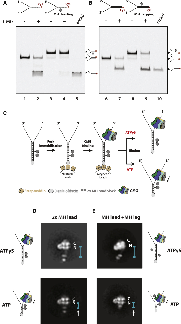
Isolation of Drosophila melanogaster CMG Engaged in Robust DNA Unwinding (A) Gel-based helicase assay showing that CMG cannot translocate past a DNA fork substrate containing a covalent HpaII methyltr...
Figureu00a02
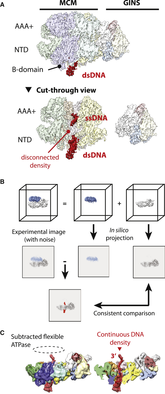
Signal Subtraction Reveals Flexibility in the AAA+ Domain of the CMG (A) Side view and cut-through side view of CMG in state 1. Duplex DNA enters the N-terminal side of the CMG and single-stranded DNA...
Figureu00a03
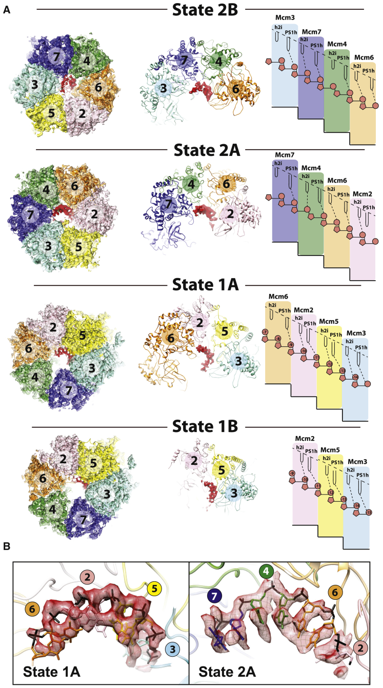
Distinct DNA Binding States around the MCM Ring in the Translocatingu00a0CMG (A) Four distinct DNA binding states for the translocating CMG. In states 2B, 2A, and 1A, four ATPase protomers contain DNA...
Figureu00a04
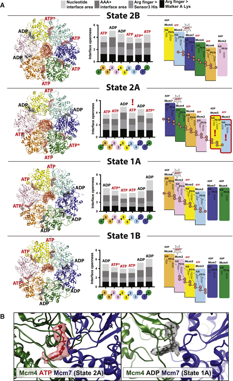
MCM Nucleotide Occupancy in Various DNA Binding States of the CMG Helicase (A) ATP at inter-protomer interfaces is shown in red. ADP is shown in black. ATP binding is observed in tighter ATPase interf...
Figureu00a05
Asymmetry of DNA Binding in Two Subsequent CMG Rotational States (A) In state 2B, Mcm3 touches single-stranded DNA at the 3u2032 end via the PS1h pore loop, while h2i is disengaged. (B) In state 2A, M...
Figureu00a06
Reconstitution of a CMG-Pol Epsilon-Mrc1-Csm3-Tof1 Complex by DNA Fork-Affinity Purification (A) Purification scheme for the CMG-Pol epsilon-Mrc1-Csm3-Tof1 complex. (B) Silver-stained gels of a comple...
Figureu00a07
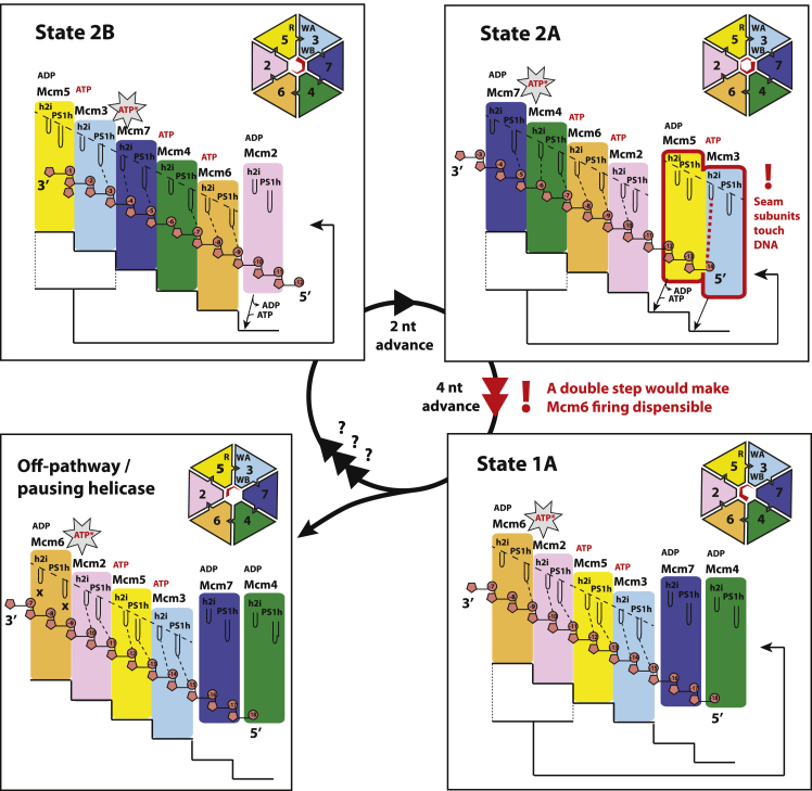
An Asymmetric Hand-over-Hand Rotational Mechanism for CMG Translocation Transition from states 2B to 2A involves release of 3u2032 DNA by Mcm3 PS1h and ATP binding by Mcm2, which binds to the N-termin...
Figure images are served from the NIH/NLM PubMed Central Open Access Subset or Europe PMC; copyright remains with the publishers and authors.
💬 Discussion
0 commentsNo comments yet. Be the first to start a discussion!
Leave a Comment