Abstract
Metabotropic γ-aminobutyric acid G protein-coupled receptors (GABAB) represent one of the two main types of inhibitory neurotransmitter receptors in the brain. These receptors act both pre- and postsynaptically by modulating the transmission of neuronal signals and are involved in a range of neurological diseases, from alcohol addiction to epilepsy. A series of recent cryo-EM studies revealed critical details of the activation mechanism of GABAB Structures are now available for the receptor bound to ligands with different modes of action, including antagonists, agonists, and positive allosteric modulators, and captured in different conformational states from the inactive apo to the fully active state bound to a G protein. These discoveries provide comprehensive insights into the activation of the GABAB receptor, which not only broaden our understanding of its structure, pharmacology, and physiological effects but also will ultimately facilitate the discovery of new therapeutic drugs and neuromodulators.
🔬 Techniques
✨ Fluorophores
🧪 Sample Preparation
🔬 Cell Lines
💾 Data Repositories
🏛️ Research Organizations (ROR)
Affiliated research institutions:
📊 Figures
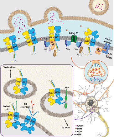
Fig. 1
GABA B trafficking, downstream effectors, and their physiological function.
GB1-GB2 heterodimer assembles in the endoplasmic reticulum (ER) of neurons, facilitated by the formation of C-terminal coiled-coil that masks the GB1 ER retention motif (bottom left). GABA B heterodim...
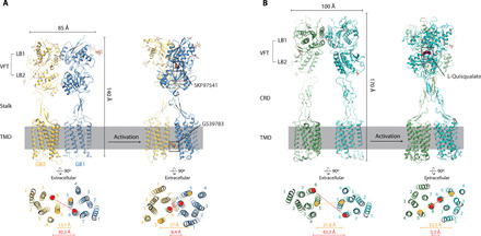
Fig. 2
Comparison of GABA B structure and activation with mGluR5.
( A ) Structures of GABA B in apo inactive [Protein Data Bank (PDB) ID 6VJM, left] and agonist-bound active (6UO8, right) states are shown in side views (top) and looking on TMDs from the extracellula...
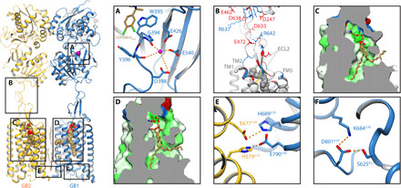
Fig. 3
Structural features of GABA B .
Left: Overall view of apo GABA B in the inactive conformation (PDB ID 6WIV). ( A ) Representative model of Ca 2+ -binding site in GB1 bound to antagonist CGP55845 (6W2V). ( B ) In each subunit, VFT is...
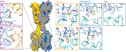
Fig. 4
Structural details of orthosteric ligands and PAM binding to GABA B .
( A and B ) Binding of PAMs at the heterodimeric TMD interface: GS39783 (PDB ID 6UO8) (A) and (+)-BHFF (7C7Q) (B). ( C and D ) Binding of agonists in the orthosteric site of GB1 VFT: SKF97541 (6UO8) (...
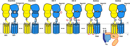
Fig. 5
Schematic mechanism of GABA B inhibition and activation.
In the apo state, the receptor adopts an inactive conformation with both VFTs being open and TMDs contacting each other on the intracellular side via an intersubunit lock between TM3 and TM5. Antagoni...
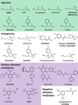
Fig. 6
Chemical structures of select GABA B ligands.
Compounds used for structural studies described in this review are highlighted in bold, and, in the following, ChEMBL ( 126 ) identifiers or PubChem ( 127 ) identifiers are given where available. Clin...
Figure images are served from the NIH/NLM PubMed Central Open Access Subset or Europe PMC; copyright remains with the publishers and authors.
💬 Discussion
0 commentsNo comments yet. Be the first to start a discussion!
Leave a Comment