Abstract
Potentially premalignant oral epithelial lesions (PPOELs) are a group of clinically suspicious conditions, of which a small percentage will undergo malignant transformation. PPOELs are suboptimally diagnosed and managed under the current standard of care. Dysplasia is the most well-established marker to distinguish high-risk PPOELs from low-risk PPOELs, and performing a biopsy to establish dysplasia is the diagnostic gold standard. However, a biopsy is limited by morbidity, resource requirements, and the potential for underdiagnosis. Diagnostic adjuncts may help clinicians better evaluate PPOELs before definitive biopsy, but existing adjuncts, such as toluidine blue, acetowhitening, and autofluorescence imaging, have poor accuracy and are not generally recommended. Recently, in vivo microscopy technologies, such as high-resolution microendoscopy, optical coherence tomography, reflectance confocal microscopy, and multiphoton imaging, have shown promise for improving PPOEL patient care. These technologies allow clinicians to visualize many of the same microscopic features used for histopathologic assessment at the point of care.
🔬 Techniques
✨ Fluorophores
🧪 Sample Preparation
🔬 Cell Lines
🏭 Microscope Brands
🏛️ Research Organizations (ROR)
Affiliated research institutions:
📊 Figures
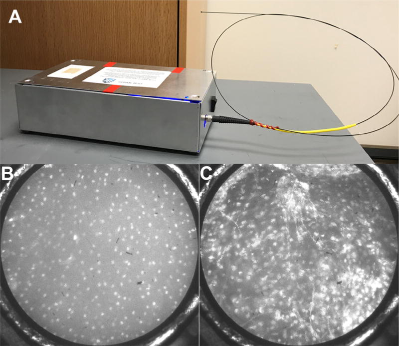
Fig. 1
High-resolution microendoscopy. A, Photo of the high-resolution microendoscope (HRME) with attached probe. HRME images of histopathologically diagnosed (B) normal oral mucosa showing small, evenly spa...
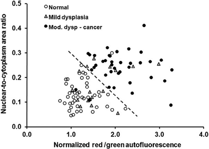
Fig. 2
Classification of imaged sites based on autofluorescence imaging and high-resolution microendoscope (HRME). The dashed line represents a linear threshold to distinguish normal sites from moderate dysp...
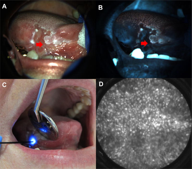
Fig. 3
Multimodal imaging example. White light (A) and autofluorescence image (B) of red and white ulcerated lesion on the right lateral tongue in a 75-year-old female presenting at the University of Texas S...
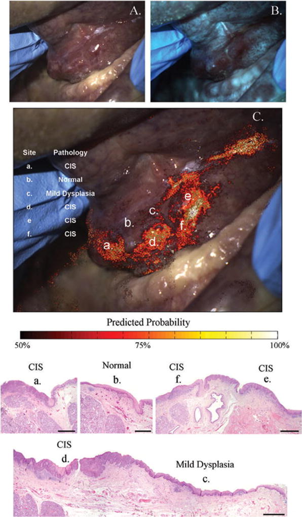
Fig. 4
Risk heat map based on autofluorescence imaging. White light (A) and autofluorescence image (B) of floor of mouth lesion containing histopathologically confirmed dysplasia and carcinoma in situ. C, Ri...
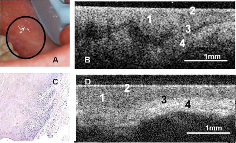
Fig. 5
Optical coherence tomography (OCT) in the oral mucosa. Au2013C, Photograph (A), in vivo OCT image (B), and u00d710 hematoxylin and eosin (H&E slide) (C) of dysplastic buccal mucosa. D, In vivo OCT ima...
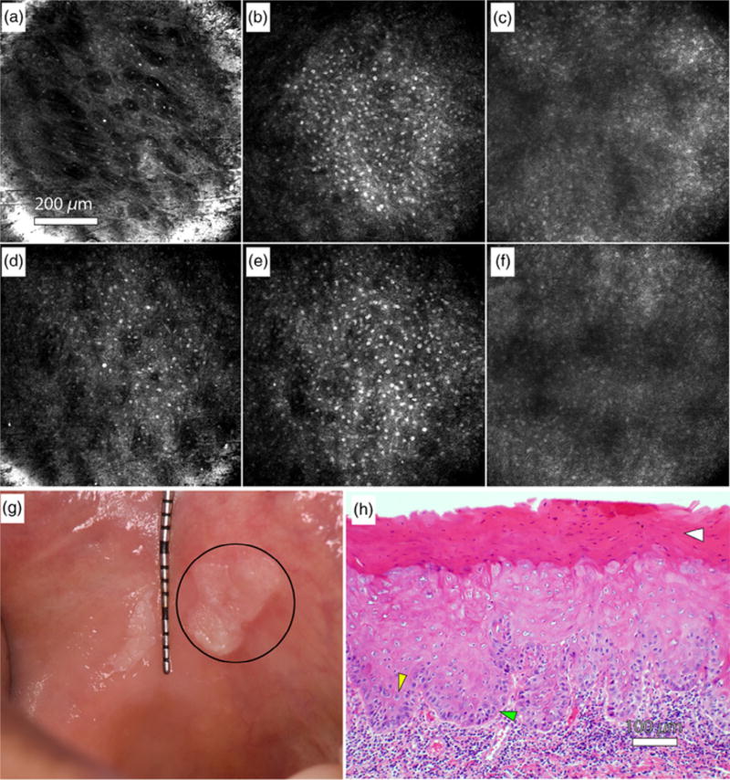
Fig. 6
Reflectance confocal microscopy in the oral mucosa. Au2013C, In vivo reflectance confocal microscopy images of clinically normal buccal mucosa at depths of approximately (A) 10 u00b5m (superficial epi...
Figure images are served from the NIH/NLM PubMed Central Open Access Subset or Europe PMC; copyright remains with the publishers and authors.
💬 Discussion
0 commentsNo comments yet. Be the first to start a discussion!
Leave a Comment