Abstract
Microtubules (MTs) and actin filaments (F-actin) function cooperatively to regulate plant cell morphogenesis. However, the mechanisms underlying the crosstalk between these two cytoskeletal systems, particularly in cell shape control, remain largely unknown. In this study, we show that introduction of the MyTH4-FERM tandem into KCBP (kinesin-like calmodulin-binding protein) during evolution conferred novel functions. The MyTH4 domain and the FERM domain in the N-terminal tail of KCBP physically bind to MTs and F-actin, respectively. During trichome morphogenesis, KCBP distributes in a specific cortical gradient and concentrates at the branching sites and the apexes of elongating branches, which lack MTs but have cortical F-actin. Further, live-cell imaging and genetic analyses revealed that KCBP acts as a hub integrating MTs and actin filaments to assemble the required cytoskeletal configuration for the unique, polarized diffuse growth pattern during trichome cell morphogenesis. Our findings provide significant insights into the mechanisms underlying cytoskeletal regulation of cell shape determination.
🔬 Techniques
🔭 Microscopes
🧬 Organisms
💻 Software
✨ Fluorophores
🧪 Sample Preparation
🔬 Cell Lines
🏭 Microscope Brands
🧪 Reagent Suppliers
📷 Detectors
🔎 Objectives
💻 Software Details
📋 Protocols
💾 Data Repositories
🏛️ Research Organizations (ROR)
Affiliated research institutions:
📋 Methods
Plant materials and growth conditions
A. thaliana ecotype Columbia (Col-0) was used as the genetic background in this study. Among the zwichel ( zwi ) alleles, the zwi-3 allele and zwi-w2 allele were frequently used in previous studies. The zwi-3 allele in Columbia ecotype background, which is expected to produce a truncated ZWI protein (KCBP) at 522 amino acids position lacking the coiled-coil and motor domains, shows the typical, strong zwi chel trichome phenotype ( Oppenheimer et al., 1997 ; Krishnakumar and Oppenheimer, 1999 ). And the zwi-w2 allele in RLD ecotype background shows weaker zwichel trichome phenotype, containing a small portion (about 15.9%) of three-branched trichomes, because sequencing analysis revealed that although a C to T transition results in a stop codon at amino acid position 72, re-initiation of translation likely occurs using the in-frame AUG ∼20 bp downstream from that mutation site as the start codon ( Folkers et al., 2002 ). To find a strong or null KCBP/ZWICHEL allele in Columbia ecotype background, we searched the Salk collection in the Arabidopsis Biological Resource Center, and finally selected the accession of Salk_031704, which contains a T-DNA insertion in the third exon (22 exons in the KCBP/ZWICHEL gene in total) and shows typical, strong zwi trichome phenotype. Coincidently, the Salk_031704 strong allele was used in recent studies and was designated either as kcbp-1 ( Humphrey et al., 2015 ) or as zwiA (accession is N531704), which was ordered from the Nottingham Arabidopsis Stock Centre ( Buschmann et al., 2015 ). The ABD2-GFP marker line is kindly provided by Prof. Shanjin Huang (Tsinghua University). The GFP-TUB6 marker line was described in our recent study ( Liu et al., 2014 ), and the mCherry-TUB6 marker line was generated using the identical method, only replacing the GFP-encoding sequence with the mCherry-encoding sequence. Plasmid construction and generation of GFP-KCBP and rigor-KCBP lines can be found in the following parts, correspondingly. Various cross combinations (the GFP-KCBP line and the mCherry-TUB6 marker line; the kcbp-1 mutant and the GFP-TUB6 marker line; the rigor-KCBP line and the GFP-TUB6 marker line; the kcbp-1 mutant and the ABD2-GFP marker line; the rigor-KCBP line and the ABD2-GFP marker line) were performed to obtain corresponding materials to observe the dynamics of KCBP, MTs, and F-actin, respectively. Plant growth conditions and transformation procedures were as described previously ( Liu et al., 2014 ). The tiny first or second true leaves in the seedlings at 10-day-old stage were dissected and used for live-cell imaging of trichomes at various developmental stages.
Show full methods section
Plant materials and growth conditions
A. thaliana ecotype Columbia (Col-0) was used as the genetic background in this study. Among the zwichel ( zwi ) alleles, the zwi-3 allele and zwi-w2 allele were frequently used in previous studies. The zwi-3 allele in Columbia ecotype background, which is expected to produce a truncated ZWI protein (KCBP) at 522 amino acids position lacking the coiled-coil and motor domains, shows the typical, strong zwi chel trichome phenotype ( Oppenheimer et al., 1997 ; Krishnakumar and Oppenheimer, 1999 ). And the zwi-w2 allele in RLD ecotype background shows weaker zwichel trichome phenotype, containing a small portion (about 15.9%) of three-branched trichomes, because sequencing analysis revealed that although a C to T transition results in a stop codon at amino acid position 72, re-initiation of translation likely occurs using the in-frame AUG ∼20 bp downstream from that mutation site as the start codon ( Folkers et al., 2002 ). To find a strong or null KCBP/ZWICHEL allele in Columbia ecotype background, we searched the Salk collection in the Arabidopsis Biological Resource Center, and finally selected the accession of Salk_031704, which contains a T-DNA insertion in the third exon (22 exons in the KCBP/ZWICHEL gene in total) and shows typical, strong zwi trichome phenotype. Coincidently, the Salk_031704 strong allele was used in recent studies and was designated either as kcbp-1 ( Humphrey et al., 2015 ) or as zwiA (accession is N531704), which was ordered from the Nottingham Arabidopsis Stock Centre ( Buschmann et al., 2015 ). The ABD2-GFP marker line is kindly provided by Prof. Shanjin Huang (Tsinghua University). The GFP-TUB6 marker line was described in our recent study ( Liu et al., 2014 ), and the mCherry-TUB6 marker line was generated using the identical method, only replacing the GFP-encoding sequence with the mCherry-encoding sequence. Plasmid construction and generation of GFP-KCBP and rigor-KCBP lines can be found in the following parts, correspondingly. Various cross combinations (the GFP-KCBP line and the mCherry-TUB6 marker line; the kcbp-1 mutant and the GFP-TUB6 marker line; the rigor-KCBP line and the GFP-TUB6 marker line; the kcbp-1 mutant and the ABD2-GFP marker line; the rigor-KCBP line and the ABD2-GFP marker line) were performed to obtain corresponding materials to observe the dynamics of KCBP, MTs, and F-actin, respectively. Plant growth conditions and transformation procedures were as described previously ( Liu et al., 2014 ). The tiny first or second true leaves in the seedlings at 10-day-old stage were dissected and used for live-cell imaging of trichomes at various developmental stages.
Plasmid construction for genetic complementation experiments
In general, the Phusion DNA polymerase with high-fidelity (New England Biolabs, Beverly, MA, United States) was used to amplify all the required gene products in this study, Fusion PCR was applied to get the GFP-KCBP fusion fragment and various constructs containing individual domain truncations ( Szewczyk et al., 2006 ), and the gateway-based technology was applied to get final expression constructs, please refer to our recent study for detailed description ( Liu et al., 2014 ). To get the GFP-KCBP construct, the genomic fragment of the KCBP gene, including its coding sequence and the 761 bp upstream fragments from the translation initiation codon ATG, was amplified from the genomic DNA with primers of KCBPP-F and GA-KCBP-R. The amplified fragment was cloned into the pENTR/D-TOPO vector by a TOPO-based cloning strategy to get the Entry 1 clone according to manufacturer's instruction (Invitrogen, Carlsbad, CA, United States). Then, a VisGreen version ( Teerawanichpan et al., 2007 ; Liu et al., 2014 ) of GFP tag was added to the NT of KCBP by the following manipulations. The promoter region of the KCBP gene was amplified with primers of KCBPP-F and KCBPP-R; the GFP-encoding sequence was amplified with primers of PGFP-F and GFP-R; the first part of KCBP -coding sequence (1–2050 bp) was amplified with primers of KCBP1X-F and KCBP1X-R. The above-mentioned three PCR fragments were further linked together by Fusion PCR using primers of KCBPP-F and KCBP1X-R. The resulting PCR fragment was subsequently cloned into the pENTR/D-TOPO vector to get the Entry 2 clone. Finally, both the Entry 1 vector and Entry 2 vector were digested by Not I and Xba I, and further ligation reaction was conducted between the gel-purified fragment containing the latter part of KCBP (1951–6023 bp) and the fragment containing the promoter, GFP, and the first part of KCBP genomic sequence, the resulting Entry 3 clone was delivered into pEarleyGate302 by recombination reaction to get the final GFP-KCBP construct. A series of constructs containing various domain truncations were made by the following manipulations. To get the KCBP-∆MyTH4 (KCBP lacking 117–275 amino acids) construct, the fragment containing promoter and 1–435 bp KCBP genomic sequence was amplified with primers of KCBPP-F and KCBP-∆MyTH4-R, and the 992–6023 bp of the KCBP genomic sequence was amplified with primers of KCBP-∆MyTH4-F and GA-KCBP-R, then the two PCR fragments were linked together by Fusion PCR using primers of KCBPP-F and GA-KCBP-R. Finally, the resulting PCR fragment was cloned into pENTR/D-TOPO vector and was then delivered into pEarleyGate302 to get the final KCBP-∆MyTH4 construct. The same strategy was used to generate constructs of KCBP-∆NT (KCBP lacking 2–121 amino acids), KCBP-∆NT-MyTH4 (KCBP lacking 2–275 amino acids), KCBP-∆FERM (KCBP lacking 275–497 amino acids), KCBP-∆MyTH4-FERM (KCBP lacking 116–505 amino acids), KCBP-∆NT-MyTH4-FERM (KCBP lacking 2–505 amino acids), KCBP-∆CC-Motor-CBD (KCBP lacking 532–1266 amino acids), and KCBP-∆CBD (KCBP lacking 1210–1266 amino acids) primers can be found in the Supplementary file 1 . To get the rigor-KCBP construct, we introduced threonine (ACT) 982-to-asparagine (AAC) mutation by the PCR-based mutagenesis. In detail, we designed a pair of overlapping primers (KCBP-T982N-R and KCBP-T982N-F) with the desired nucleotide changes at the target site, and amplified the front half with primers of KCBP2X-F and KCBP-T982N-R, and that latter half with primers of KCBP-T982N-F and GA-KCBP-R, then the two fragments were linked together by Fusion PCR using primers of KCBP2X-F and GA-KCBP-R. The resulting PCR fragment was cloned into the pENTR/D-TOPO vector to get the Entry 4 clone. Finally, both the Entry 1 vector and the Entry 4 vector were digested by Not I and Xba I, and further ligation reaction was conducted between the gel-purified fragment containing the latter part of KCBP containing the threonine 982-to-asparagine mutation and the fragment containing the promoter and the first part of KCBP genomic sequence, the resulting Entry 5 clone was delivered into pEarleyGate302 to get the final rigor-KCBP construct. Genetic complementation and trichome phenotype identification The KCBP (At5G65930) genomic fragment comprising its endogenous promoter and coding region was used for genetic complementation tests. Plasmid construction for the GFP-KCBP construct, the rigor-KCBP construct, and other constructs containing various domain truncations was all based on the above-mentioned KCBP fragment. The constructs were introduced into the kcbp-1 mutant. The T3 homozygous transgenic lines were used to observe trichome phenotype under a fully automated Stereo Microscope (Leica M205 FA) with Leica Application Suite V4.2. The images of trichomes were a maximum z-projection of image series acquired by the Leica LAS Multifocus program.
Spinning-disc confocal microscopy and motor motility analysis
Live-cell imaging was carried out under a spinning disk confocal microscope (UltraView VoX, Perkin Elmer, Beaconsfield, Buckinghamshire, UK) equipped with the Yokogawa Nipkow CSU-X1 spinning disk scanner, Hamamatsu EMCCD 9100-13, Nikon TiE inverted microscope with the Perfect Focus System as described previously ( Liu et al., 2014 ). Acquired images were processed and analyzed using Volocity (Perkin Elmer), Image J ( http://rsbweb.nih.gov/ij ), MetaMorph (Molecular Devices, Sunnyvale, CA, United States). The run-length distribution and the velocity distribution of GFP-KCBP were calculated in Origin software (OriginLab) by frequency counts. The mean values and 95% confidence interval were calculated in SAS (SAS Software), as described previously ( Kong et al., 2015 ).
Purification of GFP-tagged motor-less KCBP with various domain truncations
The full-length cDNA fragments of KCBP were amplified from the Arabidopsis cDNA with primers of KCBP1X-F and GA-KCBP-R, and the GFP-coding sequence amplified with primers of GFP-F and GFP-R, then the two PCR fragments were linked together by Fusion PCR using primers of GFP-F and GA-KCBP-R. The resulting PCR fragment was cloned into the pENTR/D-TOPO vector to get the Entry 6 clone. The cDNA fragments encoding polypeptide of GFP-NT-MyTH4-FERM-CC (1–749 amino acids), GFP-NT-MyTH4-FERM (1–614 amino acids), GFP-NT-MyTH4 (1–275 amino acids), GFP-MyTH4 (116–275 amino acids), GFP-FERM (276–503 amino acids), GFP-NT(1–115 amino acids), and GFP were generated based on the Entry 6 clone. The truncated fragments were digested with Sal I and Not I and were reconstructed into the pET-28a vector to get final expression constructs. Primers can be found in the Supplementary file 1 . Finally, the expression constructs were transformed into Escherichia coli strain Transetta (DE3, TransGen Biotech, Beijing, China) to induce expression. The recombinant proteins were purified using nickel-nitrilotriacetic acid (Ni-NTA) resin following procedures described by the manufacturer (Qiagen, Hilden, Germany). Fractions containing the protein were collected, combined, and dialyzed overnight against PEM buffer (80 mM PIPES, 1 mM EGTA (ethylene glycol tetraacetic acid), and 1 mM MgSO 4 , pH 6.9). Protein concentration was determined by a Bio-Rad protein assay kit. Protein samples of 3 μg were analyzed by SDS-PAGE (Sodium Dodecyl Sulfate Polyacrylamide Gel Electropheresis). Single-molecule (particle) imaging assay The purified porcine brain tubulin labeled with NHS-rhodamine was kindly provided from Prof. Tonglin Mao (China Agricultural University). Taxol-stabilized NHS-rhodamine MTs were incubated with 1 μM GFP-NT, 1 μM GFP-MyTH4, 1 μM GFP-FERM, 1 μM GFP-NT-MyTH4, 1 μM GFP-NT-MyTH4-FERM, 1 μM GFP-NT-MyTH4-FERM-CC, and 1 μM control GFP, respectively, in PEM buffer at equal molar ratios for 30 min at room temperature, modified from a previous study ( Liu et al., 2013 ).
Fluorescent images of MTs and various
GFP-KCBP truncated proteins were visualized using a Zeiss inverted fluorescence microscope (Axio Observer Z1) with a Zeiss Plan-Apochromat 100× oil immersion objective (NA = 1.4). The rabbit skeletal muscle actins were provided by Prof. Shanjin Huang (Tsinghua University). F-actin (3 µM) was incubated with 1 μM GFP-NT, 1 μM GFP-MyTH4, 1 μM GFP-FERM, 1 μM GFP-NT-MyTH4, 1 μM GFP-NT-MyTH4-FERM, 1 μM GFP-NT-MyTH4-FERM-CC, and 1 μM control GFP at the indicated concentrations at room temperature for 30 min and then labeled with 3 µM Alexa561-phalloidin (Invitrogen). Actin filaments were subsequently diluted to a final concentration of 10 nM in fluorescence buffer (10 mM imidazole, pH 7.0, 50 mM KCl, 2 mM MgCl 2 , 1 mM EGTA, 100 mM DTT (Dithiothreitol), 100 µg/ml Glucoxidase, 15 mg/ml Glucose, 20 µg/ml catalase, and 0.5% methylcellulose), modified from a previous study ( Wu et al., 2010 ). The diluted samples were visualized using a Zeiss inverted fluorescence microscope (Axio Observer Z1) with a Zeiss Plan-Apochromat 100× oil immersion objective (NA = 1.4).
Identification of T-DNA insertion in kcbp-1 mutant
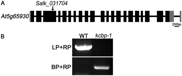
A standard PCR-based method was used to identify the T-DNA insertion in kcbp-1 ( Humphrey et al., 2015 ), as described by Kong et al. (2015) . Gene-specific primers (031704-LP and 031704-RP) were used to amplify an approximate 1000 bp DNA fragment in KCBP . The T-DNA insertion was detected using 031704-RP and the left-border primer (LBb1.3). For RT-PCR analysis of transcription level of KCBP in the kcbp-1 mutant, total RNA was extracted using Trizol reagent Invitrogen and was used for first strand cDNA synthesis by the SuperScript III First-Strand Synthesis System (Life Technologies, Carlsbad, CA, United States) with oligo (dT)18 primers. Then, the cDNA was used as a template for PCR reactions using primers shown in the primer list. UBQ5 was used as control.
Plant materials and growth conditions
A. thaliana ecotype Columbia (Col-0) was used as the genetic background in this study. Among the zwichel ( zwi ) alleles, the zwi-3 allele and zwi-w2 allele were frequently used in previous studies. The zwi-3 allele in Columbia ecotype background, which is expected to produce a truncated ZWI protein (KCBP) at 522 amino acids position lacking the coiled-coil and motor domains, shows the typical, strong zwi chel trichome phenotype ( Oppenheimer et al., 1997 ; Krishnakumar and Oppenheimer, 1999 ). And the zwi-w2 allele in RLD ecotype background shows weaker zwichel trichome phenotype, containing a small portion (about 15.9%) of three-branched trichomes, because sequencing analysis revealed that although a C to T transition results in a stop codon at amino acid position 72, re-initiation of translation likely occurs using the in-frame AUG ∼20 bp downstream from that mutation site as the start codon ( Folkers et al., 2002 ). To find a strong or null KCBP/ZWICHEL allele in Columbia ecotype background, we searched the Salk collection in the Arabidopsis Biological Resource Center, and finally selected the accession of Salk_031704, which contains a T-DNA insertion in the third exon (22 exons in the KCBP/ZWICHEL gene in total) and shows typical, strong zwi trichome phenotype. Coincidently, the Salk_031704 strong allele was used in recent studies and was designated either as kcbp-1 ( Humphrey et al., 2015 ) or as zwiA (accession is N531704), which was ordered from the Nottingham Arabidopsis Stock Centre ( Buschmann et al., 2015 ). The ABD2-GFP marker line is kindly provided by Prof. Shanjin Huang (Tsinghua University). The GFP-TUB6 marker line was described in our recent study ( Liu et al., 2014 ), and the mCherry-TUB6 marker line was generated using the identical method, only replacing the GFP-encoding sequence with the mCherry-encoding sequence. Plasmid construction and generation of GFP-KCBP and rigor-KCBP lines can be found in the following parts, correspondingly. Various cross combinations (the GFP-KCBP line and the mCherry-TUB6 marker line; the kcbp-1 mutant and the GFP-TUB6 marker line; the rigor-KCBP line and the GFP-TUB6 marker line; the kcbp-1 mutant and the ABD2-GFP marker line; the rigor-KCBP line and the ABD2-GFP marker line) were performed to obtain corresponding materials to observe the dynamics of KCBP, MTs, and F-actin, respectively. Plant growth conditions and transformation procedures were as described previously ( Liu et al., 2014 ). The tiny first or second true leaves in the seedlings at 10-day-old stage were dissected and used for live-cell imaging of trichomes at various developmental stages.
Additional files 10.7554/eLife.09351.027 Supplementary file 1. List of primer sequences used in this study. DOI: http://dx.doi.org/10.7554/eLife.09351.027
📊 Figures
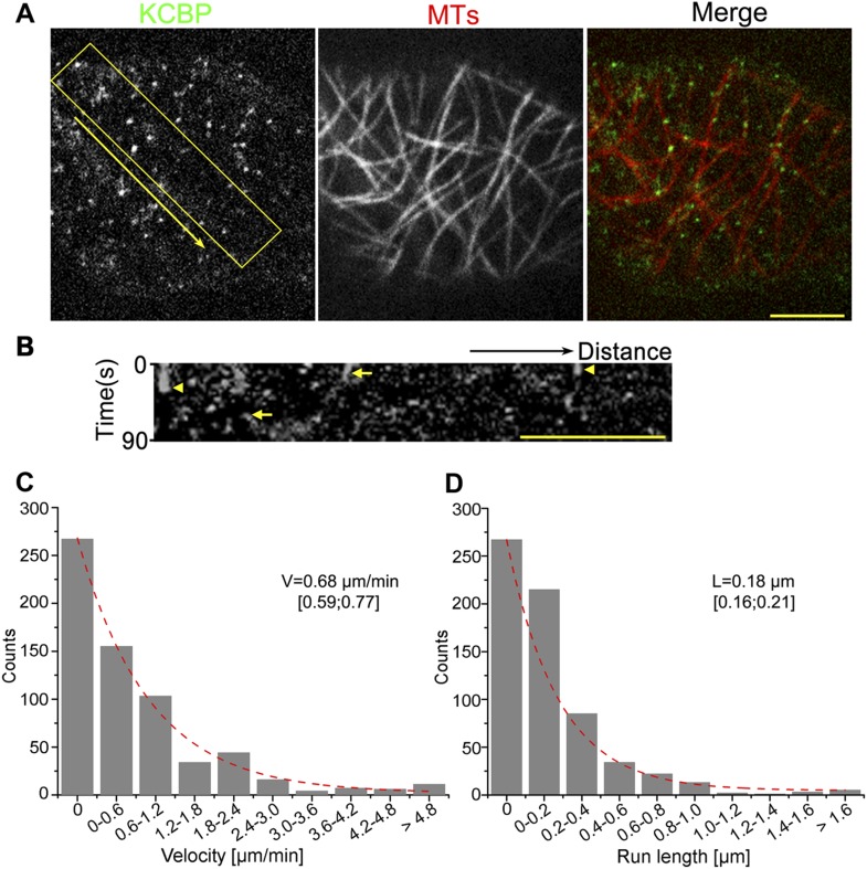
Figure 1.
KCBP colocalizes with cortical MTs in vivo.
( A ) GFP-labeled kinesin-like calmodulin-binding protein (KCBP) localizes along cortical microtubules (MTs) (mCherry-TUB6) in a punctate pattern in Arabidopsis epidermal pavement cells. The yellow bo...
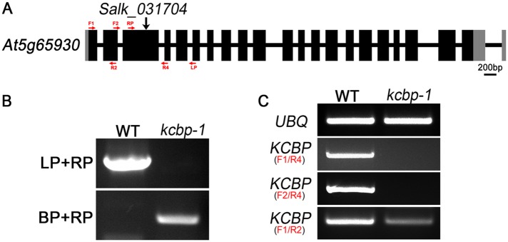
Figure 1u2014figure supplement 1.
Genetic identification of the kcbp-1/zwiA mutant.
( A ) Gene structure of the KCBP gene. Black rectangles indicate exons, gray rectangles indicate the 5u2032 and 3u2032 untranslated regions, and thick lines indicate introns. The arrow indicates the l...
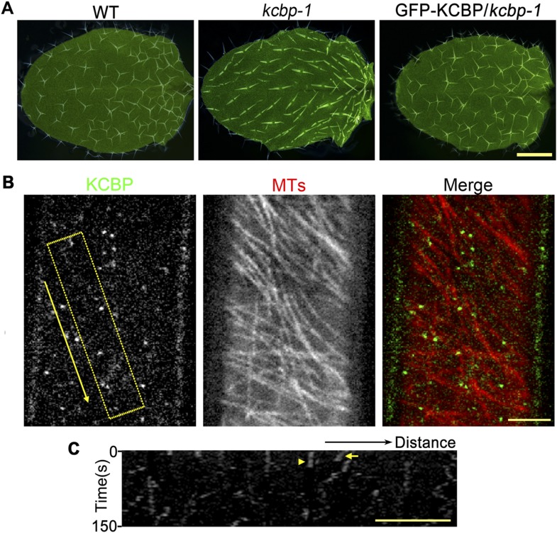
Figure 1u2014figure supplement 2.
KCBP colocalizes with cortical MTs in Arabidopsis hypocotyl cells.
( A ) The genomic GFP-KCBP fusion complements the trichome defects of the kcbp-1 mutant. Scale bar, 1 mm. ( B ) GFP-labeled KCBP localizes along cortical MTs (mCherry-TUB6) in a punctate pattern in Ar...
Video 1.
Localization and dynamicity of kinesin-like calmodulin-binding protein (KCBP) on cortical microtubules (MTs) in Arabidopsis epidermal pavement cells.
Images were obtained at 3-s intervals. A total of 30 time lapse images were applied to make the video. Scale bar, 5 u03bcm. DOI: http://dx.doi.org/10.7554/eLife.09351.007
Video 2.
Localization and dynamicity of KCBP on cortical MTs in Arabidopsis hypocotyl cells.
Images were obtained at 3-s intervals. A total of 35 time lapse images were applied to make the video. Scale bar, 5 u03bcm. DOI: http://dx.doi.org/10.7554/eLife.09351.008
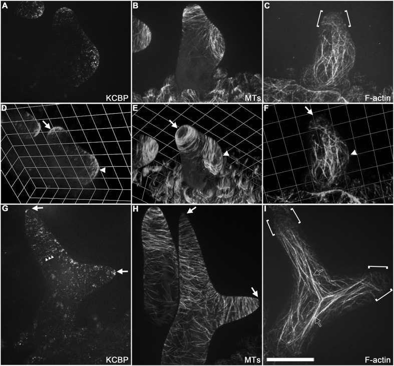
Figure 2.
Spatio-temporal distribution of GFP-KCBP in developing wild-type trichomes.
( A u2013 F ) Localization of KCBP and spatial organization of the cytoskeleton in stage 2/3 trichomes. GFP-KCBP particles form a cortical gradient with the highest expression at the branching site an...
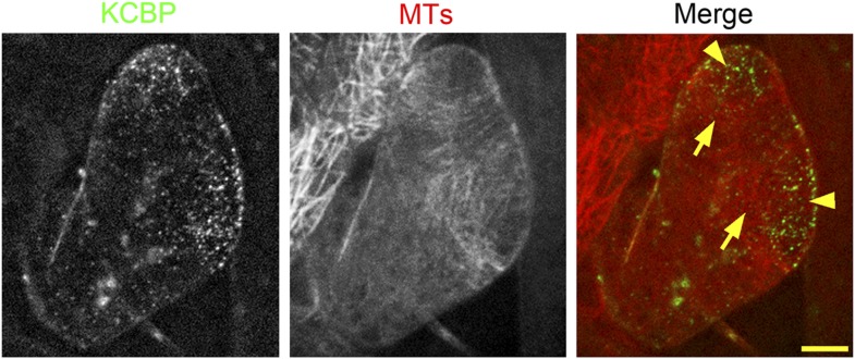
Figure 2u2014figure supplement 1.
Colocalization of KCBP with cortical MTs in stage 2 trichomes.
GFP-KCBP particles form a cortical gradient with the highest expression at the branching site and the tip region of the main stem. The arrowheads highlight the strongest accumulation of GFP-KCBP at th...
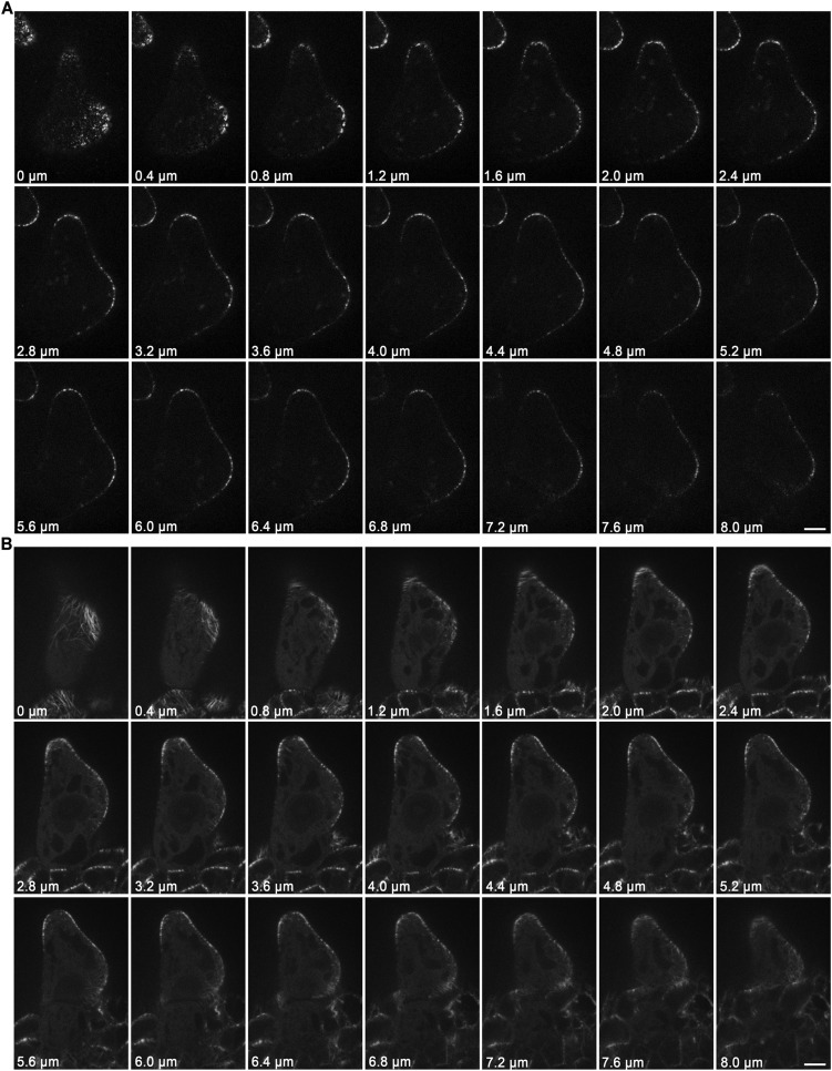
Figure 2u2014figure supplement 2.
Localization of KCBP and spatial organization of MTs in stage 2/3 wild-type trichomes.
( A ) The GFP-KCBP images, which were used to make the z-projection in Figure 2A , were sequentially illustrated at 0.4-u03bcm intervals. GFP-KCBP was observed to strongly accumulate at apexes of elon...
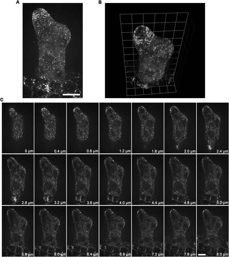
Figure 2u2014figure supplement 3.
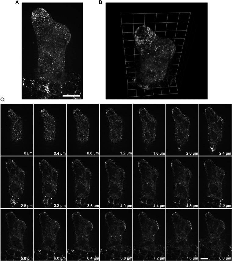
Localization of GCP2 in stage 2/3 wild-type trichomes.
( A ) The Z-projection image, which was acquired from a high-resolution stack of 46 planes at 0.2-u03bcm intervals, shows a tip-oriented cortical gradient of GCP2-3u00d7GFP particles in the elongating...
Video 3.
Spatio-temporal distribution of GFP-KCBP, MTs, and actin filaments is highlighted by 3-D reconstitution in stage 2/3 trichomes.
DOI: http://dx.doi.org/10.7554/eLife.09351.013
Video 4.
The spatio-temporal dynamics and distribution of GFP-KCBP in developing trichomes.
Images were obtained at 3-s intervals. A total of 8 time lapse images were applied to make the video. Scale bar, 5 u03bcm. DOI: http://dx.doi.org/10.7554/eLife.09351.014
Video 5.
Spatio-temporal distribution of GCP2-3XGFP is highlighted by 3-D reconstitution in stage 2/3 trichomes.
DOI: http://dx.doi.org/10.7554/eLife.09351.015
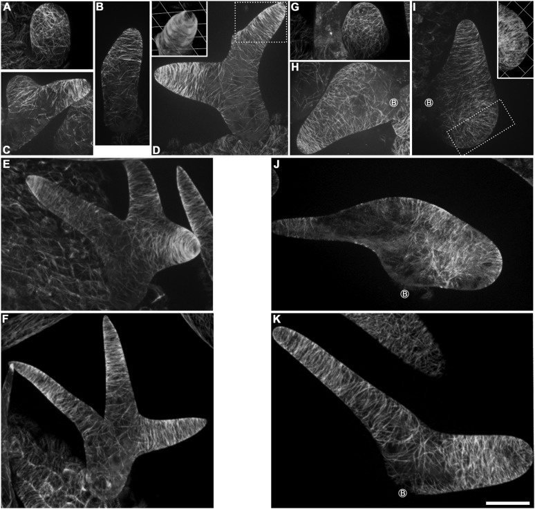
Figure 3.
Abnormal MT organization in kcbp-1 trichomes.
( A u2013 F ) Spatio-temporal MT organization in wild-type trichomes during development. The stage 1 trichomes exhibit random MT networks ( A ). In stage 2/3 trichomes, the random MT network shifts in...
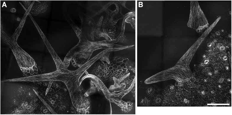
Figure 3u2014figure supplement 1.
Cortical MT organization in wild-type and kcbp-1 mature trichomes.
In stage 6 wild-type mature trichomes ( A ), fully elongated branches form a fine and pointed tip, and cortical MTs exhibit an oblique or longitudinal configuration. By contrast, the stage 6 kcbp-1 ma...
Video 6.
The 3-D reconstructed cortical MT configuration in stage 3/4 wild-type trichomes.
DOI: http://dx.doi.org/10.7554/eLife.09351.018
Video 7.
The 3-D reconstructed cortical MT configuration in stage 3/4 kcbp-1 trichomes.
DOI: http://dx.doi.org/10.7554/eLife.09351.019
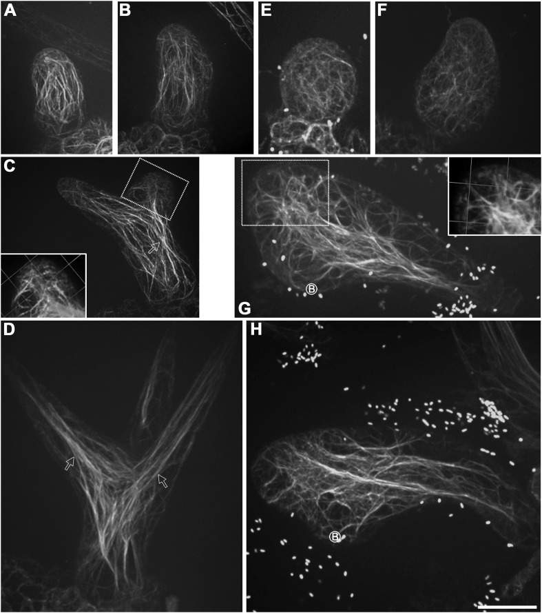
Figure 4.
Aberrant organization of F-actin in kcbp-1 trichomes.
( A u2013 D ) Spatio-temporal organization of F-actin in wild-type trichomes during development. In stage 1/2 trichomes, a population of cytoplasmic actin cables align with the growth axis ( A , B ). ...
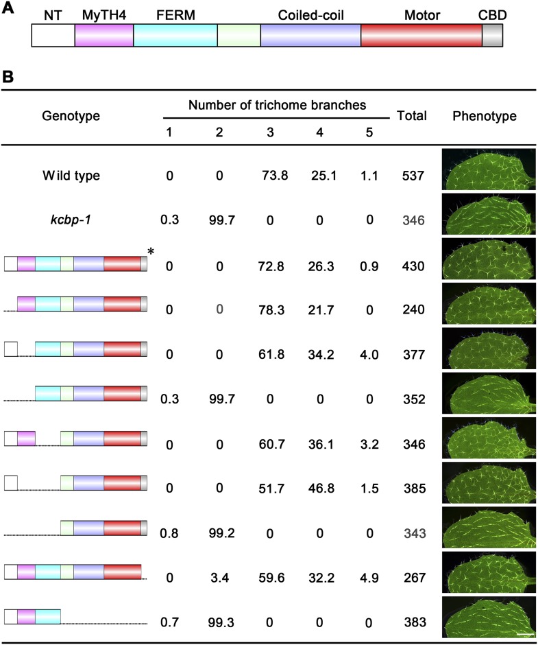
Figure 5.
Genetic analyses on the role of individual domains of KCBP in trichome development.
( A ) Schematic diagram of the domain organization of KCBP. ( B ) Genetic complementation test using various truncated versions of KCBP. The genotype column shows the individual constructs containing ...
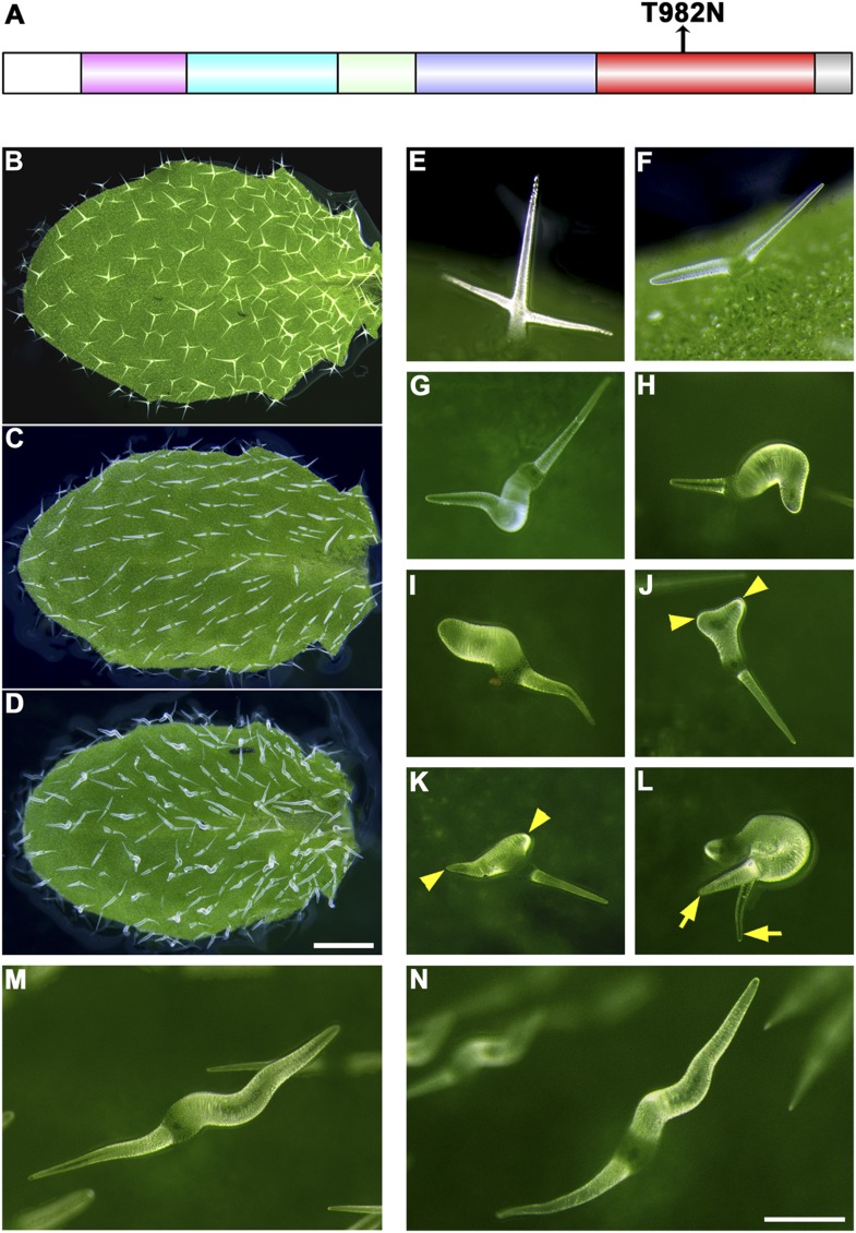
Figure 6.
The trichome phenotype of rigor-KCBP transformants.
( A ) A point mutation was introduced into genomic KCBP including its native regulatory elements generate the rigor-KCBP with a threonine 982-to-asparagine substitution in its ATP-binding motif. The r...
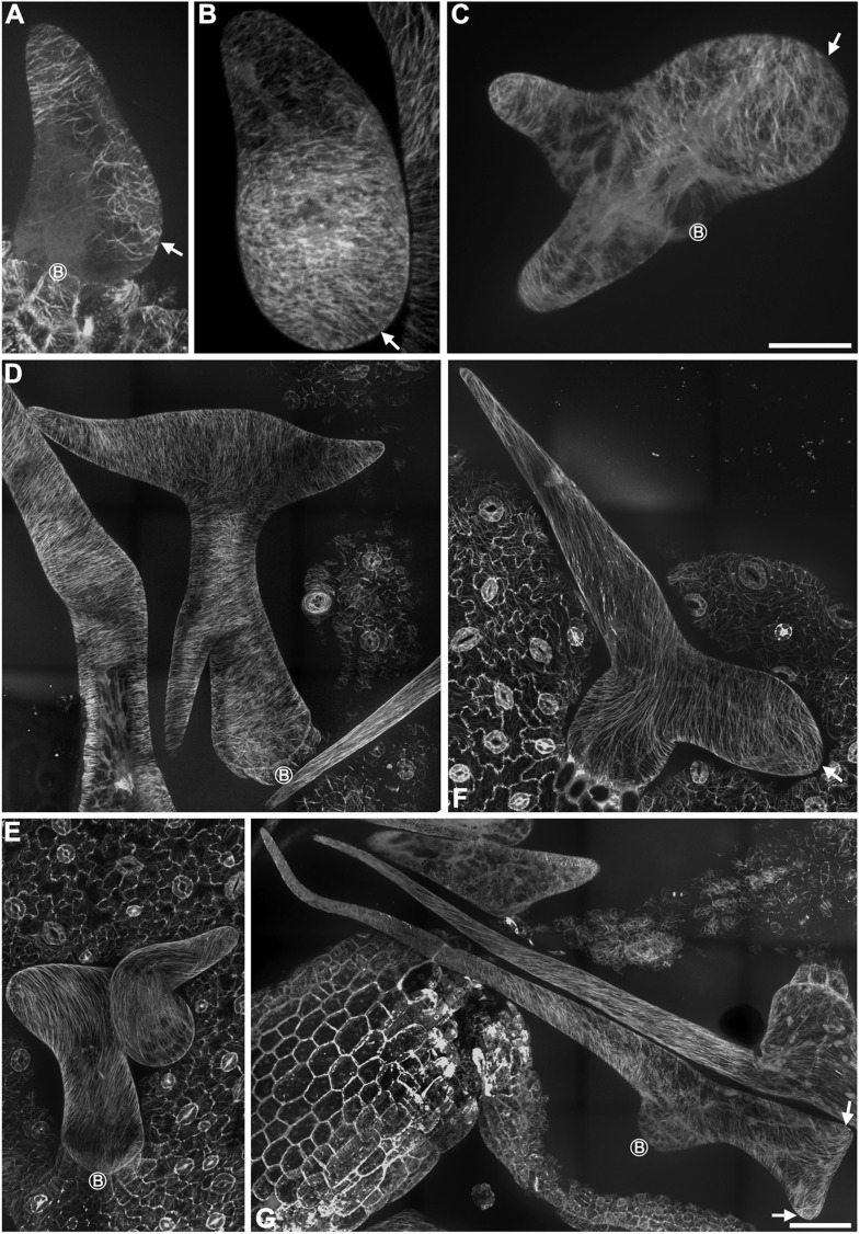
Figure 6u2014figure supplement 1.
Abnormal MT organization in rigor-KCBP trichomes.
( A ) The formation of the transverse MT rings that encircles the incipient primary branch (indicated by the arrow) is impaired in a stage 2/3 trichome. ( B ) Transverse MT rings and the MT-depleted z...
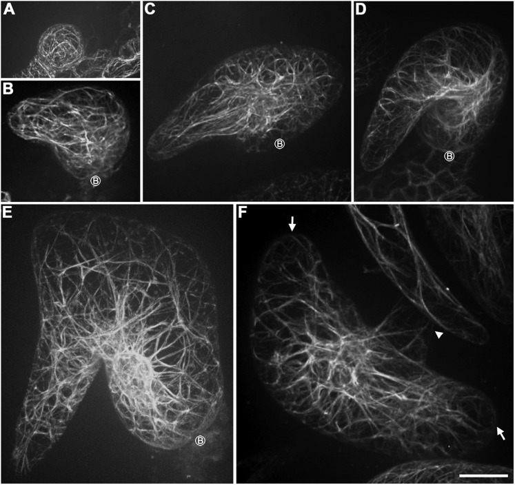
Figure 6u2014figure supplement 2.
Abnormal organization of actin filaments in rigor-KCBP trichomes.
( A ) The stage 1 trichomes display random meshworks of thick actin bundles. ( B u2013 F ) The curly, intertwined, thick actin bundles dominate inside developing rigor-KCBP trichomes, and no parallel-...
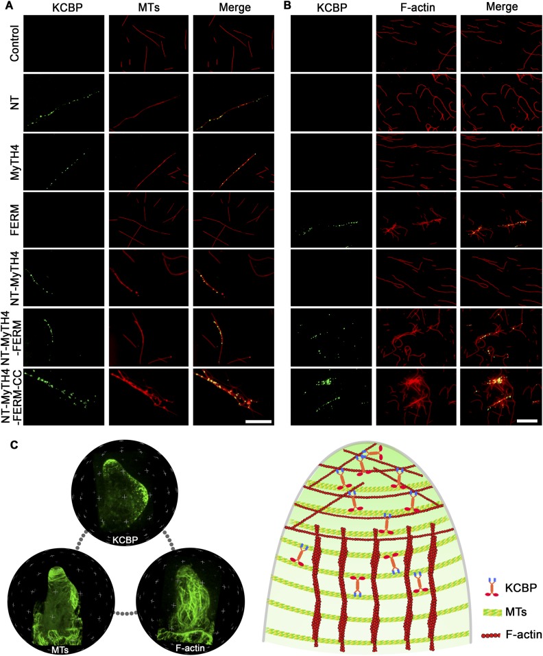
Figure 7.
In vitro MT- and F-actin-binding activity of the motorless KCBP and a working model for KCBP during trichome morphogenesis.
( A ) The KCBP N-terminal tail containing the MyTH4 domain binds to MTs in vitro. Rhodamine-labeled MTs were incubated with GFP-NT, GFP-MyTH4, GFP-FERM, GFP-NT-MyTH4, GFP-NT-MyTH4-FERM, GFP-NT-MyTH4-F...
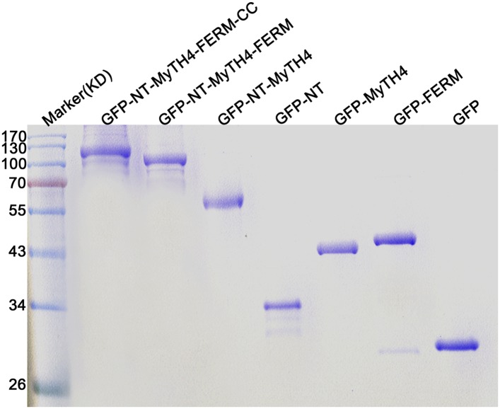
Figure 7u2014figure supplement 1.
Coomassie blue-stained SDS-PAGE gel of the purified GFP-KCBP recombinant proteins with various truncations.
KD, kiloDalton, indicates the mass of molecular markers. DOI: http://dx.doi.org/10.7554/eLife.09351.026
Author response image 1.
Localization of GCP2 in stage 2/3 wild-type trichomes. (A) The Z-projection image, which was acquired from a high-resolution stack of 46 planes at 0.2 u03bcm intervals, shows a tip-oriented cortical g...
Author response video 1.
The spatial distribution of GCP2-3u00d7GFP is highlighted by 3-D reconstitution in a stage 2/3 wild-type trichome. DOI: http://dx.doi.org/10.7554/eLife.09351.031
Author response image 2.
Genetic identification of the kcbp-1 mutant. (A) Gene structure of the KCBP gene. Black rectangles indicate exons, gray rectangles indicate the 5u2019 and 3u2019 untranslated regions, and thick lines ...
Figure images are served from the NIH/NLM PubMed Central Open Access Subset or Europe PMC; copyright remains with the publishers and authors.
💬 Discussion
0 commentsNo comments yet. Be the first to start a discussion!
Leave a Comment