Abstract
During inflammation polymorphonuclear neutrophils (PMNs) traverse venular walls, composed of the endothelium, pericyte sheath and vascular basement membrane. Compared to PMN transendothelial migration, little is known about how PMNs penetrate the latter barriers. Using mouse models and intravital microscopy, we show that migrating PMNs expand and use the low expression regions (LERs) of matrix proteins in the vascular basement membrane (BM) for their transmigration. Importantly, we demonstrate that this remodeling of LERs is accompanied by the opening of gaps between pericytes, a response that depends on PMN engagement with pericytes. Exploring how PMNs modulate pericyte behavior, we discovered that direct PMN-pericyte contacts induce relaxation rather than contraction of pericyte cytoskeletons, an unexpected response that is mediated by inhibition of the RhoA/ROCK signaling pathway in pericytes. Taking our in vitro results back into mouse models, we present evidence that pericyte relaxation contributes to the opening of the gaps between pericytes and to the enlargement of the LERs in the vascular BM, facilitating PMN extravasation. Our study demonstrates that pericytes can regulate PMN extravasation by controlling the size of pericyte gaps and thickness of LERs in venular walls. This raises the possibility that pericytes may be targeted in therapies aimed at regulating inflammation.
🔬 Techniques
🔭 Microscopes
💻 Software
✨ Fluorophores
🧪 Sample Preparation
🔬 Cell Lines
🏭 Microscope Brands
🧪 Reagent Suppliers
📷 Detectors
💻 Software Details
🏛️ Research Organizations (ROR)
Affiliated research institutions:
📋 Methods
Ethics Statement
All experiments were approved by the Institutional Animal Care Committee of the University of North Carolina School of Medicine and Public health and performed according to the legislation for the protection of animals (IACUC ID: 12–038.0).
Experimental Animals
Wild type (WT) C57BL/6 mice were purchased from the Jackson lab. Platelet-endothelial cell adhesion molecule–deficient (PECAM-1 −/− ) mice on a C57BL/6 background were kindly provided by Dr P. Newman (Blood Research Institute, Blood Center of Wisconsin, Milwaukee). C57BL/6 immorto-mice expressing a SV40 large T antigen were obtained from Charles River Laboratories (Wilmington, MA). Inflammatory Response in Mouse Cremaster Muscles or Ear Skin and Intravital Microscopy WT or PECAM-1 −/− male mice (∼25 g) were injected intrascrotally (i.s.) with saline, interleukin-1β (IL-1β) (50 ng/mouse) or tumor necrosis factor-α (TNF-α) (500 ng/mouse) (R&D Systems, Minneapolis, MN). In IL-1β-injected mouse cremaster muscles, the responding leukocytes (>90%) were previously verified to be PMNs [14] . In selected experiments where PMNs need to be depleted, 100 µg of anti-GR1 antibodies (Abs) [15] (RB6–8C5, BD Biosciences, Chicago, IL) were injected intraperitoneally (i.p.) into each mouse 24 hours before i.s. administration of IL-1β. To block PMN extravasation, in some experiments each mouse was intra-splenically pre-injected with 100 µg of Abs against either mouse intercellular adhesion molecule-1 (ICAM-1) (YN1/1.7.4, Biolegend, San Diego, CA) [16] or α 6 integrin (GoH3, Biolegend, San Diego, CA) [17] , [18] 15 minutes before IL-1β administration. Control animals received isotype-matched control Abs (Biolegend, San Diego, CA). Two hours after IL-1β injection, the cremaster muscles of fully anaesthetized mice were exteriorized and transferred to the stage of an Olympus FluoView FV1000 upright microscope and the cremaster muscles were constantly superfused with warm buffered solution. The numbers of rolling or static adherent leukocytes were quantified starting from the administration of superfusion buffer as described before [14] . In some experiments, cremaster muscles were clamped using a micro aneurysm clip (Harvard Apparatus, Holliston, MA) and superfused with buffer containing Norepinephrine (NE, 10 µM, Sigma-Aldrich, Saint Louis, MO) or Tolazoline (100 µg/ml, Sigma-Aldrich, Saint Louis, MO). Fifteen min later, the inflammatory response was stopped immediately by perfusion of 4% paraformaldehyde (PF). The cremaster muscles of all experimental mice were finally harvested and fixed in PF for further immunostaining. In some experiments, IL-1β (5 ng/mouse) or TNF-α (50 ng/mouse) in 50 µl saline was subcutaneously injected into mouse ears for two hours to induce an inflammatory response in skin.
Show full methods section
Ethics Statement
All experiments were approved by the Institutional Animal Care Committee of the University of North Carolina School of Medicine and Public health and performed according to the legislation for the protection of animals (IACUC ID: 12–038.0).
Experimental Animals
Wild type (WT) C57BL/6 mice were purchased from the Jackson lab. Platelet-endothelial cell adhesion molecule–deficient (PECAM-1 −/− ) mice on a C57BL/6 background were kindly provided by Dr P. Newman (Blood Research Institute, Blood Center of Wisconsin, Milwaukee). C57BL/6 immorto-mice expressing a SV40 large T antigen were obtained from Charles River Laboratories (Wilmington, MA). Inflammatory Response in Mouse Cremaster Muscles or Ear Skin and Intravital Microscopy WT or PECAM-1 −/− male mice (∼25 g) were injected intrascrotally (i.s.) with saline, interleukin-1β (IL-1β) (50 ng/mouse) or tumor necrosis factor-α (TNF-α) (500 ng/mouse) (R&D Systems, Minneapolis, MN). In IL-1β-injected mouse cremaster muscles, the responding leukocytes (>90%) were previously verified to be PMNs [14] . In selected experiments where PMNs need to be depleted, 100 µg of anti-GR1 antibodies (Abs) [15] (RB6–8C5, BD Biosciences, Chicago, IL) were injected intraperitoneally (i.p.) into each mouse 24 hours before i.s. administration of IL-1β. To block PMN extravasation, in some experiments each mouse was intra-splenically pre-injected with 100 µg of Abs against either mouse intercellular adhesion molecule-1 (ICAM-1) (YN1/1.7.4, Biolegend, San Diego, CA) [16] or α 6 integrin (GoH3, Biolegend, San Diego, CA) [17] , [18] 15 minutes before IL-1β administration. Control animals received isotype-matched control Abs (Biolegend, San Diego, CA). Two hours after IL-1β injection, the cremaster muscles of fully anaesthetized mice were exteriorized and transferred to the stage of an Olympus FluoView FV1000 upright microscope and the cremaster muscles were constantly superfused with warm buffered solution. The numbers of rolling or static adherent leukocytes were quantified starting from the administration of superfusion buffer as described before [14] . In some experiments, cremaster muscles were clamped using a micro aneurysm clip (Harvard Apparatus, Holliston, MA) and superfused with buffer containing Norepinephrine (NE, 10 µM, Sigma-Aldrich, Saint Louis, MO) or Tolazoline (100 µg/ml, Sigma-Aldrich, Saint Louis, MO). Fifteen min later, the inflammatory response was stopped immediately by perfusion of 4% paraformaldehyde (PF). The cremaster muscles of all experimental mice were finally harvested and fixed in PF for further immunostaining. In some experiments, IL-1β (5 ng/mouse) or TNF-α (50 ng/mouse) in 50 µl saline was subcutaneously injected into mouse ears for two hours to induce an inflammatory response in skin.
Immunostaining of Mouse Cremaster Muscles and Ear Skin
Fixed mouse cremaster muscles and ear skin were immunostained for multiple proteins as described previously [9] . An Alexa 488- or 647-labeled rat anti-mouse PECAM-1 Ab (MEC13.3 BioLegend, San Diego, CA) was applied to label ECs. The vascular BM was displayed by a rabbit anti–mouse collagen IV polyclonal Ab (Abcam, Cambridge, MA) plus appropriate secondary Abs conjugated to Alexa 405 or 488 (Invitrogen, Carlsbad, CA). To mark the PMNs, a goat anti–mouse MRP-14 Ab (Calgranulin B (M-19), Santa Cruz Biotechnology, Santa Cruz, CA) or Allophycocyanin-conjugated rat anti-mouse CD11b Ab (M1/70, eBioscience, San Diego, CA) was used. To stain the pericytes, tissues were further incubated with a Cy3-labeled anti-mouse α-SMA Ab (1A4, Sigma-Aldrich, St. Louis, MO) or a goat IgG against mouse Platelet-derived growth factor receptor-β (PDGFR-β) (R&D Systems, Minneapolis, MN) plus fluorescent secondary Abs. In all studies, appropriate control IgG (BioLegend, San Diego, CA) was used in parallel with the specific primary Abs.
Imaging and Analysis of Immunostained Cremaster Muscles and Ear Skin
Immunostained samples were imaged in three-dimensions (3D) using a Zeiss 510 Meta confocal microscope with lasers excited at 488, 545, 633, and 740 nm respectively, and with a x40 water-dipping objective (numerical aperture = 1.25). The imaged tissues were quantified for transmigrating leukocytes, defined as the number of leukocytes within the immunostained venular walls (per 200 µm vessel segment) and extravasated leukocytes, defined as the number of leukocytes in the interstitium of the whole tissues (per mm 2 cremaster muscle) or in the interstitial tissues within 100 µm around a 200 µm vessel segment. 3D images of vessels were split in the middle along the longitudinal axis using the LSM software and fluorescence intensity of “hemi-vessels” was analyzed. The area of gaps between adjacent pericytes, the area of Collagen IV LERs, and the protein contents of Collagen IV in LERs (fluorescence intensity, pixels/unit area), were measured as previously described [9] .
Isolation of Mouse Peritoneal PMNs Mouse
PMNs were harvested from the thioglycollate-stimulated peritoneal cavity as previously described [19] . Briefly 1 ml of 4% thioglycollate solution (Sigma-Aldrich, St. Louis, MO) was injected into the mouse peritoneal cavity. Four hours later, exudate cells were harvested and purified for PMNs by Percoll density gradient centrifugation, yielding PMN preparations of around 90% purity. Collection of Mouse Bone Marrow PMNs and Culture of Mouse Retinal Pericytes Bone marrow neutrophils were harvested from wash-outs of femurs of mice using three-layer Percoll gradients, yielding neutrophil preparations of around 85% purity [20] . Primary pericytes were isolated from retinas of WT Immorto mice (6–7 pups, 4 week old). Twelve to fourteen retinas were collected and digested at 37°C for 45 min in serum-free DMEM containing collagenase type II (1 mg/ml, Worthington, Lakewood, NJ) and 0.1% BSA. After washes, digested tissue was resuspended in DMEM supplied with 10% FBS, 2 mM L-glutamine, 100 g/ml streptomycin, 100 U/ml penicillin, and murine recombinant interferon-γ (at 44 U/ml, R&D Systems, Minneapolis, MN) and cultured at 33°C with 5% CO 2 . Cells were progressively passed to larger plates [21] and used for different assays within 20 passages. Collection of Transmigrated PMNs in vitro Within a 6-well Boyden transwell, mouse primary retinal endothelial cells were seeded on the top surface of each FN-pre-coated insert (with 3 µm pores) and grown to confluence. Then the endothelial cell layer was incubated with serum-free DMEM containing TNF-α (10 ng/ml) overnight and freshly isolated mouse bone marrow PMNs were added on the top of the endothelial cells. The inserts were put back into the wells containing serum-free DMEM in the presence of 3.0×10 −8 M of interleukin-8 (IL-8, R&D Systems, Minneapolis, MN) and incubated at 37°C for 4 hours. Transmigrated PMNs were harvested and washed for further experiments.
Retroviral Packing and Infection
Retroviral packaging and infection were performed as previously described [22] . Briefly, human 293 FT cells (Invitrogen, Carlsbad, CA) were placed on poly (L-Lysine)-pre-coated dish and transfected with the packing plasmids pRSV-Rev, pMDLg/pRRE, pCMV-VSV-G (Addgene, Cambridge, MA) and the pLentiLox 5.0 shuttle plasmid encoding WT, constitutively active (CA) Q63L, or dominant negative (DN) T19N human RhoA. Virus-containing medium, together with 6 µg/ml of polybrene, was added to pericytes which were plated at 50% confluence. Forty eight hours later, protein expression was checked by using fluorescence microscopy and western blot.
Plasmids and shRNA Construction
To generate a lentiviral construct encoding short-hairpin RNA (shRNA) specific to mouse RhoA, forward hairpin oligos 5′-[Phos] TGTCAAGCATTTCTGTCCAATTCAAGAGATTGGACAGAAATGCTTGACTTTTTTC -3′ and reverse oligos 5′-[Phos] TCGAGAAAAAAGTCAAGCATTTCTGTCCAATCTCTTGAATTGGACAGAAATGCTTGACA -3′ were annealed and ligated into the Hpa I and Xho I sites of the pLentiLox5.0 (pLL5.0) vector (Addgene, Cambridge, MA). The dual cassette vector (pLL5.0-shRNA) co-expresses enhanced green fluorescent protein (EGFP) as a reporter gene and shRNA targeting mouse RhoA under the control of CMV and the U6 promoter, respectively. For expression of exogenous RhoA, plasmids encoding human RhoA (GenBank Accession number: NM_001664 ), were directly constructed into the SacII and BamHI sites of the pLL5.0 vector with an EGFP reporter under the control of CMV. All plasmid constructions were verified by sequencing.
RhoA Activity Assays
RhoA activity assays were performed as described previously [23] . Primary pericytes or cells expressing GFP vector or GFP-tagged WT, CA or DN RhoA were cultured in serum free medium containing TNF-α (15 ng/ml). Next day, cells were stimulated at 37°C for 1 hour by serum-free DMEM containing DMSO, Forskolin (100 µM, Ascent Scientific, Princeton, NJ) or lysophosphatidic acid (LPA, Enzo Life Sciences, Plymouth Meeting, PA) (1 µg/ml)) or mouse PMNs, which were pretreated with DMSO or phorbol 12-myristate 13-acetate (PMA, Sigma-Aldrich, Saint Louis, MO) (100 ng/ml) and washed for four times with PBS. Cells were lysed and incubated with glutathione S-transferase (GST)-Rho binding domain (RBD) beads. Samples were washed and then resolved by SDS-page gels. The endogenous RhoA in the bound fraction (active) and in the whole cell lysates (total) was immune-blotted with an anti-RhoA Ab (26C4, Santa Cruz Biotechnology, CA). The exogenous active or total RhoA was detected with an anti-GFP monoclonal Ab (Roche Applied Science, Indianapolis, IN). The results were quantified using Image J software. The relative amount of active RhoA was determined by taking the ratio of RhoA sedimented by GST-RBD beads to the amount of total RhoA. Impedance-based Assay of Pericyte Responses to Either Reagents or PMNs Mouse primary pericytes were grown in serum free medium containing TNF-α (15 ng/ml). Next day, 4.0×10 4 cells were seeded onto gold electrodes in each well (surface area of each well is equal to 19.2 mm 2 /well) of a RTCA analyzer (Roche Applied Science, Indianapolis, IN) and electrical impedance was monitored over time. Changes in impedance may be used to measure a number of characteristics of cells in a real-time manner including cell spreading, degree of cell confluence, cell invasion, and permeability of a cell layer, depending on the experimental design ( https://www.roche-applied-science.com/sis/xcelligence/index.jsp?&id=xcect_020000 ). Here we use the RTCA analyzer to monitor the spreading and morphology of pericytes within a confluent culture and their response to a number of factors. To verify our experimental results collected by the RTCA analyzer, in some selected experiments, changes of cell morphology at indicated time-points were analyzed in parallel, by imaging techniques as detailed below. In an equivalent experiment using 96-well plates, we found that 4.0×10 4 pericytes were able to make a confluent cell monolayer and cover a surface of 19.2 mm 2 2 hours after spreading. Our preliminary experiments indicated that the impedance for each well reached a plateau from 2 to 6 hours after cell seeding. Within this time frame, reagents such as DMSO, PBS, Forskolin (100 µM), C3 Transferase (C3) (2 µg/ml, Cytoskeleton Inc, Denver, CO), Y27632 (10 µM, Calbiochem, Billerica, MA), Blebbistatin (10 µM, Sigma-Aldrich, Saint Louis, MO), Tolazoline (100 µg/ml), NE (10 µM), or LPA (1 µg/ml), or 5.0×10 4 mouse PMNs, were added to the pericyte layers and the impedance was recorded in real-time. In the case of adding PMNs, these cells were pretreated with DMSO, saline, PMA (100 ng/ml), or MnCl 2 (2 mM) and extensively washed using 10 ml PBS at least four times before adding to the pericyte monolayers.
Image-based Analysis of Pericyte
Responses to Reagents or PMNs Coverslips (12 mm in diameter) placed in the wells of a 24 well plate were pre-coated with human Fibronectin (FN, 10 µg/ml). Mouse pericytes (primary cells or cells expressing GFP vector or GFP-tagged WT, CA or DN RhoA) were grown in serum free medium containing TNF-α (15 ng/ml). On the next day cells were seeded onto each well at 37°C for 4 h, allowing cells to fully spread. During this time period, PMNs were freshly isolated from mouse bone marrow and treated with DMSO, saline, PMA (100 ng/ml), MnCl 2 (2 mM) or recombinant murine CXCL1 (KC) (1×10 −7 M) at 37°C for 15 min. After being extensively washed, 5.0×10 5 pretreated PMNs were added onto the pericyte layer at 37°C for 1 hour and cells were fixed in 4% PF. Alternatively, chemicals such as DMSO, PBS, Forskolin (100 µM), C3 (2 µg/ml), Y27632 (10 µM), Blebbistatin (10 µM), Tolazoline (100 µg/ml), NE (10 µM), or LPA (1 µg/ml) were added at 37°C for 1 hour to the cultures. In some selected experiments, pericytes were co-incubated for 1 hour with mouse peritoneal PMNs or PMNs which had passed across an endothelial cell layer and migrated to IL-8 in the bottom well of a Boyden chamber transwell system. Fixed cells were stained with Alexa-labeled phalloidin (Invitrogen, Carlsbad, CA), Hoechst 33342 (Invitrogen, Carlsbad, CA) and primary Abs against either paxillin (rabbit anti-paxillin Abs, Abcam, Cambridge, MA) or phosphorylated myosin light chain 2 (ser19) (anti-phospho-MLC Abs, Cell Signaling Technology, Danvers, MA) plus appropriate fluorescent second Abs and imaged using a Zeiss 510 Meta multi-photon confocal microscopy with lasers excited at 488, 545, 633 and 740 nm respectively, using a x63 oil objective (numerical aperture = 1.4). To view the behavior of pericytes, DMSO- or PMA-pretreated PMNs were added onto a TNF-α-induced pericyte layer, which had been grown on a FN-coated glass surface of culture dishes (MatTeK, Ashland, MA), and the interaction of pericytes and PMNs was recorded with a 20 x A-Plan ph1 objective and an ORCA ER CCD digital camera within a Zeiss Axiovert 200 M inverted microscope equipped with a heating stage and a CO2 supply and driven by a MetaMorph 7.1.7 software (MDS Analytical Technologies, Exton, PA).
Measurement of the Area of Gaps between Adjacent Pericytes
Grown in vitro 3.0×10 5 mouse pericytes (primary cells or cells expressing GFP vector or GFP-tagged WT, CA or DN RhoA) in serum free medium containing TNF-α were seeded on FN-coated coverslips (12 mm in diameter) placed in the wells of a 24 well plate overnight. At this density, pericytes became confluent next day and were labeled with Calcein green. 5.0×10 5 DMSO- or PMA-pretreated PMNs were labeled by Calcein blue and added on the top of each pericyte monolayer for 1 hour. Alternatively, compounds such as DMSO, Forskolin (100 µM), Tolazoline (100 µg/ml), NE (10 µM), or LPA (1 µg/ml) were added. Cells were fixed in 4% PF and 15 random fields per sample were imaged using a 20 x A-Plan ph1 objective in a Zeiss Axiovert 200 M microscopy using the setting for phase contrast or fluorescent images. The area of pericyte gaps (the area of negative staining in terms of fluorescence intensity) was measured using Image J software. Transmigration of PMNs Through a Pericyte Single Layer Within Boyden transwells, 4.0×10 4 pericytes (primary cells or cells expressing GFP vector or GFP-tagged WT or CA RhoA), which were grown in serum free medium containing TNF-α overnight, were seeded on the top surface of each FN-pre-coated insert (with 3 µm pores). Three hours later, cells in the inserts were stimulated with DMSO, Forskolin (100 µM), NE (10 µM), or NE (10 µM) plus Tolazoline (100 µg/ml) at 37°C for 1 hour and 2.0×10 5 of Calcein-green labeled resting PMNs were added into each unit. Alternatively, pericytes were stimulated with 1.0×10 5 DMSO- or PMA-pretreated PMNs for 1 hour followed by gentle washes with PBS and then 2.0×10 5 of Calcein-green labeled resting PMNs were loaded into each insert. The inserts were put back into the transwells which were filled with serum-free RP1640 medium containing 3 10 −8 M of IL-8 and incubated at 37°C for 4 h. Fluorescent PMNs which moved into the bottom wells were counted.
Statistics
All results are expressed as means ± s.e.m. Statistical significance was assessed by one-way analysis of variance (ANOVA) plus Neuman-Keuls multiple comparisons. Where two variables were analyzed a Student’s t test was used. P
📊 Figures
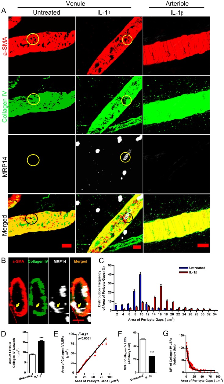
Figure 1
Expansion of pericyte gaps and collagen IV LERs in IL-1u03b2-stimulated venular walls.
( A ) 3D images of vessel walls in mouse cremaster muscles in the presence and absence of IL-1u03b2 revealed pericyte gaps and collagen IV LERs (circled areas) in venules. u03b1-SMA (a marker of venul...
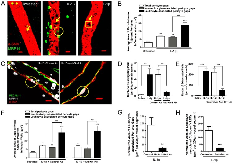
Figure 2
Depletion of circulating PMNs decreases the expansion of pericyte gaps and collagen IV LERs during inflammation.
( A ) 3D images showing the association of pericyte gaps and PMNs in mouse cremaster muscles that were untreated (left) or treated with IL-1u03b2 (middle and right). Arrows indicated pericyte gaps and...
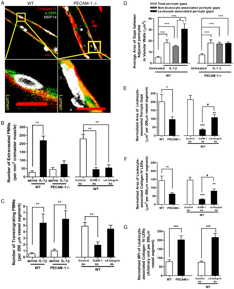
Figure 3
Direct cell-cell contact between transmigrating PMNs and pericytes mediates expansion of pericyte gaps and collagen IV
LERs. ( A ) 2D images of IL-1u03b2-stimulated venules along the longitudinal axis. The areas outlined in the top panels are shown magnified in the bottom panels, and reveal that transmigrating PECAM-1...
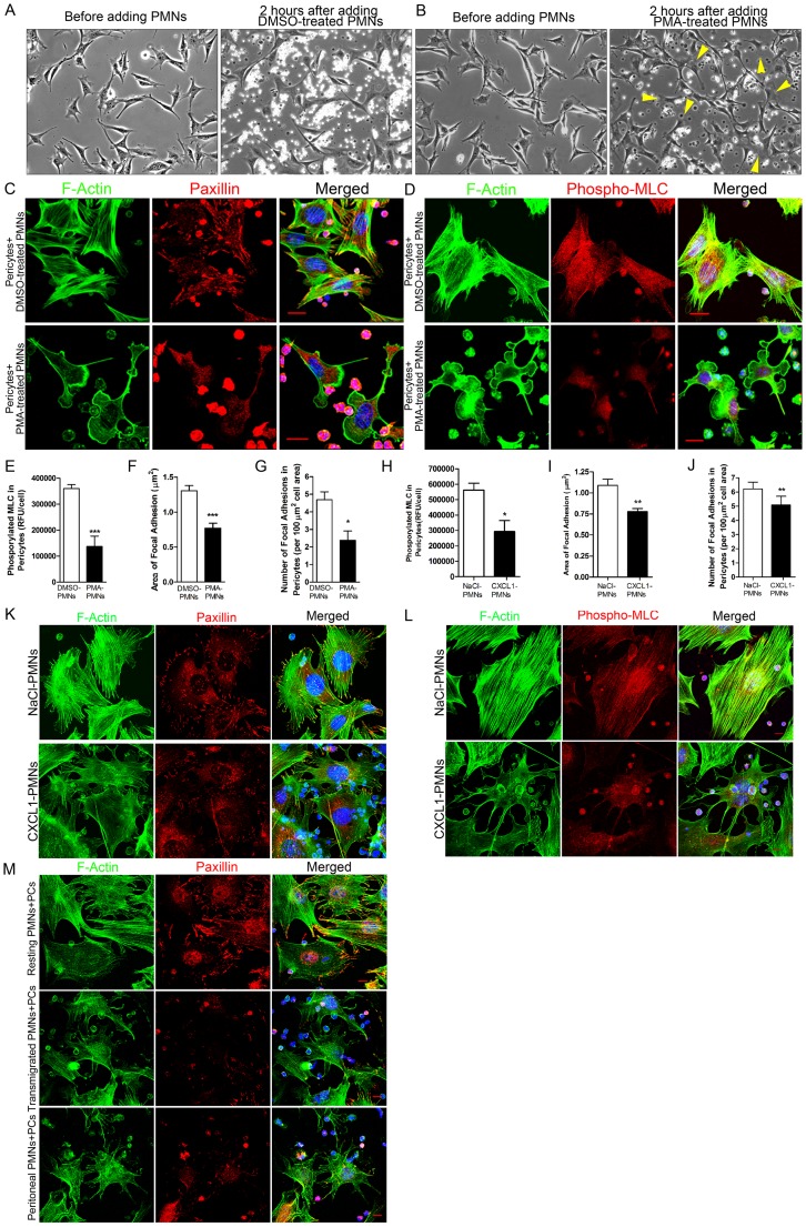
Figure 4
Engagement of activated PMNs induces pericyte relaxation.
( A ) Changes in mouse pericyte morphology before and after adding DMSO-treated PMNs or ( B ) before and after adding PMA-treated PMNs. ( C ) After incubation of DMSO- or PMA-treated PMNs with mouse p...
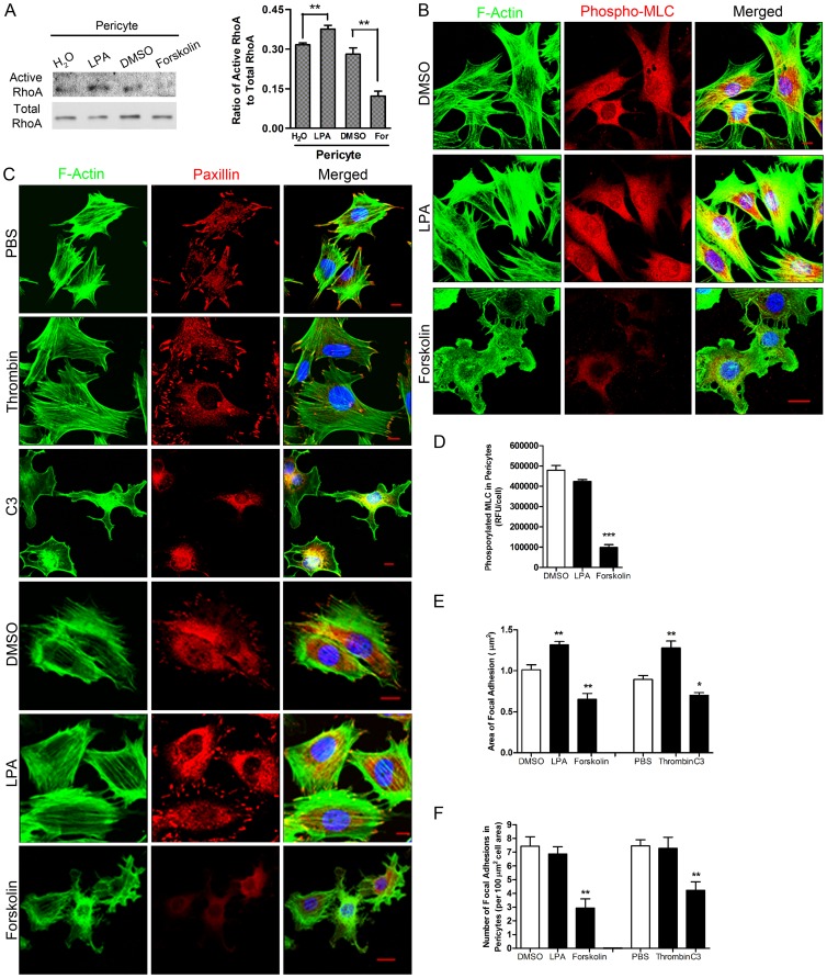
Figure 5
Inhibition of endogenous RhoA leads to a relaxation phenotype in pericytes.
( A ) Mouse primary pericytes were treated with LPA, Forskolin (For) or their vehicles. RhoA activity in cell lysates was detected using a pull-down assay. nu200a=u200a5, t test and **P<0.01 as ind...
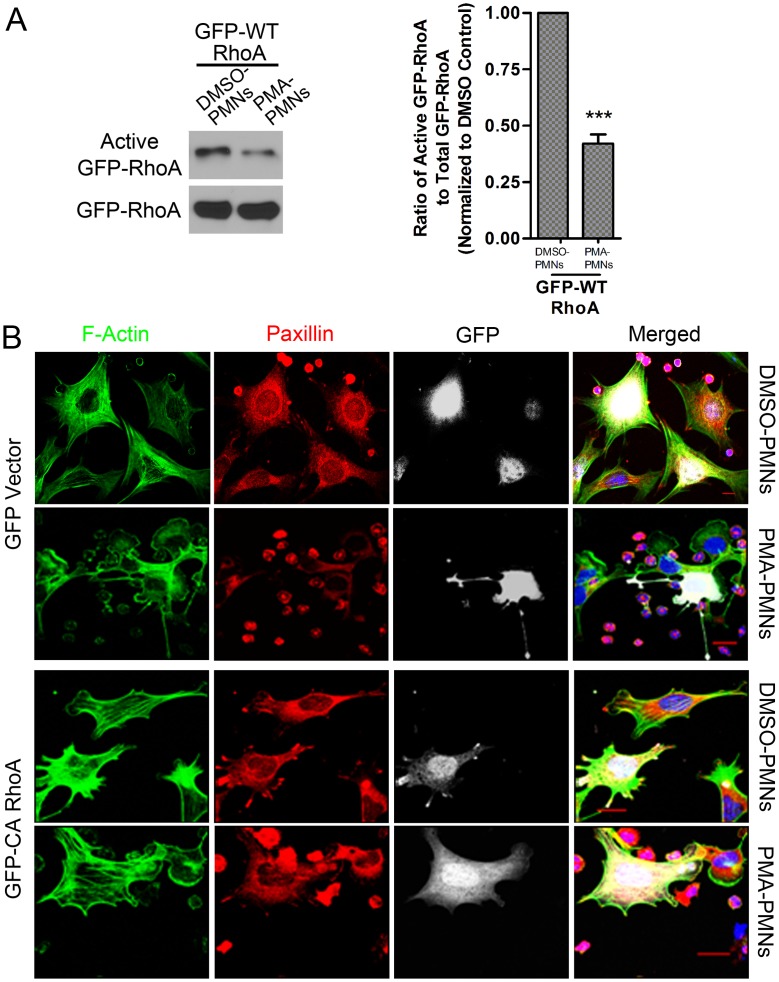
Figure 6
Engagement of activated PMNs inhibits RhoA activity in pericytes.
( A ) Pericytes expressing GFP-tagged WT human RhoA were exposed to DMSO-treated or PMA-treated PMNs. Activity of GFP-tagged RhoA was detected. nu200a=u200a3, t test. ***P<0.001. ( B ) Pericytes ex...
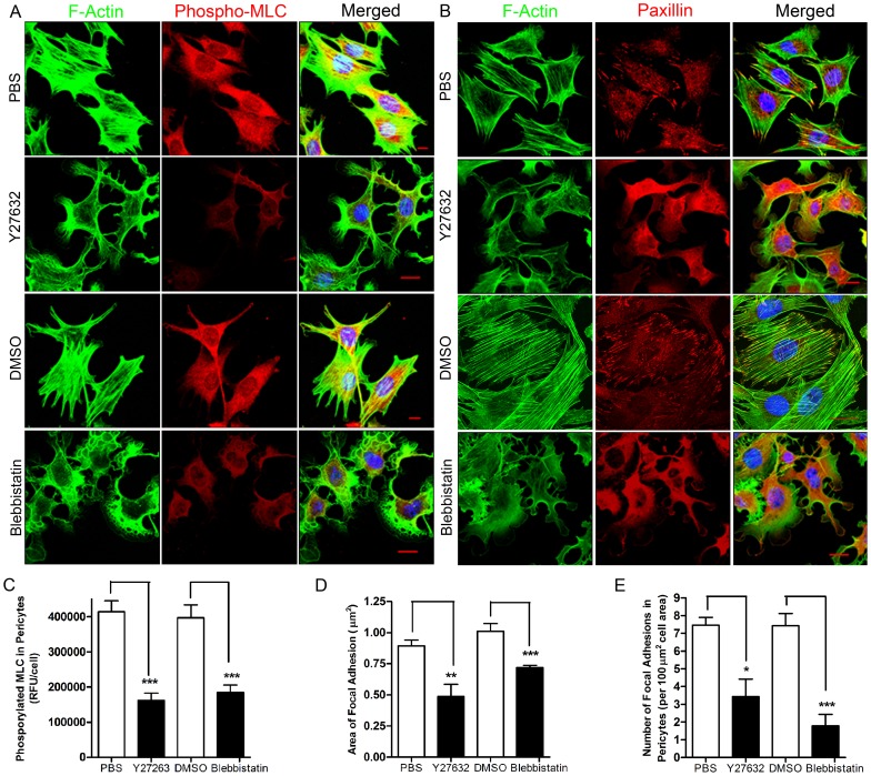
Figure 7
Inhibition of ROCK or myosin ATPase, downstream of RhoA, induces a relaxation phenotype in pericytes.
( A ) Mouse primary pericytes were treated with the inhibitors Y27632 or Blebbistatin. Cells were fixed and stained for F-actin, phospho-MLC and nuclei. ( B ) Mouse primary pericytes were treated with...
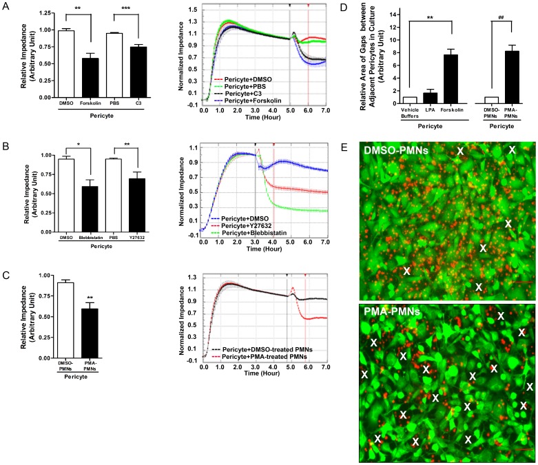
Figure 8
Relaxation of pericytes expands pericyte gaps in a pericyte monolayer.
( A )u2013( C ) On the left, the relative impedance is shown for pericytes responding to different treatments. Representative real-time read-outs are shown on the right. The relative impedance at 1 ho...
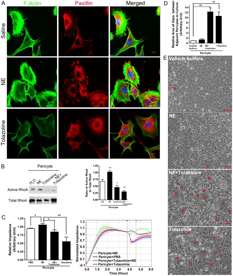
Figure 9
Effect of vasoactive drugs, NE and Tolazoline, on pericyte morphology and permeability of pericyte monolayers.
( A ) Mouse primary pericytes were treated with buffer, NE or Tolazoline. Cells were fixed and stained for F-actin, paxillin and nuclei. Baru200a=u200a10 u00b5m. ( B ) NE increased RhoA activity in pe...
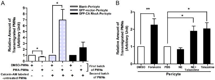
Figure 10
Relaxation of pericytes facilitates PMN transmigration in vitro .
( A ) Pericyte engagement with PMA-activated PMNs enhanced PMN passage across a pericyte monolayer. nu200a=u200a5. Note: Due to strong adhesion to the pericytes and the filters, migration of PMA-activ...
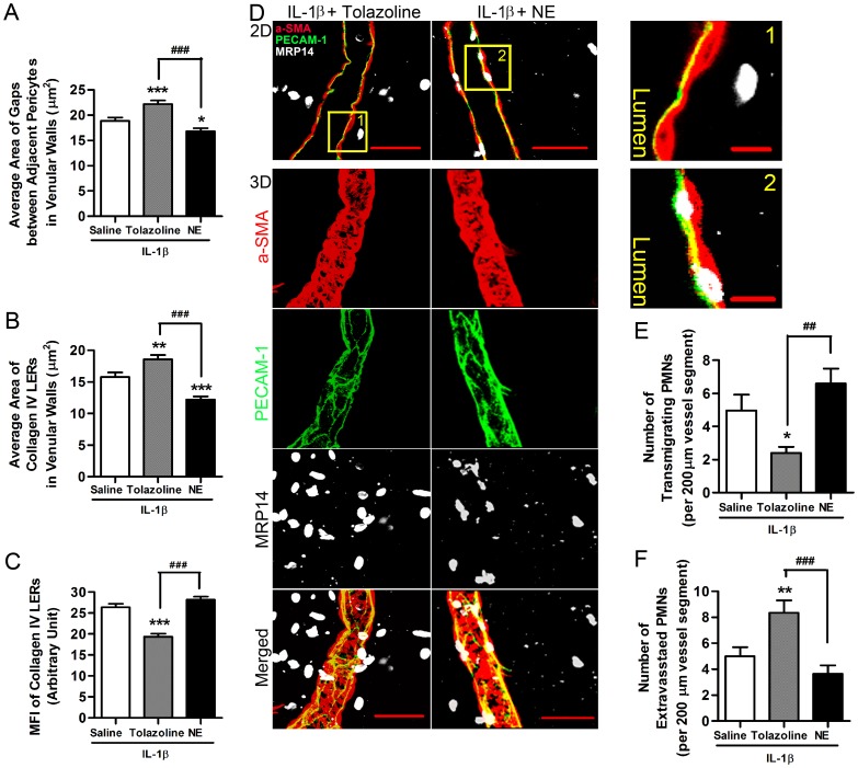
Figure 11
Relaxation of pericytes facilitates PMN transmigration in vivo .
( A ) Change in area of pericyte gaps in IL-1u03b2-stimulated venular walls exposed to Tolazoline or NE. ( B ) Change in area of collagen IV LERs in IL-1u03b2-stimulated venules treated with Tolazolin...
Figure images are served from the NIH/NLM PubMed Central Open Access Subset or Europe PMC; copyright remains with the publishers and authors.
💬 Discussion
0 commentsNo comments yet. Be the first to start a discussion!
Leave a Comment